
I am moderating a class on physiological changes in pregnancy from a critical care perspective . Help me in making sure students dont leave the seminar not understanding the bare minimum ; as an example i want to approach it with a case who is pregnant and in code
Reading File
Now let me search the medical library and web in parallel for the key content areas.
Finding Sources
Finding Sources
Searching the Web
cardiac arrest in pregnancy CPR ACLS 2023 2024 guidelines physiological changes critical care
Good - found the key textbook section. Let me read it and search for more specific physiological systems simultaneously.
Reading File
Finding Sources
Finding Sources
Excellent content coming in. Let me read more of the key textbook sections now.
Reading File
Reading File
Finding Sources
Reading File
Reading File
Searching Images
physiological changes pregnancy cardiovascular respiratory diagram
Reading File
Finding Sources
Searching Images
aortocaval compression pregnant uterus inferior vena cava supine
I now have comprehensive source material. Let me fetch the cardiac output graph from Murray & Nadel and compile the full seminar guide.
Loading Image
I now have all the material I need to build a comprehensive, case-driven seminar guide. Let me compile it.
Seminar Guide: Physiological Changes in Pregnancy - A Critical Care Perspective
Format: Case-based teaching session - a pregnant patient in cardiac arrest anchors every system discussion.
THE ANCHOR CASE
"Mrs. K, 28 years old, G2P1, 34 weeks gestation. She collapses in the antenatal clinic. No pulse, no respirations. The team activates a code."
Use this case as the thread that connects every physiological system. After teaching each system, come back to the case: "So what does this mean for Mrs. K right now?"
SYSTEM 1 - CARDIOVASCULAR
What changed and why it matters in the code
By term, pregnancy produces the following changes (Morgan & Mikhail's Clinical Anesthesiology, 7e):
| Parameter | Change |
|---|---|
| Blood volume | +35% |
| Plasma volume | +55% |
| Cardiac output | +40% |
| Stroke volume | +30% |
| Heart rate | +20% |
| Systolic BP | -5% |
| Diastolic BP | -15% |
| Peripheral vascular resistance | -15% |
| Pulmonary vascular resistance | -30% |
The mechanism: Peripheral vascular resistance drops due to the low-resistance uteroplacental circuit and progesterone-mediated vasodilation. Cardiac output rises early (by 8 weeks) driven first by increased stroke volume, then by rising heart rate as well.
The graph below shows why position matters critically during resuscitation:
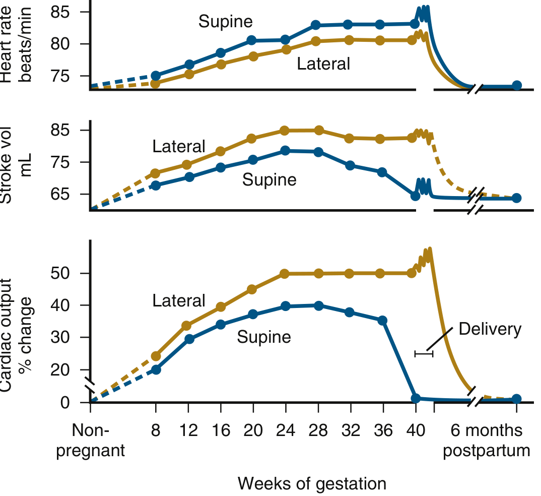
Lateral positioning raises cardiac output by up to 50% above supine at the same gestational age. In the supine pregnant patient, the gravid uterus compresses the IVC, slashing venous return. - Murray & Nadel's Textbook of Respiratory Medicine
The aortocaval compression problem - your first code modification
Ask students: "You start chest compressions. She is supine. What is the gravid uterus doing right now?"
- At ≥20 weeks, the uterus compresses the IVC when the patient is supine, reducing venous return and cardiac output dramatically.
- Standard CPR in a supine pregnant patient is partially futile if aortocaval compression is not relieved.
Code action - manual left uterine displacement (LUD):
- One dedicated team member places hands on the right side of the uterus and displaces it leftward.
- This is preferred over tilting the patient (tilting impairs compression quality, pad placement, and airway management, and it is unknown how tilt changes intrathoracic heart position).
- Chest compressions are placed on the lower half of the sternum (same as non-pregnant), not higher. The uterus does not displace the heart cranially enough to change hand placement.
Postpartum surge: At delivery, cardiac output spikes a further 60-80% due to autotransfusion from uterine contraction, relief of IVC compression, and catecholamine release. This is why resuscitative hysterotomy can rescue the mother - it removes the obstacle to venous return and dramatically improves CPR effectiveness.
SYSTEM 2 - RESPIRATORY
What changed and why it matters in the code
- O2 consumption: +20-50% (fetal demands + increased maternal metabolic rate)
- Minute ventilation: +50% (driven by tidal volume +40%, RR +15%)
- FRC: -20% (diaphragm pushed up 4 cm by the gravid uterus)
- PaCO2: 28-32 mmHg (normal in pregnancy - do not overcorrect)
- HCO3: -15% (compensatory metabolic acidosis - base ~20 mEq/L is normal)
- PaO2: +10%
(Morgan & Mikhail's Clinical Anesthesiology, 7e)
The critical interaction: Decreased FRC + markedly increased O2 consumption = rapid oxygen desaturation during apnea. A pregnant woman at term will desaturate from 100% to 95% in roughly 3 minutes - compared to ~8 minutes in a non-pregnant person. Pre-oxygenation time is shorter and apnea is less tolerated.
Ask students: "You need to intubate Mrs. K. What three airway problems are working against you?"
- Difficult intubation: Upper airway mucosal engorgement and friability from estrogen-driven hyperemia. Mallampati scores are higher. Use a video laryngoscope as first-line. Use a smaller ETT (6.0-6.5 mm).
- Aspiration risk: Gastric emptying is delayed, lower esophageal sphincter tone is reduced, and intra-abdominal pressure is increased by the gravid uterus. She is never "nil by mouth" enough. Rapid sequence induction (RSI) is mandatory.
- Fast desaturation: She has minimal apnea tolerance. Pre-oxygenate aggressively. You have less time than you think.
(Tintinalli's Emergency Medicine; Miller's Anesthesia, 10e)
Ventilator targets in a pregnant patient:
- Target PaCO2 of 28-32 mmHg (her normal - not 40)
- If you ventilate to PaCO2 of 40, you are relatively hypercapnic for her; this impairs fetal CO2 elimination via the placenta.
SYSTEM 3 - HEMATOLOGICAL
What changed and why it matters in the code
- Hemoglobin: -20% (dilutional anemia - plasma volume rises more than RBC mass)
- Platelets: -10% (dilutional thrombocytopenia - a platelet count of 100-120k can still be normal)
- Clotting factors II, VII, VIII, IX, X, XII, fibrinogen, vWF: +30 to +250% - pregnancy is a profoundly hypercoagulable state
- Protein S and antithrombin III: decrease
- Venous thrombosis risk: 5-6 times that of non-pregnant women
(Harrison's Principles of Internal Medicine 22E; Rosen's Emergency Medicine)
Code implication: "Normal" hemoglobin values look abnormal. Hgb of 10-11 g/dL can be normal at term. Do not reflexively transfuse for this alone. Conversely, a Hgb of 9 in a pregnant woman with a true baseline of 12 represents significant hemorrhage.
Interpreting labs in the code context:
- A normal fibrinogen in your lab range (200-400) can mask DIC in pregnancy, where the baseline is 400-600 mg/dL. A "normal" level may actually represent a fall.
- Markedly elevated clotting factors mean pregnancy-related causes of arrest (amniotic fluid embolism, massive PE) must always be on the differential.
SYSTEM 4 - RENAL & METABOLIC
- GFR: +50% (renal blood flow increases substantially)
- Serum creatinine drops to ~0.4-0.5 mg/dL at term (normal non-pregnant ~0.8-1.0)
- BUN also falls
Code implication: A creatinine of 0.8 mg/dL in a pregnant patient is equivalent to 1.6 in a non-pregnant patient. Do not dismiss it as normal. Dose renally-cleared drugs accordingly.
SYSTEM 5 - NEUROLOGICAL / PHARMACOLOGICAL
- MAC (minimum alveolar concentration) for volatile anesthetics: -40% by term
- Sensitivity to local anesthetics markedly increased
- Epidural venous plexus is engorged (IVC compression pushes blood into epidural veins), reducing volume of epidural space and CSF volume, causing exaggerated cephalad spread
Drug dosing in the code:
- ACLS medications are given at standard doses - do not withhold epinephrine, amiodarone, or other drugs because of pregnancy. The life of the mother takes priority.
- IV access must be established above the diaphragm (upper extremity or central venous). IVC compression means anything given below the diaphragm may not reach the central circulation effectively.
SYSTEM 6 - GASTROINTESTINAL
- Delayed gastric emptying
- Reduced lower esophageal sphincter tone
- Elevated intragastric pressure from uterus
All pregnant patients ≥16 weeks are considered "full stomach" for airway management purposes. There is no safe fasting period in late pregnancy.
THE CODE ALGORITHM - Integrating all systems
Ask students to build this together as you go through the systems:
Immediately upon recognizing arrest:
- Call for help - activate OB and neonatal teams immediately
- Start high-quality CPR - same standard compressions (lower half of sternum, 2 inches depth, 100-120/min)
- Apply manual left uterine displacement - assign one dedicated person
- Do not tilt the patient - this impairs compression quality
Airway (expect difficult):
- Pre-oxygenate maximally - she will desaturate faster than you expect
- Video laryngoscope first line
- RSI mandatory (aspiration risk)
- Smaller ETT (6.0-6.5 mm)
- Ventilate to PaCO2 28-32 mmHg, not 40
Circulation:
- IV access above the diaphragm
- Standard ACLS drugs at standard doses - do not withhold
- Defibrillate at standard energy for shockable rhythms - remove fetal monitors, no dose adjustment needed
The "4-minute rule" - Resuscitative Hysterotomy:
- If no ROSC within 4 minutes of arrest, begin resuscitative hysterotomy
- Target delivery by 5 minutes from collapse
- Gestational age ≥23-24 weeks (viable fetus)
- This is no longer called "perimortem cesarean" - the preferred term is resuscitative hysterotomy because the procedure benefits the mother as much as the fetus (relieves aortocaval compression, dramatically improves CPR effectiveness)
- Do NOT wait for ROSC to perform it
- Do NOT delay CPR to transport to OR - this can be done at bedside
(Tintinalli's Emergency Medicine; NAEMSP guidelines; AHA 2025 CPR Highlights)
CAUSES OF ARREST IN PREGNANCY - The Obstetric H's
Help students distinguish causes unique to pregnancy. A useful mnemonic extending the ACLS H's and T's:
| Obstetric cause | Key feature |
|---|---|
| Hemorrhage (obstetric) | Placenta previa, abruption, uterine rupture |
| Hypertensive crisis / eclampsia | Seizure + arrest, MgSO4 toxicity |
| Heart disease | Now the leading cause of maternal death in high-income countries |
| Hypoxia (airway failure) | Difficult intubation + aspiration |
| Amniotic fluid embolism | Sudden cardiovascular collapse + DIC |
| PE (massive) | Pre-existing hypercoagulability |
| Sepsis | Chorioamnionitis, pyelonephritis |
| Drug / MgSO4 toxicity | Hypermagnesemia causes cardiac arrest |
KEY TEACHING TAKE-AWAYS
Frame these for your students as the "things you cannot leave this room not knowing":
-
Two patients, one priority. The best fetal resuscitation is maternal resuscitation. Never withhold a maternal intervention for fear of fetal harm.
-
The uterus is your enemy during CPR. Manual left uterine displacement is non-negotiable at ≥20 weeks. Without it, you are doing partial CPR.
-
Her airway will be harder than you expect. Hyperemic mucosa, weight gain, aspiration risk, and fast desaturation all conspire against you. Prepare the video laryngoscope before you need it.
-
Her normal labs are not your normal labs. Hgb 10 can be fine. Creatinine 0.8 may be AKI. "Normal" fibrinogen may be coagulopathy.
-
4 minutes. 5 minutes. Resuscitative hysterotomy. Know this number. Burn it in. It saves the mother AND the baby.
-
Do not withhold ACLS medications. Give epinephrine. Give the shock. Give the drug. Pregnancy is not a contraindication to life-saving interventions.
SEMINAR FLOW SUGGESTION (60-90 min)
| Time | Activity |
|---|---|
| 0-5 min | Present the anchor case. Ask: "What would you do first? What's different?" |
| 5-30 min | Walk through each system (5 min each) - always return to case implications |
| 30-45 min | Build the modified ACLS algorithm together on the board |
| 45-55 min | Small groups: "You're 4 minutes in. No ROSC. What do you do? Who does what?" |
| 55-65 min | Debrief causes of arrest in pregnancy |
| 65-75 min | MCQ rapid-fire quiz (5-6 questions from take-aways) |
| 75-90 min | Open Q&A |
Sources: Morgan & Mikhail's Clinical Anesthesiology 7e; Murray & Nadel's Textbook of Respiratory Medicine; Tintinalli's Emergency Medicine; Harrison's Principles of Internal Medicine 22E; Rosen's Emergency Medicine; NAEMSP Maternal Cardiac Arrest guidelines; AHA 2025 CPR Highlights
This is a shared conversation. Sign in to Orris to start your own chat.