
A 25 yr female 5weeks 5 days what are the development that could have happened
- The embryo is roughly 5-9 mm in size (crown-rump length)
- The heart has started beating (around 6 weeks)
- Neural tube is forming
- Limb buds are just beginning to appear
embryo development 5 weeks 6 weeks fetal development diagram
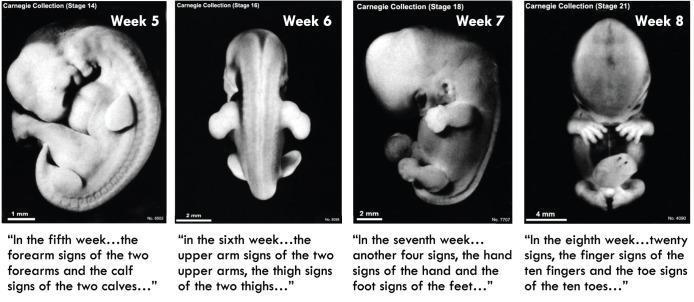
A comparative educational layout displaying four panels of human embryonic development from the Carnegie Collection, covering weeks 5 through 8. Week 5 (Stage 14) shows a C-shaped embryo with prominent branchial arches and primary limb buds for the forearms and calves. Week 6 (Stage 16), shown from a dorsal view, highlights the elongation of the body and more defined upper arm and thigh protrusions. Week 7 (Stage 18) demonstrates further straightening of the spinal axis and the emergence of distinct hand and foot plates. Week 8 (Stage 21) depicts a more upright posture where the head is proportionally smaller relative to the trunk, and digital rays have separated into ten fingers and ten toes. This series illustrates morphological milestones in embryogenesis, specifically the progression of limb morphogenesis and the transition from embryonic curvature to a more humanoid posture. Each panel includes a scale bar (ranging from 1mm to 4mm) and historical text references comparing these biological stages to ancient descriptions of gestation.
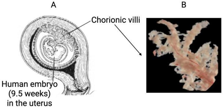
The image consists of two panels illustrating human chorionic villi. Panel A is an anatomical diagram in sagittal cross-section showing a pregnant uterus with a human embryo at approximately 9.5 weeks of gestation. The diagram highlights the spatial relationship of the gestational sac, the developing embryo, and the chorionic villi, which are concentrated at the embryonic pole (chorion frondosum) where the placenta is forming. Panel B is a high-resolution macroscopic clinical photograph of an isolated chorionic villus specimen against a dark background. The specimen exhibits a characteristic arborizing or branching morphology, resembling frond-like or tree-like structures. It appears pale cream to off-white in color, with visible fine reddish-pink vascular traces running through the core of the larger branches. This comparison demonstrates the in situ anatomical orientation of the villi versus their complex, branched physical structure, which is optimized for surface area expansion to facilitate maternal-fetal nutrient and gas exchange. The content is highly relevant to embryology, obstetrics, and prenatal diagnostic procedures such as chorionic villus sampling (CVS).
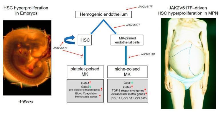
A multi-panel medical illustration comparing normal embryonic development with adult myeloproliferative neoplasms (MPN). On the left, a clinical photograph shows a human embryo at 5 weeks post-conceptus, exhibiting a characteristic C-shape and a prominent reddish liver rudiment, labeled as HSC hyperproliferation in embryos. In the center, a pathophysiology flowchart depicts the differentiation of hemogenic endothelium into hematopoietic stem cells (HSC) and megakaryocyte (MK)-primed endothelial cells. It illustrates how transcription factor balances (Gata1 vs. Gata2) determine the maturation of platelet-poised megakaryocytes versus niche-poised megakaryocytes. The diagram highlights the role of the JAK2V617F mutation in driving self-renewal and lineage-specific hyperproliferation. On the right, a clinical photograph of a patient with MPN demonstrates massive abdominal distension. Blue and red ink markings on the skin outline significant hepatosplenomegaly, representing massive extramedullary hematopoiesis. The visual comparison links embryonic developmental processes to pathological states in adult hematological malignancies, specifically illustrating how mutations in the hemogenic niche drive disease progression.
Embryonic Development at 5 Weeks 5 Days of Pregnancy
Overall Picture
System-by-System Developments
1. Cardiovascular System - Already Beating!
- The heart is the first organ to function - a primitive tubular heart has already formed and is pumping blood by around 22-23 days post-fertilization
- By 5 weeks 5 days, the heart is a 4-chamber structure in early development, beating at ~100-160 bpm
- This is the period when the heart produces a large ventral prominence visible on the embryo's surface
- Paired endocardial heart tubes have fused into a single beating heart; blood circulates through a primordial cardiovascular system that connects to the umbilical vesicle and chorion
2. Nervous System - Neural Tube Formation
- The neural tube (precursor of the entire brain and spinal cord) is forming or has just closed
- The rostral neuropore (head end) closes around day 24-26; the caudal neuropore closes around day 26-28
- The brain is dividing into three primary vesicles: prosencephalon (forebrain), mesencephalon (midbrain), rhombencephalon (hindbrain)
- The otic vesicle (primordium of the inner ear) is visible
- Rapid brain growth is the main reason the head is disproportionately large
3. Head & Face - Pharyngeal Arches
- The first pharyngeal (mandibular) arch is well developed - it will give rise to the mandible (lower jaw) and a maxillary prominence that contributes to the upper jaw
- The second pharyngeal arch is growing rapidly and beginning to overgrow the 3rd and 4th arches, forming the cervical sinus
- The face is pressing against the heart prominence due to rapid brain/face growth
- Facial prominences (frontonasal, maxillary, mandibular) are establishing the scaffolding for facial features
4. Limb Development - Buds Appearing
- Upper limb buds appear first, around Carnegie Stage 12-13 (approximately day 26-28 post-fertilization)
- Lower limb buds appear a few days later
- At this exact stage, the limb buds are small paddle-like elevations with no differentiation yet - no elbows, hands, or digits
- Regional differentiation (elbow, hand plate) begins in the 6th week (a few days ahead)
5. Gastrointestinal System
- The foregut, midgut, and hindgut are established from the primitive gut tube
- Intestinal loops are forming but will not herniate into the umbilical cord until Week 6
- The liver and pancreatic buds are emerging from the foregut endoderm
- The stomach is forming as a dilation of the foregut
6. Urinary System
- The mesonephros (interim kidney) is developing; mesonephric ridges are visible as surface elevations
- The mesonephros functions temporarily; the permanent kidney (metanephros) develops later around Week 5 post-fertilization
7. Somites (Body Segments)
- Somites are paired segmental blocks of mesoderm flanking the neural tube
- By 5 weeks 5 days LMP (~day 28-32 post-fertilization), approximately 28-35 somite pairs have formed
- Somites give rise to the vertebral column, ribs, skeletal muscles, and dermis of the back
8. Eyes
- The optic vesicles have evaginated from the forebrain
- The lens placode is forming where the optic vesicle contacts the surface ectoderm
- Retinal pigmentation is not yet visible (pigment becomes obvious in Week 6)
9. Placenta and Membranes
- Tertiary chorionic villi are established - they now contain fetal capillaries and are actively exchanging nutrients and gases with maternal blood
- The amniotic cavity surrounds the embryo
- The umbilical cord is forming, connecting the embryo to the chorion
What Can Be Seen on Ultrasound at 5 Weeks 5 Days
| Finding | Expected |
|---|---|
| Gestational sac | Clearly visible (>10 mm MSD) |
| Yolk sac | Visible (~3-5 mm) |
| Fetal pole | Just visible to small (1-5 mm CRL) |
| Cardiac activity | May just be detectable (flickering ~100 bpm) |
| Crown-rump length | ~3-9 mm |
What Has NOT Yet Developed at This Stage
- No recognizable limb segments (no hands, fingers, feet, toes)
- No external ears (auricular hillocks appear in Week 6)
- Eyes not yet pigmented / not prominent
- Intestines not yet herniated
- No eyelids, nose tip, or external genitalia
Clinical Relevance
- The Developing Human: Clinically Oriented Embryology, Moore & Persaud - Chapter 5 (Fourth to Eighth Weeks)
What to do for tvs usg
- Bladder should be empty (opposite of transabdominal USG)
- A lubricated probe is gently inserted into the vagina
- At 5w5d, you're looking for gestational sac, yolk sac, fetal pole, and cardiac activity
transvaginal ultrasound early pregnancy gestational sac yolk sac fetal pole
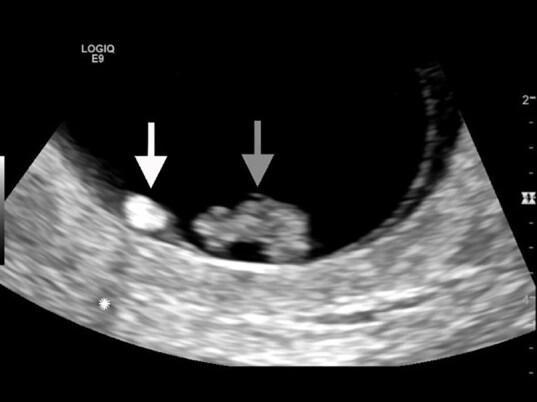
This diagnostic image is a close-up transvaginal sonogram of an intrauterine pregnancy. The grayscale ultrasound displays a gestational sac containing key embryonic structures. A fetal pole, indicated by a gray arrow, appears as an irregular mass with mixed echogenicity. Adjacent to it, a small, well-defined, and highly echogenic circular structure (white arrow) represents the yolk sac. Notable posterior acoustic shadowing, marked by an asterisk, is visible as a hypoechoic region distal to the fetal pole. This educational visual demonstrates the spatial relationship and echogenicity characteristics of early pregnancy structures, often used in obstetrics and emergency medicine to evaluate symptoms such as lower abdominal cramping and vaginal spotting. The image serves as a reference for point-of-care pelvic ultrasound interpretation, highlighting normal and abnormal sonographic markers during the first trimester.
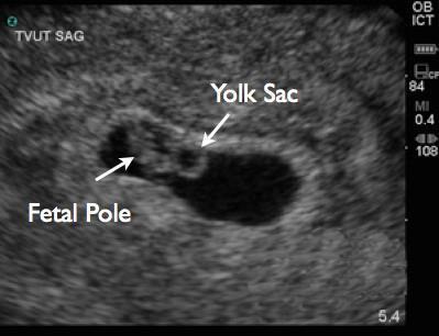
This diagnostic image is a grayscale transvaginal ultrasound (TVUS) in the sagittal plane, used for early pregnancy evaluation. It depicts a longitudinal view of a gestational sac, characterized as an anechoic (black) fluid-filled cavity within the echogenic uterine decidua. Within the gestational sac, two primary embryological structures are identified with white arrows and labels: the 'Fetal Pole' and the 'Yolk Sac'. The fetal pole appears as a small, elongated echogenic structure, representing the developing embryo. Adjacent to it, the yolk sac is visible as a distinct, circular, hyperechoic ring with an anechoic center. Technical overlays indicate the imaging modality ('TVUT SAG'), transducer type ('ICT' for intracavitary), and machine parameters (MI 0.4, PRF 84). The image provides clinical evidence of a viable intrauterine pregnancy, demonstrating the presence and spatial relationship of the yolk sac and embryo, which are critical for dating and assessing early developmental milestones.
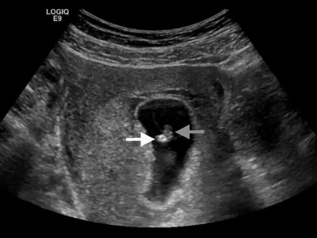
This diagnostic image is a transvaginal sonogram representing early pregnancy within the obstetric domain. The scan demonstrates a well-defined, anechoic (black) gestational sac located within the uterus. Centrally located within this sac are two distinct structures: a fetal pole, indicated by a gray arrow, appearing as a small, intermediate-echoic structure, and an adjacent echogenic yolk sac, indicated by a white arrow. The yolk sac displays higher echogenicity (hyperechoic/brighter) compared to the fetal pole. The surrounding myometrium and pelvic tissues exhibit a heterogeneous, granular echotexture typical of soft tissue. This visual serves as an educational example of early intrauterine pregnancy (IUP) and identifies the key sonographic landmarks required for dating and viability assessment. Clinical context suggests this image was captured during a point-of-care ultrasound (POCUS) evaluation in a patient presenting with symptoms of a potential threatened abortion, where the presence of these structures confirms an IUP but requires further assessment for cardiac activity.
Transvaginal Ultrasound (TVS USG) at 5 Weeks 5 Days - Complete Guide
Patient Preparation (What She Should Do Before Coming)
| Instruction | Detail |
|---|---|
| Bladder | Empty the bladder - this is the most important difference from transabdominal USG. A full bladder pushes the uterus away and worsens the image |
| Clothing | Wear comfortable, easily removable clothing from the waist down |
| Consent | Verbal consent should be taken explaining it is an internal procedure |
| Timing | No special time restriction; can be done any time |
| Diet | No fasting required |
Note: TVS gives far better resolution than transabdominal USG at this early gestational age, which is why it is preferred in the first trimester.
Procedure - What Happens During the Scan
- Patient lies in dorsal lithotomy position (on her back, knees bent, legs apart)
- A long, narrow probe (endovaginal transducer) is covered with a condom/probe cover and lubricated with gel
- Probe is gently inserted into the vagina - no pain if done correctly; slight pressure is normal
- The probe is angled to visualize the uterus, ovaries, and adnexa
- Takes about 5-10 minutes
What to Look For at 5 Weeks 5 Days
| Structure | Expected at 5w5d | Notes |
|---|---|---|
| Gestational sac | Present, well-defined, anechoic | Should be in uterine cavity |
| Yolk sac | Present (3-5 mm) | Confirms IUP; round, echogenic ring |
| Fetal pole | May be visible (~3-5 mm CRL) | A small echogenic structure next to yolk sac |
| Cardiac activity | May just be detectable | "Flickering" movement at ~100-120 bpm; most reliably seen from 6 weeks |
| Double decidual sign | May be seen | Confirms intrauterine location before yolk sac is visible |
What the Report Should Include
- Location of gestational sac (intrauterine vs. suspicious for ectopic)
- Mean Sac Diameter (MSD) - average of 3 diameters of the sac
- Crown-Rump Length (CRL) - if fetal pole visible; gives gestational age within 5 days
- Yolk sac - present/absent, size
- Cardiac activity - present/absent; rate in bpm
- Uterus - shape, any fibroids, anomalies
- Adnexa - ovaries, any mass or free fluid (rules out ectopic)
Why TVS is Done at This Stage (Indications)
- Confirm intrauterine pregnancy - rule out ectopic
- Confirm viability - cardiac activity
- Accurate dating - CRL is the most accurate dating method in the first trimester
- Vaginal bleeding / threatened abortion - assess viability
- Hyperemesis - rule out molar pregnancy
- Confirm number of embryos - twins?
Warning Signs to Report Immediately
- No gestational sac in uterus with positive urine pregnancy test - suspect ectopic
- Sac >25 mm with no yolk sac, or sac >20 mm with no fetal pole - suggests missed/blighted ovum
- No cardiac activity with CRL >7 mm - suggests embryonic demise
- Free fluid in pouch of Douglas - suspect ectopic with hemoperitoneum
- Adnexal mass alongside an empty uterus - high suspicion for ectopic
Safety
- Tintinalli's Emergency Medicine, Table 98-7
- Roberts and Hedges' Clinical Procedures in Emergency Medicine