
Anatomy of skeletal system
Reading File
The skeletal system forms the framework of the body, with 206 bones in adults organized into axial (skull, vertebral column, thoracic cage) and appendicular (limbs and girdles) divisions. Bones are classified by shape (long, short, flat, irregular, sesamoid) and serve functions including support, protection, movement, mineral storage, and hematopoiesis.
Searching sources...
Finding Sources
Finding Sources
Finding Sources
Finding Sources
Good - there are several relevant anatomy books. Let me search for skeletal system content in Gray's Anatomy and the THIEME Atlas in parallel.
Finding Sources
Finding Sources
Reading File
Finding Sources
Reading File
Finding Sources
Reading File
I now have comprehensive content from Gray's Anatomy for Students. Let me also fetch one of the relevant images to include in the answer.
Loading Image
I now have rich content from Gray's Anatomy for Students. Here is the full, well-structured answer:
Anatomy of the Skeletal System
Source: Gray's Anatomy for Students
1. What is Bone?
Bone is a calcified, living connective tissue that forms the majority of the skeleton. It consists of an intercellular calcified matrix containing collagen fibers, along with several types of cells embedded within that matrix.
Functions of Bone
- Structural support for the body
- Protection of vital organs
- Reservoir of calcium and phosphorus
- Levers on which muscles act to produce movement
- Containers for blood-producing (hematopoietic) cells
2. Types of Bone Tissue
There are two structural types of bone:
| Type | Description | Location |
|---|---|---|
| Compact (cortical) bone | Dense bone; forms the outer shell of all bones | Surrounds spongy bone |
| Spongy (trabecular/cancellous) bone | Spicules of bone enclosing cavities filled with blood-forming marrow | Interior of bones |
3. Classification of Bones by Shape
| Shape | Description | Examples |
|---|---|---|
| Long bones | Tubular structure | Humerus, femur |
| Short bones | Cuboidal shape | Carpal bones (wrist), tarsal bones (ankle) |
| Flat bones | Two compact plates separated by spongy bone | Skull bones |
| Irregular bones | Variable shapes | Facial bones, vertebrae |
| Sesamoid bones | Round/oval; develop within tendons | Patella, thumb sesamoids |
Accessory and Sesamoid Bones
Extra bones not usually part of the normal skeleton can exist as normal variants. They are commonly found in the wrist, hands, ankles, and feet. Importantly, these should not be mistaken for fractures on imaging.
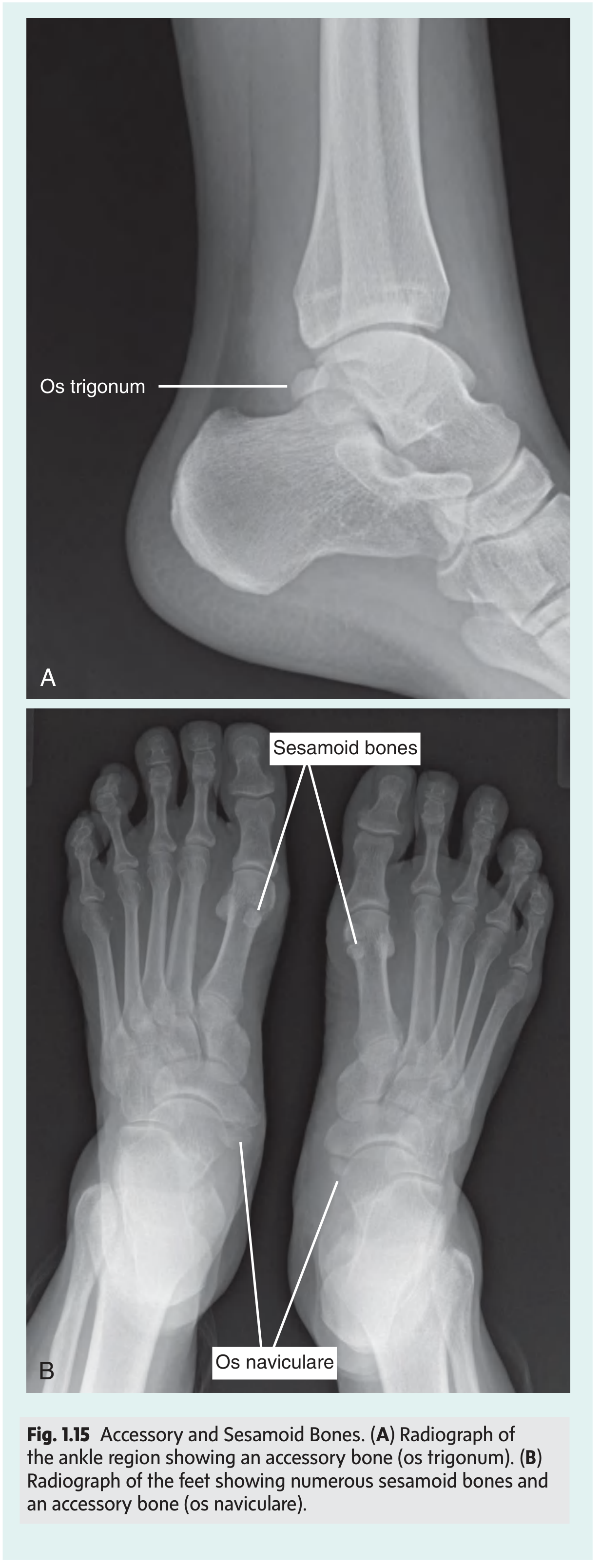
Fig. 1.15 - Accessory and Sesamoid Bones. (A) Os trigonum at the ankle. (B) Sesamoid bones and os naviculare in the feet.
4. Bone Vasculature and Innervation
- A nutrient artery (usually one per bone) from an adjacent vessel enters the internal cavity, supplying the marrow, spongy bone, and inner compact bone layers.
- The periosteum - a fibrous connective tissue membrane covering all external bone surfaces (except at articular cartilage areas) - receives blood vessels supplying the outer compact bone layers.
- A bone stripped of its periosteum will not survive.
- Nerves accompany blood vessels; most fibers entering the marrow cavity are vasomotor (regulate blood flow). The periosteum itself is densely supplied with sensory fibers and is very sensitive to injury.
5. Bone Marrow
Two types of marrow exist:
| Type | Contents | Location with Age |
|---|---|---|
| Red marrow (myeloid tissue) | Produces red blood cells, platelets, most white blood cells | Widespread at birth; progressively converts to yellow marrow |
| Yellow marrow | Dominated by fat globules; produces few white cells | Replaces red marrow in medullary cavities of long/flat bones with aging |
Stem Cells in Marrow
- Hematopoietic stem cells - give rise to blood cells and platelets
- Mesenchymal stem cells - differentiate into bone, cartilage, and muscle
6. Ossification (Bone Development)
All bones develop from mesenchyme via one of two pathways:
- Intramembranous ossification - mesenchymal models of bones undergo direct ossification (e.g., flat bones of the skull)
- Endochondral ossification - cartilaginous models first form from mesenchyme, then undergo ossification (e.g., long bones)
Skeletal Age
- Skeletal maturity typically occurs between ages 20-25 years.
- Bone age can be assessed by radiographing the non-dominant (left) hand and comparing to standard reference radiographs.
- Conditions such as malnutrition and hypothyroidism can delay skeletal maturity.
7. Joints (Articulations)
Joints connect bones and are classified into three types by the tissue that binds them and the degree of movement permitted:
| Joint Type | Binding Tissue | Movement | Examples |
|---|---|---|---|
| Fibrous joints | Dense fibrous connective tissue | Little to no movement | Sutures of skull, syndesmoses |
| Cartilaginous joints | Cartilage (hyaline or fibrocartilage) | Limited movement | Pubic symphysis, intervertebral discs |
| Synovial joints | Synovial membrane + joint capsule | Free movement | Hip, knee, shoulder |
Synovial Joints - Key Features
- Articular cartilage covers the bone ends
- A joint capsule encloses the joint
- The synovial membrane lines the inner capsule and secretes synovial fluid for lubrication
- Ligaments reinforce the capsule and limit excessive motion
- Bursae (fluid-filled sacs) reduce friction between tendons/muscles and bone near joints
8. Axial vs. Appendicular Skeleton
The adult skeleton (206 bones) is divided into two major divisions:
Axial Skeleton (~80 bones)
The central axis of the body:
- Skull - cranium (8 bones) + facial bones (14 bones)
- Hyoid bone - U-shaped bone supporting the tongue
- Vertebral column - 7 cervical + 12 thoracic + 5 lumbar + sacrum + coccyx
- Thoracic cage - sternum + 12 pairs of ribs
Appendicular Skeleton (~126 bones)
The limbs and their girdles:
- Pectoral (shoulder) girdle - clavicle + scapula
- Upper limbs - humerus, radius, ulna, carpals (8), metacarpals (5), phalanges (14) per side
- Pelvic girdle - two hip bones (ilium, ischium, pubis fused) + sacrum
- Lower limbs - femur, patella, tibia, fibula, tarsals (7), metatarsals (5), phalanges (14) per side
9. Clinical Correlates
Bone Fractures
- Fractures from abnormal load on normal bone OR normal load on weakened bone (e.g., osteoporosis).
- Greenstick fractures - common in children; partial cortical disruption, like breaking a young tree branch.
- Healing involves hematoma formation, vascular ingrowth, collagen matrix formation, and calcium hydroxyapatite deposition by osteoblasts, forming a callus.
Avascular Necrosis
Cellular death of bone from loss of blood supply. A classic site is the femoral head following femoral neck fracture in elderly patients - loss of cortical/medullary blood flow renders the femoral head bloodless, leading to necrosis and collapse. Management often requires femoral head prosthesis replacement.
Epiphyseal (Growth Plate) Fractures
Growth plates are vulnerable to injury in actively growing children (ages 7-10 years and puberty). Injury can cause asymmetrical growth if the growth plate is compressed. All growth plate fractures require prompt reduction.
- Gray's Anatomy for Students, Block 1 (Sections on Bone, Joints, and Fractures)
This is a shared conversation. Sign in to Orris to start your own chat.