
Knee jerk ankle jerk planker jerk root value according to macloid
- Knee jerk (patellar reflex): L3, L4
- Ankle jerk (Achilles reflex): S1, S2
- Plantar reflex: L5, S1, S2
deep tendon reflexes root values table knee jerk ankle jerk
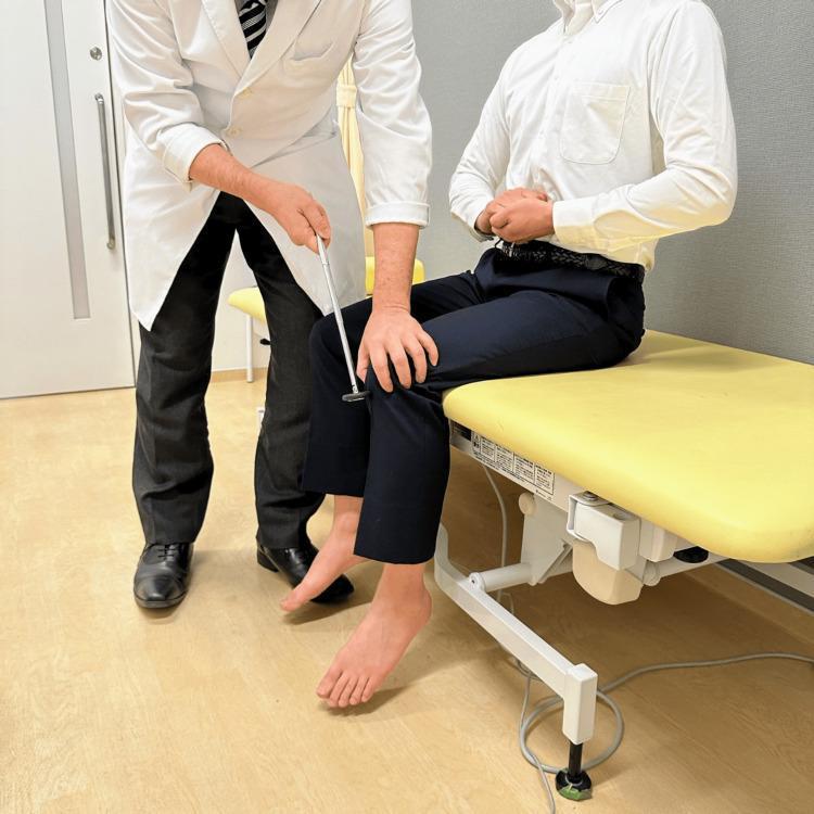
This clinical photograph demonstrates the proper technique for eliciting the patellar tendon reflex (knee jerk) in a sitting position, incorporating the Jendrassik maneuver. The patient is seated on an examination table with legs hanging freely and knees flexed at a 90-degree angle, ensuring the feet do not touch the floor to allow for unobstructed lower leg extension. Simultaneously, the patient performs the Jendrassik maneuver by interlocking their fingers and attempting to pull their hands apart, a reinforcement technique designed to facilitate deep tendon reflexes (DTR) by reducing conscious inhibition and increasing upper motor neuron activation. The examiner, standing to the patient's side, is seen using a neurological reflex hammer to strike the patellar tendon just below the patella. This procedure tests the L3-L4 spinal segments and the femoral nerve. The image serves as an educational guide for neurological physical examination, highlighting proper patient positioning, the use of reinforcement maneuvers, and the anatomical striking point for DTR assessment.
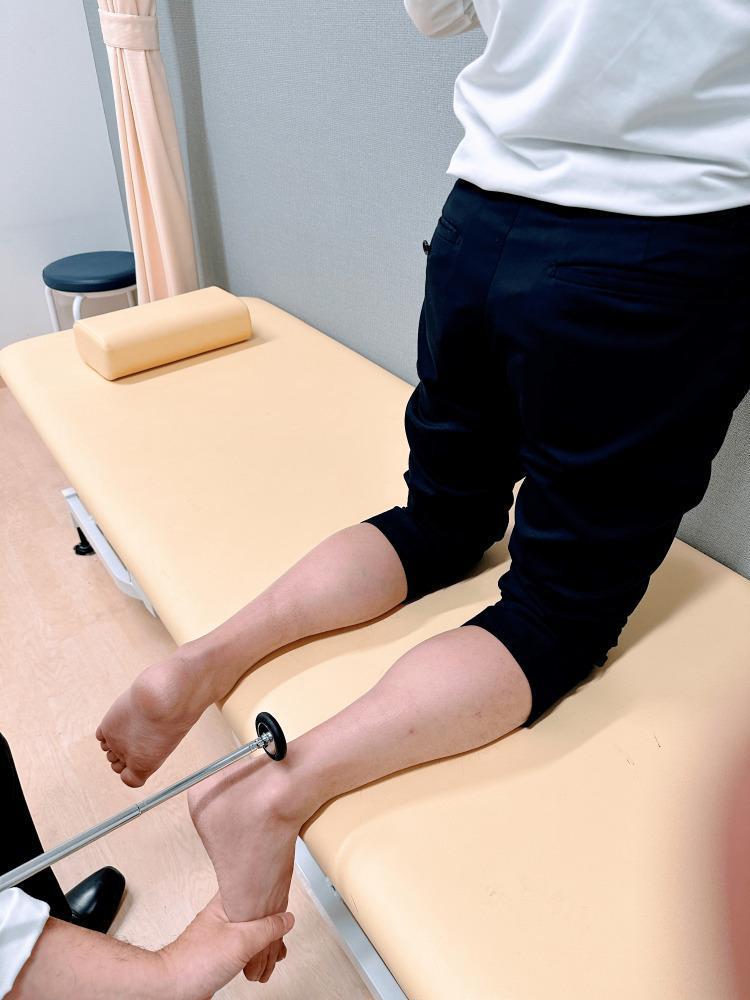
A clinical photograph demonstrating the kneeling technique for eliciting the Achilles tendon reflex (ankle jerk), which tests the S1-S2 nerve roots and the tibial nerve. The patient is positioned kneeling on an examination table with their shins supported by the table surface and their feet hanging freely over the edge. This position allows for optimal relaxation of the gastrocnemius-soleus complex. The examiner is shown performing two simultaneous actions: using one hand to apply gentle passive dorsiflexion to the plantar surface of the foot to place the Achilles tendon under slight tension, and using a standard reflex hammer in the other hand to strike the tendon just above its insertion on the calcaneus. This procedural image illustrates a standard method in a neurological physical examination to assess deep tendon reflexes and identify possible upper or lower motor neuron pathology.
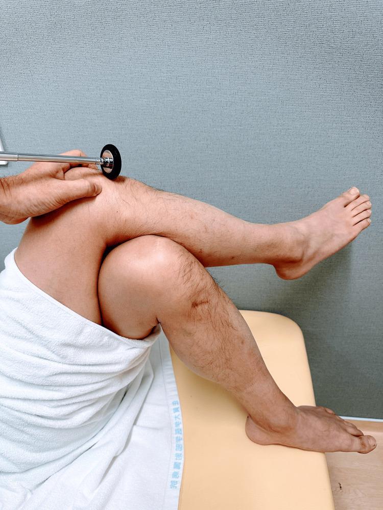
This clinical photograph demonstrates the assessment of the patellar tendon reflex (knee-jerk) in a patient positioned supine. The patient's legs are arranged with the right leg crossed over the left, placing the uppermost knee in a flexed position that naturally stretches the quadriceps tendon. An examiner’s hand is visible, stabilizing the knee while using a reflex hammer with a circular rubber head to strike the patellar ligament just distal to the patella. This procedure is used to evaluate the L3-L4 spinal nerve roots and the integrity of the femoral nerve. The image illustrates a practical modification for eliciting deep tendon reflexes when a patient is in a semi-recumbent or supine position on an examination table, emphasizing proper limb positioning and strike location for clinical diagnostic purposes.
Deep Tendon Reflexes - Root Values (Macleod's)
| Reflex | Root Value | Notes |
|---|---|---|
| Knee Jerk (Patellar reflex) | L3, L4 (mainly L4) | Tests femoral nerve; knee extension arc |
| Ankle Jerk (Achilles reflex) | S1 (S1-S2) | Tests tibial nerve; plantar flexion arc |
| Plantar reflex | L5, S1, S2 | Normal = flexion (downgoing); Abnormal upgoing = Babinski sign (UMN lesion) |
Additional Lower Limb Reflexes for Context
| Reflex | Root Value |
|---|---|
| Cremasteric reflex | L1-L2 |
| Medial hamstring | L5 |
| Bulbocavernosus | S2-S4 |
| Anal wink | S3-S4 |
Key Points
- Knee jerk (L3-L4, mainly L4): Loss indicates L3/L4 root lesion or femoral nerve injury. Seen in L4 disc prolapse.
- Ankle jerk (S1): Loss is the hallmark of S1 radiculopathy (e.g., L5-S1 disc herniation). Also lost in peripheral neuropathy (often symmetric).
- Plantar reflex (L5-S2): A flexor (downgoing) response is normal. A Babinski sign (upgoing great toe + fan of other toes) indicates upper motor neuron damage - never a root value issue per se, but the arc runs through L5-S2 cord segments.
Memory Aid
"1, 2 buckle my shoe" (ankle = S1-2); "3, 4 knock at the door" (knee = L3-4)
Knee jerk ankle jerk planter reflex exgarrated and deminish r cause
UMN LMN reflex comparison upper lower motor neuron signs diagram
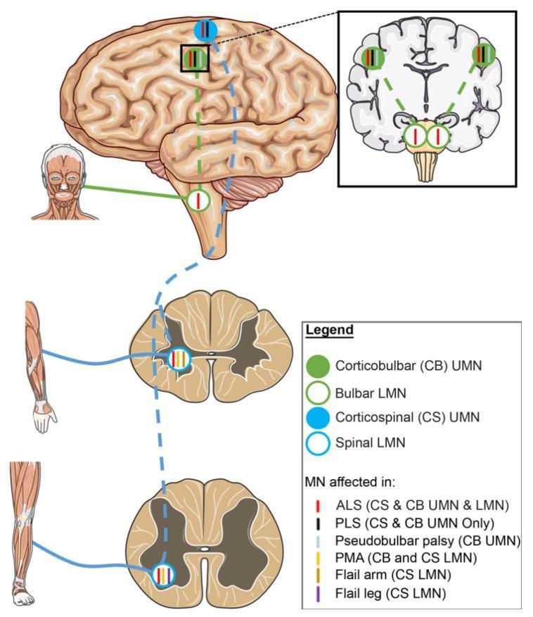
A medical schematic diagram illustrating the neuroanatomical pathways of upper motor neurons (UMN) and lower motor neurons (LMN) and their selective involvement in Amyotrophic Lateral Sclerosis (ALS) variants. The diagram features a lateral view of the human brain, a coronal section of the brain, and transverse sections of the spinal cord (cervical and lumbar levels). Two primary tracts are highlighted: the corticobulbar (CB) tract, represented by green solid circles (UMN) and open circles (LMN), and the corticospinal (CS) tract, represented by blue solid circles (UMN) and open circles (LMN). Solid circles represent UMN cell bodies in the motor cortex, while open circles denote LMNs in the brainstem (bulbar) and spinal cord. Colored vertical tick marks inside these circles indicate specific disease phenotypes: red for classic ALS (CS & CB UMN and LMN), black for Primary Lateral Sclerosis (PLS, UMN only), and yellow for Progressive Muscular Atrophy (PMA, LMN only). Other variants shown include Pseudobulbar palsy (CB UMN), Flail arm, and Flail leg (specific CS LMN segments). This illustration serves as an educational tool for differentiating motor neuron disease phenotypes based on the anatomical site of degeneration.
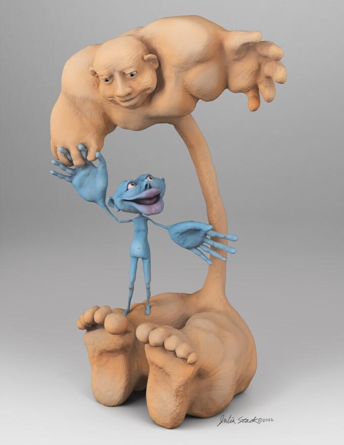
An anatomical illustration and 3D artistic model depicting a comparative visualization of the motor homunculus. The larger, flesh-colored figure represents the Upper Motor Neuron (UMN) homunculus, traditionally based on the cortical mapping of the primary motor cortex. It features massive hands, large feet, and prominent facial structures, illustrating the high density of cortical neurons dedicated to fine motor control in these regions. Juxtaposed to it is a smaller, blue-colored figure representing the Lower Motor Neuron (LMN) homunculus. The LMN homunculus is scaled relative to the UMN counterpart, with its bodily proportions determined by alpha motor neuron densities within the spinal cord and brainstem. This comparative diagram highlights the differences in somatotopic representation between the cerebral motor system and the final common pathway. The visual serves as a neuroanatomical educational tool to demonstrate how motor innervation complexity varies across different levels of the central nervous system, emphasizing the disproportionate neural representation of the hands and face in human motor control.
Grading of Deep Tendon Reflexes (DTR)
| Grade | Meaning |
|---|---|
| 0 | Absent |
| 1+ | Hypoactive (diminished) |
| 2+ | Normal (brisk) |
| 3+ | Hyperactive, no clonus |
| 4+ | Hyperactive with mild clonus |
| 5+ | Hyperactive with sustained clonus |
1. KNEE JERK (L3-L4, Patellar Reflex)
Exaggerated (Hyperreflexia) - Causes
| Cause | Mechanism |
|---|---|
| Stroke (cerebrovascular accident) | Corticospinal tract interruption |
| Multiple sclerosis | Demyelination of UMN pathways |
| Cervical/thoracic myelopathy | Cord compression above L3 |
| Spinal cord injury (above L3) | Loss of descending inhibition |
| Brain tumor | Cortical/subcortical UMN involvement |
| Amyotrophic lateral sclerosis (ALS) | Both UMN + LMN involved |
| Cerebral palsy | UMN lesion |
| Hyperthyroidism | Increased neuromuscular excitability |
| Anxiety / tension | Reduced cortical inhibition |
| Tetanus | Loss of inhibitory interneurons |
Diminished / Absent - Causes
| Cause | Mechanism |
|---|---|
| L3/L4 disc prolapse | Root compression |
| Diabetic neuropathy | Ia afferent fiber damage |
| Alcoholic neuropathy | Peripheral nerve damage |
| Guillain-Barré syndrome | Acute demyelinating polyneuropathy |
| Femoral nerve injury | LMN arc disruption |
| Tabes dorsalis (syphilis) | Posterior root/dorsal column damage |
| Hypothyroidism | Slow relaxation phase ("hung-up reflex") |
| Hypokalemia / hypomagnesemia | Neuromuscular junction dysfunction |
| Myopathy | Muscle disease (quadriceps wasting) |
| Shock/coma (acute) | Generalized depression of CNS |
| Aging | Physiologic reduction in elderly |
2. ANKLE JERK (S1, Achilles Reflex)
Exaggerated - Causes
- Stroke, spinal cord lesion (above S1), MS, myelopathy, ALS
- Hyperthyroidism - classically causes brisk reflexes with fast relaxation phase
- Anxiety, clonus present in severe UMN lesions
Diminished / Absent - Causes
| Cause | Notes |
|---|---|
| S1 radiculopathy (L5-S1 disc) | Most common cause of absent ankle jerk |
| Diabetic peripheral neuropathy | First reflex to be lost; distal > proximal |
| Alcoholic neuropathy | Length-dependent axonopathy |
| Guillain-Barré syndrome | Early bilateral absent ankle jerks |
| Charcot-Marie-Tooth disease | Hereditary neuropathy |
| Hypothyroidism | "Hung-up" or absent ankle jerk - classic sign |
| Aging (normal) | Achilles reflex often absent in elderly |
| Sciatic nerve injury | LMN arc at sciatic/tibial level |
| Vitamin B12 deficiency | Subacute combined degeneration |
| Tabes dorsalis | Posterior column + root damage |
| Carcinomatous neuropathy | Paraneoplastic |
Clinical pearl: Absent ankle jerk + upgoing plantars = combined UMN + LMN lesion → think subacute combined degeneration of cord (B12 deficiency) or motor neuron disease (ALS)
3. PLANTAR REFLEX (L5-S1-S2)
Normal Response
Exaggerated / Abnormal = BABINSKI SIGN
| Cause of Positive Babinski | Notes |
|---|---|
| Stroke | Most common cause |
| Spinal cord compression/trauma | Any level above S1 |
| Multiple sclerosis | Demyelination of CST |
| Meningitis/encephalitis | Acute CNS inflammation |
| Brain tumor | CST involvement |
| Todd's paralysis (post-seizure) | Transient Babinski |
| Hypoglycemic coma | Metabolic UMN depression |
| Deep sleep / anaesthesia | Normal in unconscious state |
| Infants < 18 months | Normal finding - corticospinal tract not yet myelinated |
Diminished / Absent Plantar Response
- Peripheral neuropathy - sensory afferents of sole damaged (cannot elicit the reflex)
- S1-S2 root lesion - efferent arc disrupted
- Heavy callus on foot - cannot elicit stimulus
Summary Table
| Reflex | Exaggerated (UMN) | Diminished (LMN/Peripheral) |
|---|---|---|
| Knee jerk | Stroke, MS, myelopathy, hyperthyroidism, ALS | L3-L4 disc, DM neuropathy, GBS, femoral nerve lesion, hypothyroidism |
| Ankle jerk | Stroke, myelopathy, MS, hyperthyroidism | S1 radiculopathy, DM neuropathy, hypothyroidism, GBS, B12 deficiency, aging |
| Plantar | Positive Babinski = stroke, MS, cord injury, ALS, encephalitis, post-ictal | Absent = peripheral neuropathy, S1-S2 root lesion |
Touch pain vibration joint position eigula loss hoi kokhn
sensory pathways spinal cord dorsal column spinothalamic tract diagram cross section
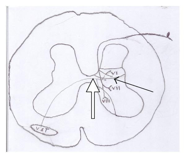
Anatomical diagram of a spinal cord cross-section illustrating the pathway of the ventral spinothalamic tract (VST). The illustration depicts the central H-shaped gray matter and surrounding white matter. Key structures labeled include Rexed laminae VI, VII, and VIII in the dorsal and intermediate horns of the gray matter. A black arrow indicates the origin of secondary sensory neurons within these deep laminae. A white arrow tracks the decussation (crossing over) of these nerve fibers through the anterior white commissure to the contralateral side. The fibers terminate in the ventral white column, labeled as the 'VST' tract. This diagram serves as a neuroanatomical representation of the ascending somatosensory pathway for crude touch and pressure, highlighting its origin in non-opioid-rich Rexed layers compared to the lateral spinothalamic tract.
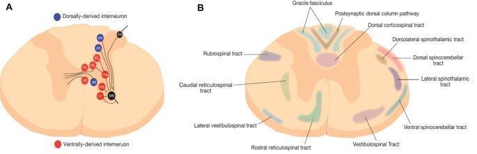
Educational neuroanatomical schematic of a rodent spinal cord cross-section, divided into two panels. Panel A illustrates the distribution and connectivity of spinal interneurons. Blue circles represent dorsally-derived interneurons (e.g., dI3, dI4) located in the dorsal horn, while red circles represent ventrally-derived interneurons (e.g., V0, V1, V2a, V3) in the ventral horn and intermediate zone. Black lines depict axonal projections from these interneuron populations synapsing onto motoneurons (MN). Panel B provides a detailed topographical map of white matter tracts. Ascending sensory pathways include the gracile fasciculus and postsynaptic dorsal column pathway medially, with the spinothalamic and spinocerebellar tracts located laterally. Descending motor pathways include the dorsal corticospinal tract, rubrospinal tract, and various reticulospinal and vestibulospinal tracts distributed throughout the ventral and lateral funiculi. This diagram serves as a foundational tool for understanding spinal circuit organization, sensorimotor integration, and neuroanatomical localization within the central nervous system.
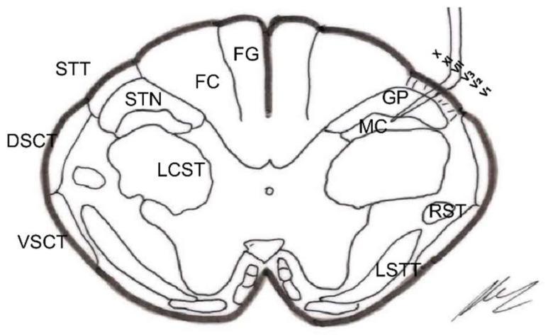
This medical anatomical diagram illustrates a cross-section of the spinal cord (medulla oblongata level, approximately 18 mm below the obex), specifically focusing on the neuroanatomical structures targeted during a trigeminal nucleotomy. The diagram highlights both white matter tracts and gray matter nuclei. In the posterior column, the Fasciculus Gracilis (FG) is medially positioned, while the Fasciculus Cuneatus (FC) is lateral to it. The lateral white matter contains the Lateral Corticospinal Tract (LCST), Dorsal Spinocerebellar Tract (DSCT), and Ventral Spinocerebellar Tract (VSCT). On the right side of the diagram, a surgical probe is shown penetrating the Spinal Trigeminal Tract (STT/STrigT) to target the Spinal Trigeminal Nucleus (STN), specifically indicating the Gelatinosa Portion (GP) and the Magnocellular Portion (MC). Other notable structures include the Rubrospinal Tract (RST) and the Lateral Spinothalamic Tract (LSTT). This illustration is designed for neurosurgical education, demonstrating the spatial relationships necessary for selective destruction of pain-transmitting nuclei in the brainstem and upper cervical spinal cord.
Sensory Modalities - When Each Is Lost (Anatomical Basis)
First: The Two Key Sensory Pathways
| Pathway | Sensation Carried | Route |
|---|---|---|
| Dorsal Column - Medial Lemniscus (DCML) | Fine touch, Vibration, Joint position sense (proprioception), 2-point discrimination | Enters spinal cord → goes UP ipsilateral dorsal column → crosses at MEDULLA |
| Lateral Spinothalamic Tract (STT) | Pain, Temperature, Crude touch | Enters spinal cord → crosses within 2-3 segments in anterior white commissure → goes UP contralateral STT |
Condition-wise Sensory Loss Pattern
1. DORSAL COLUMN LESION (Posterior column)
| Disease | Notes |
|---|---|
| Tabes dorsalis (tertiary syphilis) | Classic posterior column destruction |
| Subacute combined degeneration (Vit B12 deficiency) | Posterior + lateral columns both affected |
| Multiple sclerosis | Plaque in dorsal column |
| Friedreich's ataxia | Spinocerebellar + dorsal columns |
| Posterior cord syndrome (spinal injury) | Dorsal column spared or damaged specifically |
2. SPINOTHALAMIC TRACT (LATERAL) LESION
| Disease | Notes |
|---|---|
| Syringomyelia | Central cavity destroys crossing fibers → cape-like suspended sensory loss of pain + temperature in arms/shoulders; touch/vibration preserved |
| Anterior Spinal Artery Syndrome | Infarcts anterior cord → loss of pain + temperature BELOW lesion; vibration + proprioception PRESERVED |
| Central cord syndrome (hyperextension injury) | Crossing fibers damaged → dissociated loss; arms > legs |
| Lateral medullary syndrome (Wallenberg's) | Contralateral body pain + temperature loss |
3. COMPLETE UNILATERAL CORD LESION - BROWN-SEQUARD SYNDROME
| Side | Modality Lost |
|---|---|
| Ipsilateral (same side as lesion) | Vibration + Joint position (dorsal column) + Motor (UMN) |
| Contralateral (opposite side) | Pain + Temperature (STT crosses before ascending) |
4. ALL SENSORY MODALITIES LOST (Complete cord / peripheral lesion)
| Condition | Pattern |
|---|---|
| Complete spinal cord transection | ALL sensation lost below level (both pathways cut) |
| Peripheral neuropathy (DM, alcohol, GBS) | Loss of all modalities, distal > proximal, "glove and stocking" |
| Thalamic infarction | Contralateral loss of all sensory modalities |
| Cortical lesion | Contralateral loss, especially discriminative touch + proprioception |
5. ONLY TOUCH LOST (Rare)
- Anterior spinothalamic tract lesion - crude touch alone
- Cortical parietal lobe lesion - fine/discriminative touch, 2-point discrimination, graphesthesia lost while pain may be preserved
Master Summary Table
| Modality | Pathway | Lost In |
|---|---|---|
| Fine touch, 2-point discrimination | Dorsal column | B12 deficiency, tabes dorsalis, MS, parietal cortex lesion |
| Vibration | Dorsal column | B12 deficiency, tabes dorsalis, Friedreich's ataxia, MS, peripheral neuropathy (early loss distally) |
| Joint position (proprioception) | Dorsal column | Same as vibration; also causes sensory ataxia + Romberg positive |
| Pain | Spinothalamic (crosses early) | Syringomyelia (dissociated), anterior spinal artery syndrome, Brown-Sequard (contralateral) |
| Temperature | Spinothalamic (crosses early) | Same as pain - always lost together with pain |
| Crude touch | Anterior spinothalamic | Rarely lost in isolation; lost in complete cord/peripheral lesions |
The "Dissociated Sensory Loss" Concept (High-Yield)
Dissociated sensory loss = one type of sensation lost, another preserved at the SAME level
| Type | What's Lost | What's Preserved | Classic Disease |
|---|---|---|---|
| Spinothalamic loss | Pain + Temperature | Vibration + Position | Syringomyelia, Anterior spinal artery syndrome |
| Dorsal column loss | Vibration + Position | Pain + Temperature | Tabes dorsalis, B12 deficiency |
| Brown-Sequard | Ipsilateral vibration/position + Contralateral pain/temp | Both sides partially preserved | Penetrating cord injury |