
Pott spine findings
Pott's disease spinal tuberculosis MRI vertebral collapse gibbus deformity
"spinal tuberculosis" AND diagnosis
Pott disease spinal tuberculosis vertebral collapse gibbus deformity
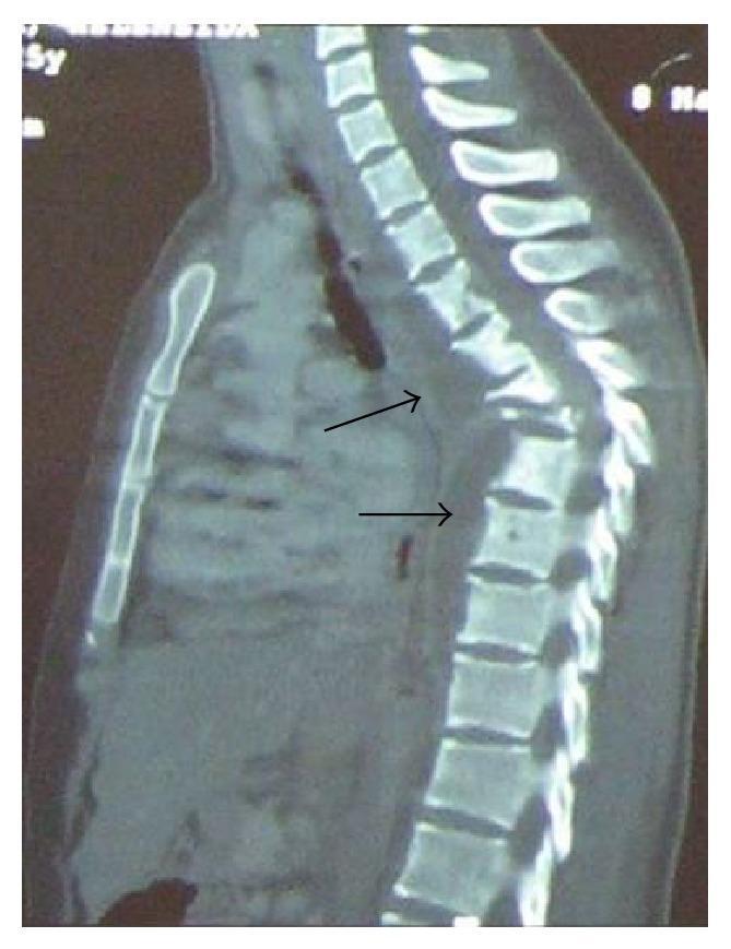
This diagnostic image is a sagittal Computed Tomography (CT) scan of the thoracic spine. It demonstrates significant osteolytic destruction of multiple mid-thoracic vertebral bodies, characteristic of spinal tuberculosis (Pott's disease). There is a prominent collapse of the anterior spinal elements leading to a severe, acute-angle kyphotic deformity (Gibbus deformity). Adjacent to the area of vertebral destruction, arrows point to a large, hypodense soft tissue mass extending along the anterior and paravertebral space, consistent with a cold abscess. The image illustrates the classic radiographic triad of spondylodiscitis: vertebral body destruction, disk space narrowing/collapse, and associated paraspinal soft tissue involvement, highlighting the risk of spinal cord compression and structural instability in advanced granulomatous infections.
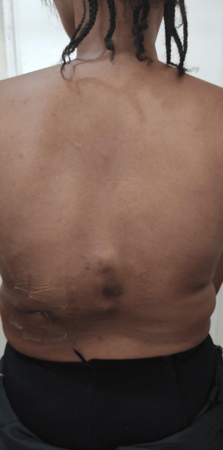
This clinical photograph displays the posterior view of a patient's back, focusing on significant spinal and cutaneous abnormalities. The primary musculoskeletal finding is a prominent thoracolumbar gibbus, a localized kyphotic deformity manifesting as a sharp posterior protrusion in the lower thoracic/upper lumbar region. Dermatological assessment reveals a visible sinus scar located on the left flank/waistline area, surrounded by hyperpigmented, darkened skin. Additional non-specific hyperpigmented patches are scattered across the mid-back region. The combination of a gibbus deformity and a chronic sinus scar is highly characteristic of Pott's disease (spinal tuberculosis), which typically involves vertebral destruction leading to spinal collapse and potential cold abscess formation with secondary skin involvement. This image serves as a teaching tool for recognizing physical manifestations of chronic spinal infections and structural spinal deformities in clinical practice.
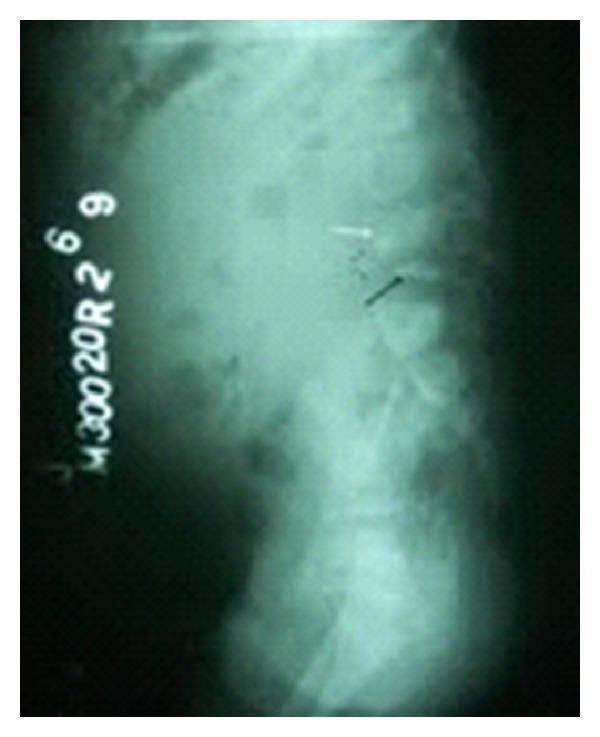
This diagnostic image is a lateral plain film X-ray of the lumbosacral spine. The radiograph reveals a significant focal kyphotic angulation, known as a gibbus deformity, centered at the L4-L5 vertebral level. A prominent wedge-shaped collapse of the L4 vertebral body is visible, indicating severe loss of anterior vertebral height and structural compromise. Additionally, radiopaque bony fragments are noted in the prevertebral space anterior to the L4 level, consistent with vertebral destruction or fragmentation. The visual findings are characteristic of advanced spinal pathology, such as Pott's disease (spinal tuberculosis), which typically manifests with vertebral body destruction, collapse, and resultant sharp angulation of the spinal column. The image demonstrates key educational markers of infectious or destructive spondylodiscitis, including vertebral wedging and secondary spinal curvature abnormalities.
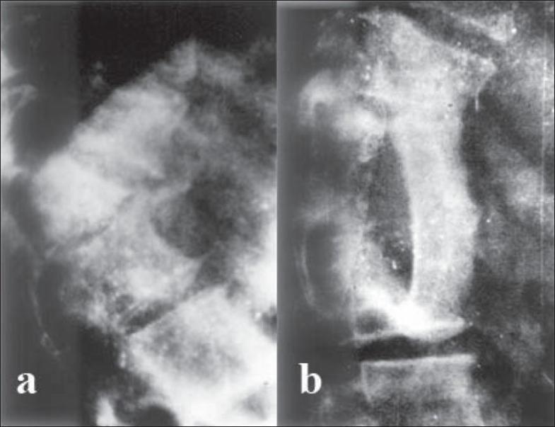
This lateral-view spinal skiagram (X-ray) comparison demonstrates the preoperative and postoperative states of a severe thoracolumbar kyphotic deformity. (a) Preoperative image: Shows a high-grade, acute angular kyphosis of the dorsal spine. A wedge-shaped 'internal gibbus' is visible, formed by the anterior collapse and subsequent posterior protrusion of vertebral remnants into the spinal canal, leading to mechanical spinal stenosis. (b) Postoperative image: Displays the result of anterior decompression via gibbus excision. The sharp curvature is noticeably corrected, and an autogenous iliac bone graft is seen bridging the intervertebral gap to provide structural stability and promote arthrodesis. The alignment of the vertebral column is significantly improved compared to the preoperative state. The images illustrate surgical management for paraplegia resulting from healed spinal tuberculosis (Pott's disease) with secondary kyphosis.
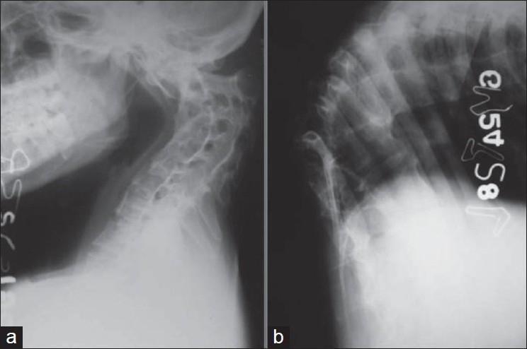
This composite diagnostic image features two lateral plain X-ray radiographs (a and b) illustrating severe spinal deformities resulting from healed spinal tuberculosis (Pott's disease). Image (a) depicts a lateral view of the cervical spine, showing significant anterior wedging and collapse of multiple mid-cervical vertebral bodies. This structural loss has resulted in a marked cervical kyphosis, reversing the normal lordotic curvature. There is visible irregularity of the vertebral endplates and narrowing of the intervertebral disc spaces, suggesting chronic degeneration and secondary fusion. Image (b) shows the thoracic (dorsal) spine, demonstrating an acute, angular kyphotic deformity (gibbus deformity). The X-ray reveals severe anterior collapse of multiple thoracic vertebrae, with the apex of the curve showing significant structural distortion and narrowing of disc spaces. Crowding of the posterior elements and ribs is evident at the apex. Together, these images illustrate the long-term musculoskeletal sequelae of spinal tuberculosis, focusing on severe sagittal plane malalignment and vertebral destruction.
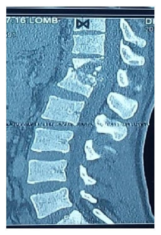
This diagnostic image is a sagittal Computed Tomography (CT) scan of the thoracolumbar spine demonstrating advanced tuberculous spondylitis (Pott's disease). The scan shows significant osteolysis and bone destruction localized to the D12 and L1 vertebral bodies. Key pathological features include the collapse of these vertebral segments and the loss of the normal intervertebral disc space. This structural failure has resulted in a marked focal kyphotic deformity (Gibbus deformity) of approximately 40 degrees, which indicates severe spinal instability and possible cord compression. There is evidence of perilesional osteosclerosis surrounding the lytic areas, reflecting a chronic inflammatory and reactive healing process. The image serves as a classic educational example of spinal tuberculosis, illustrating the progression from disc space infection to vertebral destruction and subsequent spinal malalignment.
Pott's Spine (Spinal Tuberculosis) - Key Findings
Pathogenesis
Clinical Findings
- Insidious back pain and localized tenderness over the affected spinous process, often for weeks to months
- Low-grade fever, night sweats, weight loss, malaise (constitutional TB symptoms)
- Muscle spasm/rigidity - though this is often less pronounced than in pyogenic spinal infection - S Das A Manual on Clinical Surgery, 13th Ed.
- Pain may be referred to the chest, abdomen, or as girdle pain depending on the level involved
- Progressive kyphosis from anterior vertebral body collapse
- Gibbus deformity - a sharp, angular posterior protrusion at the apex of collapse, the hallmark visible deformity of healed/advanced disease
- Paravertebral "cold" abscess (lacking the classic heat/erythema of pyogenic infection) that can track along fascial planes/cutaneous nerve branches to present at a distance - e.g., in the loin, groin (psoas abscess tracking to the thigh), posterior triangle of the neck, or chest wall
- May rupture through the skin as a chronic discharging sinus, often with surrounding hyperpigmentation and scarring
- Spinal cord compression from epidural abscess, granulation tissue mass, bony sequestra retropulsed into the canal, or mechanical angulation at the kyphotic apex
- Presents with weakness, sensory loss, and bowel/bladder dysfunction below the level of compression; can be an early complication (active disease with abscess/granulation tissue) or a late complication (mechanical, from healed kyphotic deformity)
Imaging Findings
- Plain radiographs: vertebral body height loss with anterior wedging/collapse, relative preservation of disc space early in disease, progressive angular kyphosis
- CT: vertebral destruction, bony fragmentation, paravertebral soft-tissue/abscess with possible calcification (more suggestive of TB than pyogenic infection), useful for guiding aspiration biopsy
- MRI (most sensitive): vertebral body destruction, disc involvement, paraspinal/epidural abscess, cord compression, and the classic angulated thoracic kyphotic deformity - Adams and Victor's Principles of Neurology, 12th Ed.
Histopathology
- Caseating granulomatous inflammation with epithelioid histiocytes, Langhans giant cells, and central necrosis
- Vertebral body and disc destruction with compression fractures and kyphosis - Robbins, Cotran & Kumar Pathologic Basis of Disease, Fig. 26.17
Laboratory Findings
- Elevated ESR/CRP (nonspecific but commonly elevated)
- Positive tuberculin skin test or IGRA in many, though some patients are anergic
- Definitive diagnosis: CT-guided aspiration/biopsy of the paravertebral abscess or vertebral lesion for AFB smear, culture, histology, and increasingly PCR/GeneXpert (Xpert MTB/RIF) for rapid molecular confirmation
Note on Recent Evidence
You've shared a whole-spine sagittal T2 MRI photo (screen capture) right after we discussed Pott's spine findings. What would you like me to do with it?
Pott's Spine (Spinal Tuberculosis) - Key Findings
Pathogenesis
Clinical Findings
- Insidious back pain and localized tenderness over the affected spinous process, often for weeks to months
- Low-grade fever, night sweats, weight loss, malaise (constitutional TB symptoms)
- Muscle spasm/rigidity - though this is often less pronounced than in pyogenic spinal infection - S Das A Manual on Clinical Surgery, 13th Ed.
- Pain may be referred to the chest, abdomen, or as girdle pain depending on the level involved
- Progressive kyphosis from anterior vertebral body collapse
- Gibbus deformity - a sharp, angular posterior protrusion at the apex of collapse, the hallmark visible deformity of healed/advanced disease
- Paravertebral "cold" abscess (lacking the classic heat/erythema of pyogenic infection) that can track along fascial planes/cutaneous nerve branches to present at a distance - e.g., in the loin, groin (psoas abscess tracking to the thigh), posterior triangle of the neck, or chest wall
- May rupture through the skin as a chronic discharging sinus, often with surrounding hyperpigmentation and scarring
- Spinal cord compression from epidural abscess, granulation tissue mass, bony sequestra retropulsed into the canal, or mechanical angulation at the kyphotic apex
- Presents with weakness, sensory loss, and bowel/bladder dysfunction below the level of compression; can be an early complication (active disease with abscess/granulation tissue) or a late complication (mechanical, from healed kyphotic deformity)
Imaging Findings
- Plain radiographs: vertebral body height loss with anterior wedging/collapse, relative preservation of disc space early in disease, progressive angular kyphosis
- CT: vertebral destruction, bony fragmentation, paravertebral soft-tissue/abscess with possible calcification (more suggestive of TB than pyogenic infection), useful for guiding aspiration biopsy
- MRI (most sensitive): vertebral body destruction, disc involvement, paraspinal/epidural abscess, cord compression, and the classic angulated thoracic kyphotic deformity - Adams and Victor's Principles of Neurology, 12th Ed.
Histopathology
- Caseating granulomatous inflammation with epithelioid histiocytes, Langhans giant cells, and central necrosis
- Vertebral body and disc destruction with compression fractures and kyphosis - Robbins, Cotran & Kumar Pathologic Basis of Disease, Fig. 26.17
Laboratory Findings
- Elevated ESR/CRP (nonspecific but commonly elevated)
- Positive tuberculin skin test or IGRA in many, though some patients are anergic
- Definitive diagnosis: CT-guided aspiration/biopsy of the paravertebral abscess or vertebral lesion for AFB smear, culture, histology, and increasingly PCR/GeneXpert (Xpert MTB/RIF) for rapid molecular confirmation
Note on Recent Evidence
- This is a photograph of a screen/film with glare, an obstructing hand, and no ability to adjust window/level or scroll through slices - I cannot reliably assess subtle vertebral marrow signal changes, disc space narrowing, or a paraspinal collection from this alone the way a radiologist reading the actual DICOM images could.
- What I can say from the visible anatomy: the cervical lordosis transitions into a thoracic kyphotic curve that, from this angle, doesn't show an obvious sharp angular gibbus deformity - the curve looks like a gradual sweep rather than the acute focal angulation typical of collapsed vertebrae in Pott's disease. The cord/thecal sac (bright CSF signal) appears to run continuously without an obvious focal block visible at this resolution, but I would not stake a diagnosis on that given image quality.
- I can't see a clearly demarcated area of vertebral body destruction, wedging, or a paravertebral soft-tissue/abscess collection in this image - but that absence could simply be due to photo quality, not true absence of pathology.
- Vertebral body height loss/wedging with relative disc space preservation (early) or disc destruction (later)
- Focal kyphotic angulation at the site of collapse
- Paravertebral or epidural soft-tissue signal (abscess) - often T2 hyperintense with peripheral rim enhancement on post-contrast sequences
- Cord signal change or compression at the level of collapse/abscess