
Burn in surgery
Reading File
Burns surgery is a broad topic. Let me search the medical library for comprehensive coverage across the key areas.
Searching sources...
Finding Sources
Finding Sources
Finding Sources
Reading File
Reading File
Reading File
Reading File
Finding Sources
Finding Sources
Reading File
Finding Sources
Finding Sources
Reading File
Reading File
Reading File
Reading File
Reading File
Reading File
I now have comprehensive material from the textbooks. Let me compile a thorough, well-organized answer.
Burns in Surgery - Comprehensive Overview
Sources: Bailey & Love's Short Practice of Surgery 28th Ed, Schwartz's Principles of Surgery 11th Ed, Goldman-Cecil Medicine
1. Classification of Burns
Burns are classified by depth and by total body surface area (TBSA) involved.
Depth Classification
| Grade | Layer Involved | Clinical Features | Healing |
|---|---|---|---|
| Superficial (1st degree) | Epidermis only | Erythema, pain, no blisters | 3-5 days, no scar |
| Superficial partial-thickness (2nd degree) | Epidermis + papillary dermis | Blistering, moist pink dermis, brisk capillary refill, normal pin-prick sensation | ~2 weeks, minimal scar |
| Deep partial-thickness | Reticular dermis | Moist/dry dermis, fixed capillary staining, blunted pin-prick, no blanching | >3 weeks, often requires surgery |
| Full-thickness (3rd degree) | All layers of skin | Leathery, insensate, no blanching | Does not heal spontaneously - surgery needed |
| 4th/5th degree | Subcutaneous fat, muscle, bone | Charred, deeply invasive | Requires extensive reconstruction |
Temperature-time relationship (Bailey & Love): At 65°C water - 45 seconds produces full-thickness burn; 15 seconds = deep partial-thickness; 7 seconds = superficial partial-thickness.
Depth by aetiology:
- Scalds: usually superficial (deep in infants/elderly)
- Fat burns / flame burns: deep dermal to full-thickness
- Electrical contact burns: always full-thickness
- Alkali burns (including cement): often deep dermal or full-thickness
- Acid burns: weak - superficial; strong - deep dermal
2. Estimating Burn Size (TBSA)
The Rule of Nines gives a first approximation in adults:
- Head & neck = 9%
- Each arm = 9%
- Anterior trunk = 18%
- Posterior trunk = 18%
- Each leg = 18%
- Perineum = 1%
In children, the Lund & Browder chart is preferred as it accounts for the proportionally larger head and smaller legs. The patient's palm (including fingers) = approximately 1% TBSA - useful for irregular or scattered burns.
Important: Superficial (1st-degree) burns are NOT included in TBSA calculations for resuscitation.
3. Initial Management - Primary Survey (ABCDE)
-
Airway - Signs of inhalation injury: hoarseness, stridor, singed nasal hairs, carbonaceous sputum, facial burns. Early intubation is lifesaving - do not delay. Use fiberoptic bronchoscopy as the gold standard for airway assessment. All patients with suspected inhalation injury receive 100% O₂ empirically.
-
Carbon monoxide (CO) poisoning - Pulse oximetry is falsely elevated; request ABG with carboxyhemoglobin levels. Treatment: 100% O₂.
-
Cyanide poisoning - Consider in enclosed space fires; treat empirically with hydroxocobalamin in severe cases.
-
Fluid resuscitation - Begin promptly (see below).
-
Exclude other injuries - All patients with burns >10% TBSA undergo full ATLS primary and secondary survey.
4. Fluid Resuscitation
Indications: Adults with burns >15% TBSA; children with >10% TBSA. Begin large-bore IV access (avoid burned skin if possible).
Parkland (Baxter) Formula
4 mL × weight (kg) × %TBSA burned (Ringer's lactate)
- 50% given in the first 8 hours (from time of burn, not time of presentation)
- Remaining 50% over the next 16 hours
Monitoring endpoint: Urine output of 0.5 mL/kg/hour in adults; 1 mL/kg/hour in children <30 kg.
Important caveat: The Parkland formula commonly leads to fluid creep (over-resuscitation). Modern practice titrates volumes carefully to avoid abdominal compartment syndrome and pulmonary oedema. Colloid (albumin) may be added after 8-24 hours once capillary leak decreases.
Nasogastric/nasojejunal feeding should begin within 6 hours for burns >20% TBSA to protect gut mucosal integrity and reduce the hypermetabolic response.
5. Burn Wound Management
Topical Agents
| Agent | Notes |
|---|---|
| Silver sulphadiazine 1% | Broad-spectrum, first-line for most burns |
| Mafenide acetate (5%) | Penetrates eschar well; can cause metabolic acidosis; painful |
| Silver sulphadiazine + cerium nitrate | Induces sterile eschar; especially useful in elderly; boosts cell-mediated immunity |
| Acticoat | Nanocrystalline silver; broad-spectrum antimicrobial |
| Biobrane | Biologic occlusive dressing for fresh, superficial partial-thickness burns |
- Bailey & Love's Short Practice of Surgery 28th Ed, p. 695-697
6. Burn Excision Surgery
Timing
Early (within 48-72 hours) vs. Staged excision:
Early total burn wound excision advantages:
- Reduces incidence of burn wound sepsis
- Reduces overall mortality and hospital stay
- Most surgery performed before significant lung injury develops
- Allows effective use of vasoconstrictive tumescence fluids
Staged approach: Serial debridement over first week; shorter individual operating times; reduces single blood transfusion requirements; requires managing eschar between procedures with silver dressings.
Techniques
Tangential excision (for deep partial-thickness burns):
- Burn eschar is shaved in thin layers using a Watson or Humby knife
- Excision stops when punctate bleeding is seen and dermis is free of thrombosed vessels
- Followed by split-skin grafting
Full-thickness excision (for full-thickness burns):
- All necrotic skin excised down to viable fat or fascia
- Skin graft/substitute applied immediately if possible
Haemostasis
- Subcutaneous tumescence injection of adrenaline (epinephrine) 1:500,000 to 1:1,000,000
- Tourniquets on extremities
- Topical adrenaline 1:500,000 on excised wound bed
- Warm theatre (patient temperature must not drop below 36°C to prevent coagulopathy)
7. Wound Coverage: Skin Grafts and Substitutes
Split-Skin Autograft (SSG)
The mainstay of burn wound repair. Consists of epidermis + superficial dermis.
- Sheet graft: Used over functionally/cosmetically important areas - face, neck, hands, major joints; provides better cosmetic outcome
- Meshed graft: Allows expansion (1.5:1 up to 6:1); permits fluid drainage; faster healing at cost of cosmesis (meshed pattern remains visible)
- Meek-Wall technique: 3mm x 3mm squares of graft placed on a cork board; used for massive burns where graft is scarce; minimises wastage; mesh ratios can be varied for large expansion
When TBSA >50% (donor site becomes limiting factor):
- Serial grafting operations
- Very thin harvested grafts (allows faster re-epithelialisation and earlier re-harvest of donor sites)
- Cadaver allograft skin as temporary biological cover while donor sites regenerate
- Dermal substitutes (see below)
Dermal Substitutes
Used to create a "neo-dermis" through autologous tissue in-growth (active temporisation):
- Integra (bilayer matrix of bovine collagen and glycosaminoglycan, covered by silicone) - pioneered by Jack Burke
- Biodegradable Temporising Matrix (BTM) - synthetic biodegradable polyurethane; used in extensive burns; a thin SSG is placed over it at ~3 weeks; improves scar outcome compared with immediate thin mesh graft alone
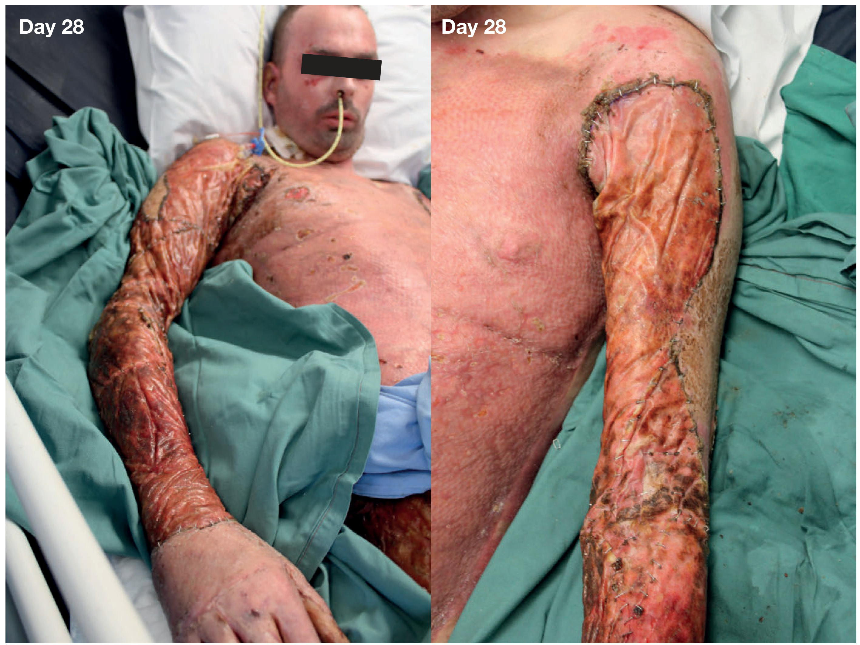
Figure: Day 28 post-full-thickness burns - BTM on arms (left), immediate split-skin graft to chest (right) - Bailey & Love's, p.698
8. Escharotomy and Fasciotomy
Escharotomy
Indication: Circumferential full-thickness burns of extremities or chest causing compartment syndrome or respiratory restriction.
Timing: Serial clinical examination is usually sufficient to determine need; do not delay if signs develop.
- Chest escharotomy: required when circumferential burns restrict ventilation
- Extremity escharotomy: longitudinal incisions through eschar on medial and lateral aspects
- Fasciotomy is added if escharotomy alone is insufficient
Electrical burns: High-voltage injury requires early fasciotomy/escharotomy given extensive deep muscle destruction and risk of myoglobinuria; urine output should be increased to 2 mL/kg/hour to protect renal tubules.
- Goldman-Cecil Medicine, p. Decompression Procedures section
9. Burn Centre Referral Criteria (ABA)
Transfer to a specialised burn centre is indicated for:
- Partial-thickness burns >10% TBSA
- Burns involving face, hands, feet, genitalia, perineum, or major joints
- Full-thickness burns (any size)
- Electrical burns (including lightning injury)
- Chemical burns
- Inhalation injury
- Burn injury with pre-existing medical conditions
- Burns in children admitted to hospitals without paediatric care
10. Nutrition in Burns
Burns >15-20% TBSA produce a profound hypermetabolic response - basal metabolic rate may increase by up to 200%.
- Nasogastric/nasojejunal feeding within 6 hours of admission for burns >20% TBSA
- Nasojejunal tube allows feeding to continue during theatre visits without fasting
- Harris-Benedict equation × 2 for large burns; Currier formula (25 kcal/kg/day + 40 kcal/% TBSA burned/day) for burns <40% TBSA
- Glutamine supplementation reduces infectious complications
- Micronutrients: vitamin C, vitamin E, selenium, zinc, copper - optimise wound healing and immune function
- Metoclopramide and prokinetics to maintain gastric motility
11. Analgesia
- Small/superficial burns: paracetamol + NSAIDs ± topical cooling
- Large burns: IV opiates initially (avoid IM - variable absorption due to oedema/poor perfusion)
- Ongoing large burns: continuous analgesia with infusions; powerful short-acting opioids before dressing changes
- Procedural analgesia: GA, midazolam + ketamine, or morphine + nitrous oxide for dressing changes
- Early involvement of pain team is beneficial
12. Late Complications
- Hypertrophic scarring and contractures - managed with compression garments, physiotherapy, steroid injections, and revisional surgery
- Heterotopic ossification - rare but recognised complication of major burns, especially around large joints
- Psychological morbidity - PTSD, depression, body image issues - specialist psychology input required from early in the admission
- Marjolin's ulcer - malignant transformation (typically squamous cell carcinoma) in old burn scars, often years/decades after injury
Key surgical principle: The goal is early stable wound cover - permanent (autograft) or temporary (allograft, dermal substitute) - to eliminate the burn wound as a source of ongoing sepsis and metabolic drain. - Bailey & Love's Short Practice of Surgery 28th Ed, p.697
This is a shared conversation. Sign in to Orris to start your own chat.