I now have rich, authoritative textbook content from three major microbiology textbooks. Let me compile the comprehensive nursing notes.
🦠 VIROLOGY — COMPREHENSIVE NURSING NOTES
Microbiology | Nursing Exam Preparation | AIIMS NORCET / Staff Nurse Exams
📌 1. INTRODUCTION
Virology is the branch of microbiology that studies viruses — the smallest infectious agents known. Viruses cause many common diseases like flu, COVID-19, measles, and HIV. Unlike bacteria, viruses cannot live or multiply on their own — they must enter a living cell to survive and reproduce. Understanding viruses is essential for nurses because viral infections are managed differently from bacterial infections.
📌 2. DEFINITION
Virus = A very small infectious particle (20–300 nm) that contains ONLY one type of nucleic acid (either DNA or RNA), surrounded by a protein coat. It can only replicate (make copies of itself) inside a living host cell.
Key terms explained:
- Virion = The complete, fully formed virus particle (outside the cell)
- Capsid = The protein shell/coat that protects the genetic material
- Envelope = A fat (lipid) layer that surrounds some viruses
- Nucleocapsid = Capsid + Nucleic acid (genetic material) together
- nm (nanometer) = 1 nm = one millionth of a millimeter (extremely tiny!)
📌 3. STRUCTURE / MORPHOLOGY OF VIRUS
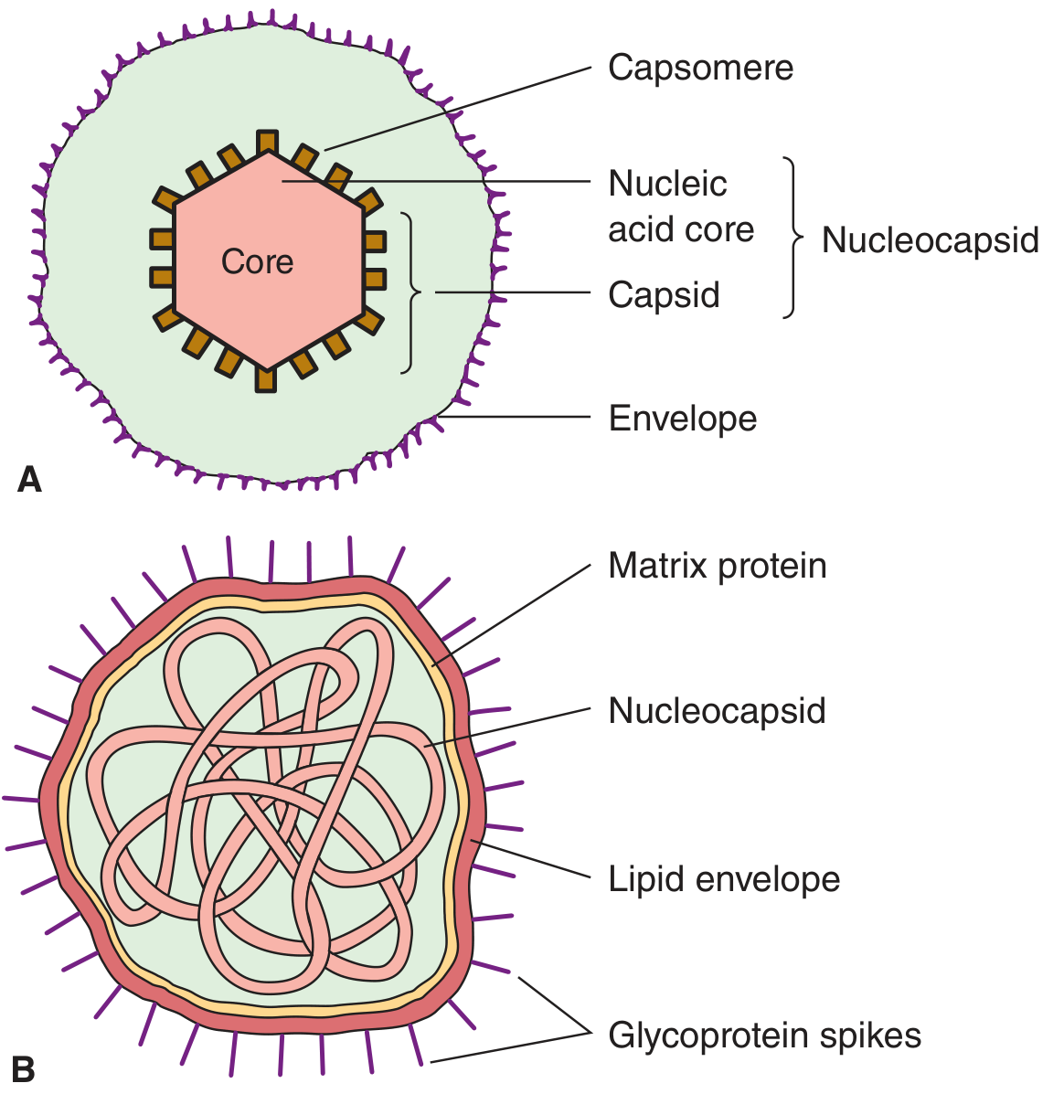
Diagram of Virus Structure
Fig. A: Non-enveloped (naked) virus — Icosahedral structure. Fig. B: Enveloped virus — Helical nucleocapsid with glycoprotein spikes
Basic Parts of a Virus:
| Part | What it is | Function |
|---|
| Nucleic Acid Core | DNA or RNA (genetic material) | Contains instructions to replicate |
| Capsid | Protein shell made of capsomeres | Protects genetic material |
| Nucleocapsid | Capsid + Nucleic acid | Packaged genome |
| Envelope | Lipid membrane (in some viruses) | Helps virus enter host cells |
| Glycoprotein Spikes / Peplomers | Sugar-protein projections on envelope | Attach to host cell receptors |
| Matrix Protein | Protein layer beneath envelope | Structural support |
Simple Diagram (Easy to draw in exams):
         ┌─────────────────────────┐
         │  GLYCOPROTEIN SPIKES    │  ← Attachment to host cell
         │  ┌─────────────────┐    │
         │  │   ENVELOPE      │    │  ← Lipid bilayer (only in some)
         │  │  ┌───────────┐  │    │
         │  │  │  CAPSID   │  │    │  ← Protein coat (capsomeres)
         │  │  │ (Protein) │  │    │
         │  │  │  NUCLEIC  │  │    │
         │  │  │  ACID     │  │    │  ← DNA or RNA
         │  │  │ (Core)    │  │    │
         │  │  └───────────┘  │    │
         │  └─────────────────┘    │
         └─────────────────────────┘
📌 4. CLASSIFICATION OF VIRUSES
A. Based on Nucleic Acid (Most Important)
| Feature | DNA Viruses | RNA Viruses |
|---|
| Genetic material | DNA (Deoxyribonucleic acid) | RNA (Ribonucleic acid) |
| Replication site | Usually nucleus | Usually cytoplasm |
| Mutation rate | Slower (more stable) | Faster (mutate often) |
| Examples | Herpes, Hepatitis B, Pox, Adeno | HIV, Influenza, Measles, Hepatitis C |
B. Based on Capsid Shape (Symmetry)
| Type | Shape | Examples |
|---|
| Icosahedral (20-sided sphere) | Round/spherical | Herpes, Adenovirus, Polio |
| Helical | Spiral/rod | Rabies, Tobacco mosaic virus |
| Complex | Irregular shape | Poxvirus, Bacteriophages |
C. Based on Envelope
| Type | Feature | Stability | Spread |
|---|
| Enveloped virus | Has lipid envelope | Fragile — killed by soap, heat, detergent, drying | Spreads via droplets, blood, secretions |
| Non-enveloped (Naked) | No lipid envelope | Stable — resistant to drying, acid, detergents | Spreads via fomites (surfaces), fecal-oral, aerosols |
Nursing Tip: Enveloped viruses (HIV, Flu, Corona) are KILLED by hand washing with soap. Non-enveloped viruses (Polio, Rota, Norovirus) are MORE resistant — need stronger disinfectants.
D. Major Virus Families (Classification Table)
| Family | Nucleic Acid | Envelope | Key Viruses |
|---|
| Herpesviridae | dsDNA | Yes | HSV 1&2, VZV (Chickenpox), CMV, EBV |
| Poxviridae | dsDNA | No | Smallpox, Monkeypox |
| Adenoviridae | dsDNA | No | Common cold, conjunctivitis |
| Papillomaviridae | dsDNA (circular) | No | HPV (causes cervical cancer) |
| Hepadnaviridae | Partial dsDNA | Yes | Hepatitis B virus |
| Parvoviridae | ssDNA | No | Parvovirus B19 |
| Picornaviridae | +ssRNA | No | Polio, Hepatitis A, Rhinovirus |
| Reoviridae | dsRNA | No | Rotavirus (diarrhea in children) |
| Flaviviridae | +ssRNA | Yes | Dengue, Hepatitis C, Zika, Yellow fever |
| Togaviridae | +ssRNA | Yes | Rubella |
| Retroviridae | +ssRNA (+RT) | Yes | HIV |
| Orthomyxoviridae | −ssRNA (segmented) | Yes | Influenza A, B |
| Paramyxoviridae | −ssRNA | Yes | Measles, Mumps, RSV, Parainfluenza |
| Rhabdoviridae | −ssRNA | Yes | Rabies |
| Filoviridae | −ssRNA | Yes | Ebola, Marburg |
| Coronaviridae | +ssRNA | Yes | SARS-CoV-2 (COVID-19), MERS |
| Bunyaviridae | −ssRNA (segmented) | Yes | Hantavirus, Crimean-Congo HF |
| Arenaviridae | −ssRNA | Yes | Lassa fever |
ss = single-stranded | ds = double-stranded | RT = Reverse transcriptase | + = positive sense | − = negative sense
📌 5. REPLICATION OF VIRUSES (Step-by-Step Flowchart)
Viruses cannot replicate outside cells. They hijack the host cell's machinery.
STEP 1: ATTACHMENT (Adsorption)
Virus spike/surface protein binds to specific receptor on host cell surface
             ↓
STEP 2: PENETRATION (Entry)
Entire virus OR just nucleic acid enters the host cell
(Enveloped viruses: membrane fusion | Non-enveloped: endocytosis)
             ↓
STEP 3: UNCOATING
Capsid is removed — nucleic acid is released into the cell
             ↓
STEP 4: BIOSYNTHESIS
Host cell machinery reads viral DNA/RNA
→ Viral mRNA made → Viral proteins synthesized
→ New viral nucleic acid copied (replicated)
             ↓
STEP 5: ASSEMBLY (Maturation)
New viral proteins + new nucleic acid come together
→ New virus particles (virions) are assembled
             ↓
STEP 6: RELEASE
New viruses exit the cell by:
• Budding (enveloped viruses — cell survives temporarily)
• Lysis — cell bursts and dies (non-enveloped viruses)
             ↓
NEW VIRIONS infect more cells → Disease spreads
Memory Trick — APUBAR:
Attachment → Penetration → Uncoating → Biosynthesis → Assembly → Release
📌 6. PATHOGENESIS OF VIRAL INFECTION (How Viruses Cause Disease)
Virus enters body
(Respiratory tract / GIT / Skin / Blood / Sexual contact)
        ↓
Primary replication at entry site
        ↓
Viremia [Virus in blood] — spreads to target organs
        ↓
Virus attacks specific cells (Cell Tropism = viruses prefer certain cells)
Example: HIV → T-lymphocytes | Poliovirus → Motor neurons | Hepatitis → Liver cells
        ↓
Cell damage by:
1. Direct lysis (cell destruction)
2. Immune-mediated damage (body attacks infected cells)
3. Transformation (virus changes cell → cancer)
        ↓
DISEASE MANIFESTATIONS:
• Fever, inflammation, organ dysfunction
• Immunosuppression (HIV)
• Malignancy (HPV → Cervical cancer)
• Latency — virus hides and reactivates later (Herpes, VZV)
Types of Viral Infection:
| Type | Description | Example |
|---|
| Acute infection | Short-lived, then resolved | Flu, Common cold |
| Chronic infection | Virus persists long-term | Hepatitis B & C, HIV |
| Latent infection | Virus hides in cells, reactivates later | Herpes simplex, Varicella-Zoster |
| Persistent infection | Low-level continuous infection | Hepatitis B (carrier) |
| Transforming infection | Virus converts normal cell to cancer cell | HPV, EBV, HTLV-1 |
📌 7. SIGNS AND SYMPTOMS OF VIRAL INFECTIONS
Symptoms vary by virus, but common features include:
| System Affected | Common Symptoms |
|---|
| General (Systemic) | Fever, malaise (weakness), fatigue, body aches |
| Respiratory | Cough, sore throat, runny nose, breathlessness |
| GIT | Nausea, vomiting, diarrhea, abdominal pain |
| Skin | Rash, vesicles (fluid-filled blisters), ulcers |
| Neurological | Headache, photophobia (light sensitivity), seizures, encephalitis |
| Lymph Nodes | Swollen glands (lymphadenopathy) |
| Eyes | Conjunctivitis (pink eye), redness |
📌 8. LABORATORY DIAGNOSIS OF VIRAL INFECTIONS
Methods in Table:
| Method | What it Tests | Examples / Details |
|---|
| 1. Direct Microscopy | See virus under electron microscope | Electron microscopy — sees virus shape/structure |
| 2. Cell Culture (Tissue Culture) | Grow virus in living cells | Most accurate; see CPE (Cytopathic Effect = cell damage) |
| 3. Antigen Detection | Find viral proteins directly | ELISA, Immunofluorescence — rapid results |
| 4. Serology (Antibody Detection) | Find patient's antibodies to virus | ELISA, Western Blot, Complement Fixation, HI test |
| 5. PCR (Polymerase Chain Reaction) | Detect viral DNA/RNA directly | Gold standard for many viruses; very sensitive |
| 6. Viral Inclusion Bodies | Abnormal structures in infected cells (Light microscopy) | Negri bodies = Rabies |
Cytopathic Effect (CPE) — [Damage visible in cell cultures]:
Virus infects cell culture
        ↓
Cells show changes:
• Cell lysis (death) / rounding
• Syncytia formation (cells fuse together) — e.g., Measles, RSV
• Inclusion bodies (abnormal deposits inside cells)
• Plaque formation (clear areas in cell sheet)
Serology Interpretation Table:
| Antibody Found | Interpretation |
|---|
| IgM positive | Acute/recent infection |
| IgG positive (rising titre) | Current or recent infection |
| IgG positive (stable, high) | Past infection or vaccination (immunity) |
| IgM + IgG both positive | Active infection |
📌 9. MEDICAL MANAGEMENT OF VIRAL INFECTIONS
A. Antiviral Drugs:
| Drug | Mechanism | Used For | Nursing Considerations |
|---|
| Acyclovir | Inhibits viral DNA polymerase | Herpes Simplex, VZV (Chickenpox) | Monitor kidney function; hydrate well |
| Oseltamivir (Tamiflu) | Neuraminidase inhibitor — stops virus release | Influenza A & B | Give within 48 hours of symptom onset |
| Zanamivir (Relenza) | Neuraminidase inhibitor | Influenza | Inhaled form — caution in asthma |
| Ribavirin | Inhibits RNA synthesis | Hepatitis C, RSV, Hantavirus | Teratogenic — contraindicated in pregnancy |
| Zidovudine (AZT) | NRTI — inhibits reverse transcriptase | HIV/AIDS | Monitor CBC (bone marrow suppression) |
| Tenofovir | NRTI | HIV, Hepatitis B | Monitor kidney function |
| Interferon-alpha | Boosts immune response, antiviral | Hepatitis B & C | Flu-like side effects; depression risk |
| Remdesivir | RNA polymerase inhibitor | COVID-19 | Monitor liver function |
| Lopinavir/Ritonavir | Protease inhibitor | HIV | Drug interactions — monitor carefully |
B. Supportive Treatment:
- Antipyretics (Paracetamol) for fever
- IV fluids for dehydration
- Oxygen for respiratory distress
- Nutrition support
- Pain management
⚠️ IMPORTANT: Antibiotics DO NOT work against viruses. Using antibiotics unnecessarily causes resistance. Nurses must educate patients about this.
📌 10. NURSING MANAGEMENT (Detailed)
A. Assessment:
- Take full history — onset, duration, contact with sick persons
- Assess vital signs — especially temperature (fever pattern)
- Check for rash, lymph node enlargement, respiratory distress
- Note vaccination history
B. Nursing Diagnoses:
- Hyperthermia related to viral infection
- Ineffective airway clearance related to respiratory viral infection
- Fluid volume deficit related to vomiting/diarrhea
- Risk for infection transmission related to communicable viral disease
- Deficient knowledge related to viral illness and prevention
C. Nursing Interventions Table:
| Problem | Nursing Action |
|---|
| High Fever | Monitor temperature every 4 hours; give antipyretics as prescribed; cool sponging; adequate fluids |
| Dehydration | Monitor intake-output; IV fluids or ORS; watch for signs of shock |
| Respiratory distress | Monitor SpO2; administer O2 as needed; position upright (semi-Fowler's); suction if needed |
| Skin rash / blisters | Keep skin clean and dry; prevent scratching; apply calamine lotion if prescribed; prevent secondary infection |
| Pain / Headache | Administer analgesics; provide rest and quiet environment; dim lights for photophobia |
| Infection spread prevention | Strict hand hygiene; PPE (gloves, mask, gown); isolation precautions based on transmission route |
| Patient education | Teach about disease, transmission, medications, signs of complications |
| Psychological support | Provide reassurance; explain disease course; address anxiety |
D. Isolation Precautions for Viral Diseases:
| Transmission Route | Precaution Type | Examples |
|---|
| Airborne (tiny droplets, travel far) | Airborne precautions — N95 mask, negative pressure room | Measles, Varicella (Chickenpox), TB |
| Droplet (large droplets, short range) | Droplet precautions — surgical mask | Influenza, Mumps, COVID-19 |
| Contact (skin, surfaces) | Contact precautions — gloves, gown | Herpes Zoster, RSV |
| Blood-borne | Blood and body fluid precautions | HIV, Hepatitis B, Hepatitis C |
E. Hand Hygiene Protocol:
Before patient contact
        ↓
Before aseptic procedure
        ↓
After body fluid exposure
        ↓
After patient contact
        ↓
After touching patient environment
(WHO 5 Moments of Hand Hygiene)
📌 11. IMPORTANT VIRAL DISEASES — QUICK SUMMARY
| Virus | Disease | Transmission | Key Feature |
|---|
| HIV | AIDS | Blood, sexual, mother-to-child | CD4 count < 200 = AIDS |
| Influenza A/B | Flu | Droplet | Antigenic drift & shift |
| SARS-CoV-2 | COVID-19 | Droplet/Airborne | Spike protein, ACE2 receptor |
| Hepatitis B | Liver disease | Blood, sexual | HBsAg = surface antigen |
| Hepatitis C | Liver disease | Blood | No vaccine available |
| Herpes Simplex 1 | Oral herpes (cold sores) | Contact | Latent in trigeminal ganglion |
| Herpes Simplex 2 | Genital herpes | Sexual | Latent in sacral ganglion |
| Varicella-Zoster | Chickenpox / Shingles | Airborne | Reactivation = Shingles |
| Measles (Rubeola) | Measles | Airborne | Koplik spots (white spots inside cheek) |
| Rubella | German measles | Droplet | Danger to fetus (congenital rubella) |
| Mumps | Parotid gland swelling | Droplet | Orchitis complication |
| Rabies | Encephalitis | Animal bite | Negri bodies; always fatal if untreated |
| Dengue | Hemorrhagic fever | Aedes mosquito | Low platelets; tourniquet test |
| Rotavirus | Watery diarrhea | Fecal-oral | Leading cause of diarrhea in children |
| HPV | Warts, cervical cancer | Sexual | Pap smear screening; HPV vaccine |
| Ebola | Hemorrhagic fever | Contact/blood | Filovirus; very high mortality |
| Polio | Paralysis | Fecal-oral | Motor neuron destruction |
📌 12. COMPLICATIONS OF VIRAL INFECTIONS
| Complication | Example |
|---|
| Secondary bacterial infection | Influenza → Pneumonia |
| Encephalitis (brain inflammation) | Herpes, Rabies, Japanese Encephalitis |
| Septicemia | Dengue hemorrhagic fever |
| Immunosuppression | HIV → Opportunistic infections |
| Cancer | HPV → Cervical cancer, EBV → Burkitt's lymphoma |
| Congenital abnormalities | Rubella in pregnancy → baby born with heart defects, blindness, deafness |
| Post-infectious syndromes | Post-COVID syndrome (Long COVID) |
| Liver failure | Hepatitis B or C — cirrhosis, liver cancer |
📌 13. PREVENTION OF VIRAL INFECTIONS
A. Vaccination (Active Immunization):
| Vaccine | Disease Prevented |
|---|
| MMR vaccine | Measles, Mumps, Rubella |
| OPV / IPV | Polio |
| Hepatitis B vaccine | Hepatitis B |
| HPV vaccine (Gardasil, Cervarix) | Cervical cancer, genital warts |
| Influenza vaccine | Flu (given yearly) |
| Varicella vaccine | Chickenpox |
| COVID-19 vaccine | COVID-19 |
| Rabies vaccine | Rabies (post-exposure prophylaxis) |
B. Other Prevention Methods:
Personal Protection:
• Hand hygiene (soap + water or alcohol sanitizer)
• PPE use (masks, gloves, gowns)
• Avoid contact with sick persons

Environmental Control:
• Disinfection of surfaces (especially for non-enveloped viruses)
• Sterilization of equipment
• Safe disposal of sharps/blood products

Vector Control:
• Mosquito nets, repellents (Dengue, Zika, Yellow fever)
• Rodent control (Hantavirus)

Blood Safety:
• Screen blood before transfusion (HIV, HBV, HCV)
• Use sterile needles only — never share needles
📌 14. QUICK REVISION POINTS ⚡
- Viruses are 20–300 nm in size — smallest infectious agents
- Viruses contain ONLY ONE type of nucleic acid — either DNA or RNA (not both)
- The complete virus particle is called a virion
- The protein coat = Capsid | Capsid units = Capsomeres
- Viruses with fat coat = Enveloped = sensitive to soap/alcohol
- Viruses without fat coat = Naked/Non-enveloped = more resistant
- Steps of replication: A-P-U-B-A-R (Attachment → Penetration → Uncoating → Biosynthesis → Assembly → Release)
- IgM = acute infection | IgG = past infection/immunity
- PCR = most sensitive method to detect viral nucleic acid
- Inclusion bodies: Negri = Rabies | Guarnieri = Smallpox | Cowdry = Herpes
- Antibiotics do NOT work against viruses
- Retroviruses (HIV) use reverse transcriptase — RNA → DNA
- Enveloped viruses spread via droplets, blood, secretions; non-enveloped via fecal-oral, fomites
- Antigenic drift = small mutation (seasonal flu) | Antigenic shift = major genetic change (pandemic flu)
- Latent viruses hide and reactivate: HSV, VZV (shingles), EBV
📌 15. CLINICAL EXAMPLES
Case 1: Herpes Zoster (Shingles)
Patient: 60-year-old woman with burning pain on left chest, followed by vesicular rash along one side
History: Had chickenpox as a child (VZV was dormant in her nerve cells for 50 years)
Observed: Unilateral vesicular rash following a dermatome (nerve pathway); pain before rash (prodrome)
Nursing Action:
- Isolate patient (airborne + contact precautions)
- Administer Acyclovir (antiviral) as prescribed
- Manage pain (analgesics, gabapentin for nerve pain)
- Prevent scratching; keep skin clean
- Educate: rash is contagious to those who never had chickenpox
Case 2: Dengue Fever
Patient: 25-year-old male with 5 days of high fever, severe headache, bone pain ("breakbone fever"), and skin rash
Investigations: Platelet count = 40,000/μL (normal: 1,50,000–4,00,000), NS1 antigen positive
Observed: Positive tourniquet test, petechiae (tiny red spots under skin)
Nursing Action:
- Monitor vitals every 2–4 hours; watch for signs of shock
- Monitor platelet count and hematocrit daily
- IV fluids as prescribed (avoid over-hydration)
- Avoid aspirin/NSAIDs (increase bleeding risk) — give only paracetamol
- Watch for warning signs: abdominal pain, bleeding, restlessness
- No specific antiviral — management is supportive
📌 16. EXAM WRITING FORMAT
For a 5-Mark Answer: "Classification of Viruses"
Introduction (1–2 lines): Viruses are smallest infectious agents classified based on nucleic acid type, capsid symmetry, and presence of envelope.
Body (3 marks):
- Based on nucleic acid: DNA viruses vs RNA viruses (table with examples)
- Based on capsid shape: Icosahedral / Helical / Complex
- Based on envelope: Enveloped vs Non-enveloped
Conclusion (1 line): Classification helps understand viral transmission, pathogenesis, and treatment.
For a 10-Mark Answer: "Describe the Structure, Replication, and Pathogenesis of Viruses"
- Introduction — Definition, size, importance
- Structure — Diagram + components explained (capsid, envelope, glycoprotein spikes)
- Classification — Brief table (DNA/RNA; enveloped/non-enveloped)
- Replication — Flowchart (APUBAR steps)
- Pathogenesis — Types of infection (acute, chronic, latent); how viruses cause cell damage
- Lab Diagnosis — PCR, ELISA, cell culture, inclusion bodies
- Conclusion — Clinical significance
📌 17. PRACTICE QUESTIONS
LONG QUESTIONS (10 marks each):
- Describe the structure of a virus with a neat labeled diagram. Classify viruses based on nucleic acid type and envelope, giving examples of each.
- Explain the replication cycle of a virus with a flowchart. How does the replication strategy differ between DNA and RNA viruses?
- Describe the pathogenesis of viral infections. Explain the different types of viral infections with examples.
- Write in detail about the laboratory diagnosis of viral infections. Include methods of antigen detection, serology, and molecular techniques.
- Explain the nursing management of a patient with a viral fever. Include assessment, nursing diagnoses, interventions, and patient education.
SHORT QUESTIONS (5 marks each):
- Write a note on the Baltimore Classification of viruses.
- Describe cytopathic effects (CPE) in viral diagnosis.
- Explain the difference between enveloped and non-enveloped viruses with nursing relevance.
- Write about antiviral drugs with mechanism of action and nursing considerations.
- Explain viral latency with two examples.
VERY SHORT QUESTIONS (2 marks each):
- Define virion.
- What is a capsid?
- What does PCR stand for? What does it detect?
- Define antigenic drift and antigenic shift.
- Name two RNA viruses and two DNA viruses.
- What is cytopathic effect (CPE)?
- Name the inclusion bodies seen in Rabies and Smallpox.
- What is a retrovirus? Give one example.
- State the difference between IgM and IgG in serology.
- Name the precautions used for airborne viral infections.
MCQs (with Answer Key):
1. Which of the following is the complete virus particle?
- A) Capsid
- B) Virion ✅
- C) Nucleocapsid
- D) Capsomere
2. Viruses differ from bacteria because they:
- A) Have both DNA and RNA
- B) Replicate by binary fission
- C) Contain only ONE type of nucleic acid ✅
- D) Can grow on artificial media
3. HIV belongs to which family?
- A) Flaviviridae
- B) Herpesviridae
- C) Retroviridae ✅
- D) Paramyxoviridae
4. The FIRST step in viral replication is:
- A) Uncoating
- B) Biosynthesis
- C) Attachment ✅
- D) Assembly
5. Negri bodies are inclusion bodies seen in:
- A) Smallpox
- B) Herpes
- C) Rabies ✅
- D) Chickenpox
6. Which virus has the ability to remain latent in sensory ganglia?
- A) Influenza virus
- B) Herpes Simplex Virus ✅
- C) Rotavirus
- D) Poliovirus
7. Which of the following is NOT an antiviral drug?
- A) Acyclovir
- B) Oseltamivir
- C) Amoxicillin ✅
- D) Ribavirin
8. IgM antibodies in a viral infection indicate:
- A) Past immunity
- B) Vaccination response
- C) Acute/recent infection ✅
- D) Chronic carrier state
9. Dengue fever is transmitted by which vector?
- A) Anopheles mosquito
- B) Culex mosquito
- C) Aedes mosquito ✅
- D) Sandfly
10. Which virus causes reactivation as "Shingles" in adults?
- A) Measles virus
- B) Varicella-Zoster Virus ✅
- C) Rubella virus
- D) Cytomegalovirus
📌 18. ANSWER KEY
Very Short Questions:
- Virion = complete, fully formed virus particle outside the host cell
- Capsid = protein shell/coat that protects viral nucleic acid
- PCR = Polymerase Chain Reaction; detects viral DNA/RNA (molecular method)
- Antigenic drift = small mutation in surface proteins (seasonal flu); Antigenic shift = major genetic reassortment (pandemic flu)
- RNA viruses: HIV, Influenza | DNA viruses: Herpes simplex, Hepatitis B
- CPE = visible changes (damage/death) in host cells caused by viral infection in cell culture
- Negri bodies = Rabies | Guarnieri bodies = Smallpox
- Retrovirus = virus using reverse transcriptase to convert RNA → DNA; Example: HIV
- IgM = acute/recent infection | IgG = past infection or immunity
- Airborne precautions — N95 mask, negative pressure room, door kept closed
MCQ Answers:
1-B | 2-C | 3-C | 4-C | 5-C | 6-B | 7-C | 8-C | 9-C | 10-B
📌 SOURCES
- Jawetz, Melnick & Adelberg's Medical Microbiology, 28th Edition
- Medical Microbiology, 9th Edition (Murray)
- Sherris & Ryan's Medical Microbiology, 8th Edition
Study Tip: Read once slowly. Then cover and recall using Quick Revision Points. Practice MCQs at the end. These notes replace the textbook for revision — everything important is here. Good luck! 🌟