create a image for algorithm to how to approach for rif mass including abdominal wall mass intraabdominal and retroperitoneal on the basis of clinical examination and investigations
Medical algorithm flowchart for approaching Right Iliac Fossa (RIF) mass. The flowchart starts with "RIF MASS" at the top, then branches into three main categories based on clinical examination and investigations: 1. ABDOMINAL WALL MASS - includes lipoma, sebaceous cyst, desmoid tumor, hernia (inguinal/femoral), hematoma, abscess. Clinical features: superficial, moves with abdominal wall, reducible if hernia. Investigations: Ultrasound, CT scan. 2. INTRA-ABDOMINAL MASS - subdivided into: - Appendicular: appendicular mass/abscess, carcinoma cecum - Ileocecal: Crohn's disease, TB ileocecal, actinomycosis - Ovarian/Gynecological: ovarian cyst, ectopic pregnancy, fibroid - Vascular: iliac artery aneurysm Clinical features: moves with respiration, dull on percussion. Investigations: USG, CT abdomen, colonoscopy, barium enema, pregnancy test, tumor markers (CEA, CA-125). 3. RETROPERITONEAL MASS - includes psoas abscess, lymphoma, iliac lymph node enlargement, retroperitoneal sarcoma, kidney (pelvic kidney). Clinical features: fixed, does not move with respiration, resonant on percussion (bowel anterior). Investigations: CT/MRI abdomen, FNAC, biopsy, IVP. Flow: History & Physical Examination → Determine Location (Superficial vs Deep) → Further Classification → Targeted Investigations → Diagnosis. Include a decision box: "Does mass disappear on tensing abdominal muscles?" Yes = Intra-abdominal/Retroperitoneal, No = Abdominal Wall. Color coded: blue for abdominal wall, green for intra-abdominal, orange for retroperitoneal. Clean medical diagram style with arrows and boxes.
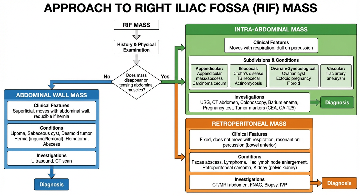
Key Decision Point
- No (persists) → Abdominal Wall Mass
- Yes (disappears) → Intra-abdominal or Retroperitoneal
1. Abdominal Wall Mass
- Lipoma, sebaceous cyst, desmoid tumor, hernia (inguinal/femoral), hematoma, abscess
- Investigations: Ultrasound, CT scan
2. Intra-abdominal Mass
- Appendicular: appendicular mass/abscess, carcinoma cecum
- Ileocecal: Crohn's disease, TB ileocecal
- Gynecological: ovarian cyst, ectopic pregnancy
- Vascular: iliac artery aneurysm
- Investigations: USG abdomen, CT abdomen, colonoscopy, barium enema, β-hCG, CA-125, CEA
3. Retroperitoneal Mass
- Psoas abscess, lymphoma, iliac lymphadenopathy, retroperitoneal sarcoma, pelvic kidney
- Clinical clue: Resonant on percussion (bowel loops lie anterior)
- Investigations: CT/MRI abdomen, FNAC/biopsy, IVP