
create a image of algorithm for how to approach for rif mass
Medical algorithm flowchart for approaching Right Iliac Fossa (RIF) mass in clinical practice. The flowchart should show: Start with "RIF Mass" at the top, then branch into History and Physical Examination, followed by investigations (CBC, ESR, CRP, Ultrasound abdomen, CT scan). Then differential diagnosis branches including: Appendicular mass/abscess, Carcinoma cecum, Crohn's disease, Ileal TB (ileocecal tuberculosis), Ovarian cyst/tumor (in females), Psoas abscess, Lymphoma, Mesenteric cyst. Further branches showing management pathways: Conservative vs Surgical, with specific steps for each diagnosis. Include key distinguishing features and decision points. Clean professional medical flowchart style with boxes, arrows, and color coding.
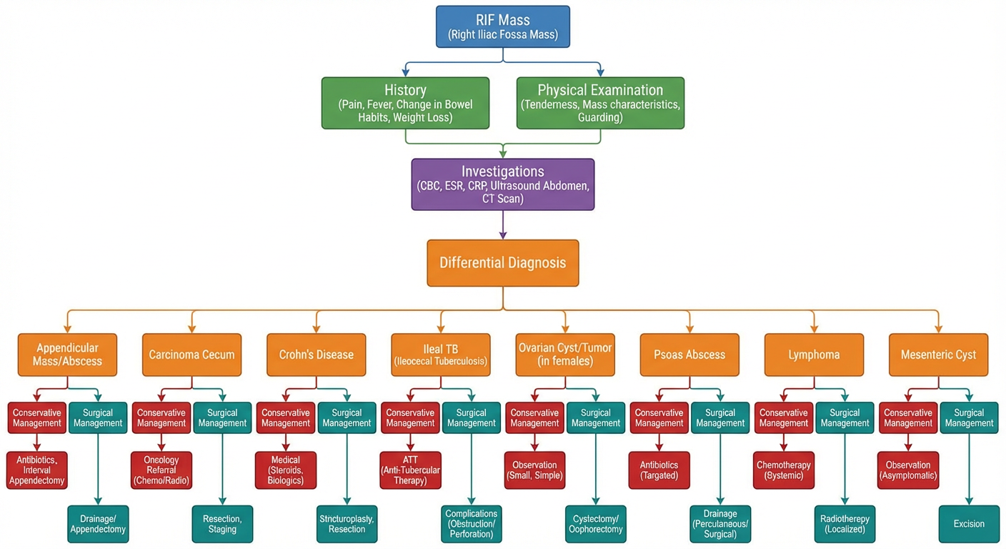
- History & Examination — starting point for assessment
- Investigations — bloods (CBC, ESR, CRP) and imaging (USG, CT scan)
- Differential Diagnoses — including:
- Appendicular mass/abscess
- Carcinoma cecum
- Ileocecal tuberculosis
- Crohn's disease
- Ovarian pathology (females)
- Psoas abscess
- Lymphoma / Mesenteric cyst
- Management pathways — conservative vs. surgical decision branches