
Fever
Reading File
The query "Fever" is broad. Let me clarify what you're looking for before diving into research.
Asking for Preferences
What aspect of fever are you interested in?
Searching sources...
Finding Sources
Finding Sources
Reading File
Reading File
Reading File
Reading File
Loading Image
Here is a thorough breakdown of the pathophysiology and mechanisms of fever, drawn from Guyton & Hall Medical Physiology, Janeway's Immunobiology, and Cellular & Molecular Immunology.
Pathophysiology of Fever
1. Normal Temperature Regulation
The anterior hypothalamic-preoptic area acts as the body's "thermostat," maintaining core temperature around 98.6°F (37°C). It does this by balancing heat production (metabolism, shivering, brown fat catabolism) with heat loss (vasodilation, sweating, radiation).
2. Triggering Fever: Pyrogens
Exogenous pyrogens are foreign substances - classically bacterial lipopolysaccharide (LPS/endotoxin) from gram-negative bacteria cell walls, as well as other microbial products. LPS acts partly through TLR-4 (Toll-like receptor 4) signaling.
Endogenous pyrogens are cytokines produced by the host's own immune cells (macrophages, monocytes, dendritic cells, NK cells) in response to infection, tissue injury, or inflammation. The key endogenous pyrogens are:
- IL-1β (Interleukin-1 beta) - also historically called "leukocyte pyrogen"
- TNF-α (Tumor necrosis factor-alpha)
- IL-6 (Interleukin-6)
Note: Even exogenous LPS ultimately causes fever by stimulating production of these endogenous cytokines. The historical distinction (exogenous vs. endogenous) is largely conceptual now.
3. The Core Mechanism: Cytokines → PGE2 → Hypothalamus
The pathway works as follows:
Infection / Tissue damage
        ↓
Macrophages/Dendritic cells phagocytize pathogens
        ↓
Release of IL-1β, TNF-α, IL-6 (endogenous pyrogens)
        ↓
These cytokines induce COX-2 (cyclooxygenase-2) expression
in hypothalamic vascular endothelial cells and glial cells
        ↓
COX-2 converts arachidonic acid → Prostaglandin E2 (PGE2)
        ↓
PGE2 acts on the hypothalamic preoptic area
        ↓
Raises the temperature "set point"
        ↓
Body responds to the new set point with heat-conserving + heat-generating mechanisms
        ↓
FEVER
This is why aspirin and NSAIDs reduce fever - they inhibit COX (cyclooxygenase), blocking PGE2 synthesis from arachidonic acid. - Guyton and Hall Medical Physiology
4. How the Body Raises Its Temperature
Once the hypothalamic set point is elevated, the body perceives itself as "too cold" relative to the new target. It activates heat-gain mechanisms:
| Mechanism | Effect |
|---|---|
| Peripheral vasoconstriction | Reduces heat loss through skin (causes pallor/cold skin) |
| Shivering | Rapid skeletal muscle contractions generate heat |
| Piloerection | Traps air near skin (less effective in humans) |
| Epinephrine secretion | Increases metabolic rate |
| Increased brown fat catabolism | Thermogenesis |
The patient experiences chills during this rising phase because the actual body temperature is still below the new set point.
5. The Fever State
Once core temperature matches the new set point (~103°F in the diagram), chills stop. The patient no longer feels hot or cold. Temperature is maintained at the elevated level as long as the pyrogenic stimulus persists.
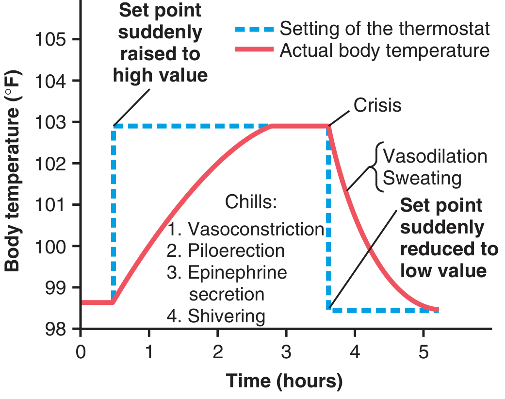
Figure: Hypothalamic thermostat set point vs. actual body temperature over time. (Guyton & Hall)
6. Resolution: The "Crisis" (Flush)
When the pyrogenic stimulus is removed (e.g., antibiotic clears infection), PGE2 levels fall and the hypothalamic set point resets to normal. Now the body temperature is above the set point, so heat-loss mechanisms activate:
- Vasodilation (flushing, hot skin)
- Profuse sweating
This rapid defervescence is called the crisis or flush. - Guyton and Hall Medical Physiology, p. 901
7. Broader Systemic Effects of the Pyrogen Cytokines
IL-1β, TNF-α, and IL-6 do more than just cause fever - they coordinate the entire acute-phase response:
- Liver: Hepatocytes produce acute-phase proteins (CRP, fibrinogen, SAP, complement proteins) - useful opsonins
- Bone marrow: Release of neutrophils and monocytes (→ leukocytosis)
- Muscle/fat: Increased catabolism to fuel immune cells
- Brain: Fever plus sickness behavior (fatigue, anorexia, sleepiness)
At very high TNF-α levels (as in severe sepsis), the response becomes pathological: myocardial depression, vasodilation, shock, and disseminated intravascular coagulation (DIC). - Janeway's Immunobiology 10e
8. Is Fever Beneficial?
Fever is generally considered beneficial to host defense:
- Higher temperatures impair bacterial and viral replication
- Adaptive immune cells (lymphocytes, neutrophils) function more efficiently at elevated temperatures
- Behavioral changes (rest, reduced food intake) conserve energy for the immune response
However, extreme hyperpyrexia (>105-108°F / 40.5-42°C) is damaging - causing neuronal death, hepatic and renal injury, and can be fatal if not treated promptly. - Cellular and Molecular Immunology, p. 282
Summary Table
| Step | Key Players | Result |
|---|---|---|
| Pathogen exposure | LPS, viral proteins | Macrophage/DC activation |
| Cytokine release | IL-1β, TNF-α, IL-6 | Endogenous pyrogen flood |
| COX-2 induction | Hypothalamic cells | PGE2 synthesis |
| Set point raised | Hypothalamus | Body "thinks" it's cold |
| Heat conservation + production | Vasoconstriction, shivering | Temperature rises (chills phase) |
| Fever maintained | Set point held high | Regulation at new temperature |
| Resolution | Pyrogen removed, PGE2 falls | Vasodilation, sweating (flush/crisis) |
This is a shared conversation. Sign in to Orris to start your own chat.