Essentials of Oral Histology and Embryology: A Clinical Approach – Chapter Connective Tissue, especially Figures 2-6 and 2-7 (loose and dense connective tissue

Reading File
Finding Sources
Finding Sources
Finding Sources
Finding Sources
Reading File
Reading File
Reading File
Reading File
Loading Image
Loading Image
Reading File
Loading Image
I now have everything needed for a thorough, well-illustrated answer. Let me compile it.

Connective Tissue - Loose and Dense Types

(Essentials of Oral Histology and Embryology: A Clinical Approach - Chapter: Connective Tissue, Figs. 2-6 & 2-7)
Note: "Essentials of Oral Histology and Embryology" (Chiego) is not in the library's collection. The content and figures below are sourced from Histology: A Text and Atlas with Correlated Cell and Molecular Biology (Pawlina), which contains the same connective tissue plates and equivalent figures. The histology is identical in content and the images directly correspond to what Figs. 2-6 and 2-7 illustrate in the Chiego textbook.

Overview: Classification of Connective Tissue Proper

Connective tissue proper divides into two broad categories based on the ratio of fibers to cells and the organization of collagen:
FeatureLoose (Areolar) CTDense CT
CellsMany, diverse typesFew, mainly fibroblasts
Collagen fibersThin, sparse, loosely arrangedThick, abundant
Ground substanceAbundant, gel-likeRelatively little
StrengthLowHigh
LocationBeneath epithelia, around glands, vesselsDermis, submucosa, tendons, ligaments

Loose Connective Tissue (Areolar Tissue)

Corresponds to Fig. 2-6 in Chiego
Loose connective tissue is characterized by loosely arranged, thin collagen fibers and an abundance of cells of many types embedded in a gel-like ground substance. The ground substance occupies more volume than the fibers and is viscous, playing a key role in diffusing oxygen and nutrients from small vessels and removing CO2 and metabolic waste.
Cellular population includes:
  • Fibroblasts - produce and maintain the ECM
  • Lymphocytes, macrophages, plasma cells, eosinophils, mast cells - immune/defense cells
Typical locations in oral tissues:
  • The lamina propria beneath oral epithelium
  • Connective tissue papillae
  • Surrounding minor salivary glands and blood vessels
  • Gingival connective tissue (superficial zones)
At low magnification, loose connective tissue is recognizable by the large number of nuclei visible - reflecting the cellular richness - and the pale, wispy appearance of the sparse collagen network.

Dense Connective Tissue

Dense CT contains abundant, thick collagen fiber bundles with very few cells (almost exclusively fibroblasts). Ground substance is minimal. It exists in two subtypes:

Dense Irregular CT

Also illustrated in the comparison figures
Collagen fiber bundles run in multiple, interlacing directions ("irregular"). This multi-directional weave allows the tissue to resist stress from many angles.
  • Found in: reticular layer of the dermis, submucosa of hollow organs, periosteum, organ capsules
  • In the oral region: the deeper connective tissue of the gingiva and palatal submucosa

Dense Regular CT

Collagen bundles are arranged in strict parallel arrays, maximizing tensile strength along one axis. Fibroblasts (called tendinocytes) lie compressed between fiber bundles and appear as rows of flattened nuclei on longitudinal section.
  • Found in: tendons, ligaments, aponeuroses
  • Oral relevance: periodontal ligament (PDL) is a prime example - the principal fibers run in organized groups (oblique, horizontal, apical, alveolar crest, transseptal groups) from cementum to alveolar bone

Histological Comparison - Key Images

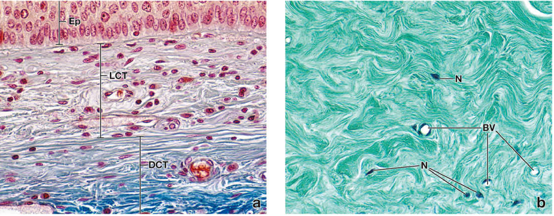
Figure 4.2 / equivalent to Chiego Figs. 2-6 & 2-7 — Epiglottis section, Mallory-Azan stain, ×540
Loose (LCT) and Dense (DCT) connective tissue comparison - epiglottis, Mallory-Azan stain
(a) Left panel: Section through the epiglottis showing:
  • Ep (top) = stratified epithelium
  • LCT (middle zone) = loose connective tissue - note the many nuclei of various sizes and shapes; elongated nuclei belong to fibroblasts; the collagen stains pale/thin
  • DCT (bottom zone) = dense connective tissue - intensely blue-stained thick collagen bundles, markedly fewer nuclei
(b) Right panel: Dense connective tissue alone at higher magnification:
  • Numerous, densely packed interlacing collagen fibers (blue-green with Mallory stain)
  • Only scattered fibroblast nuclei (N) visible
  • Small blood vessels (BV) present but sparse

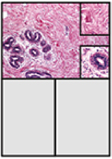
Plate 6.1 — Loose and Dense Irregular CT, Mammary Gland, H&E ×175 (with ×350 insets)
Plate 6.1: Loose and dense irregular connective tissue, mammary gland H&E
This plate directly parallels what Chiego shows in the oral context:
  • Glandular epithelium (GE) is surrounded by loose connective tissue (LCT) - numerous mixed cell nuclei, sparse wispy fibers
  • Peripheral areas show dense irregular CT (DICT) - thick collagen bundles, very few cells
  • Upper inset (higher mag of DICT): few nuclei relative to massive collagen expanse
  • Lower inset (higher mag of LCT): abundant cells, very few fibers

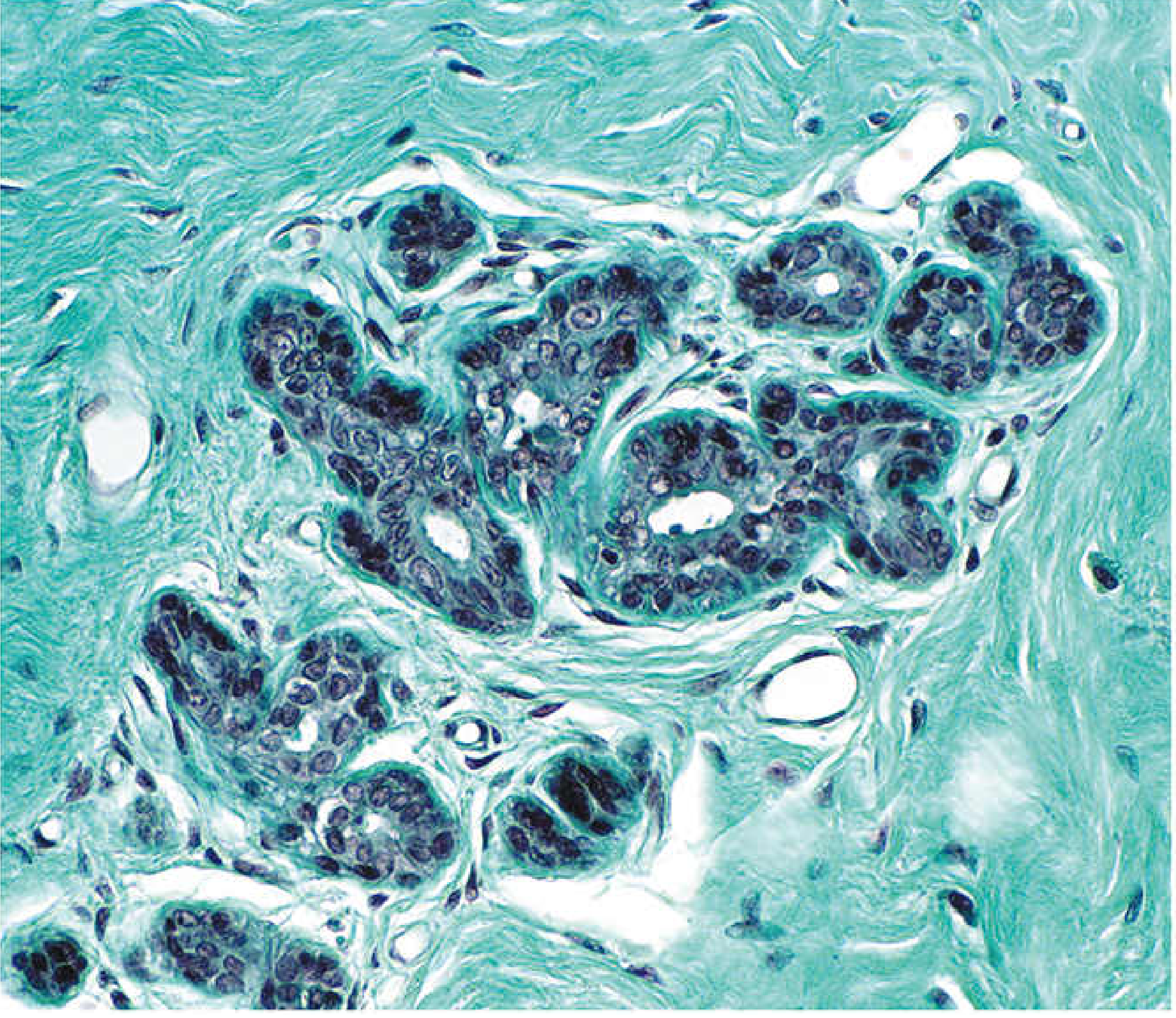
Figure 6.3 — Masson Trichrome comparison, Mammary Gland
Figure 6.3: Masson trichrome showing loose CT surrounding glandular epithelium vs. dense CT peripherally
With Masson trichrome, collagen stains blue-green. The loose connective tissue around the glands is pale and cellular; the dense irregular CT at the edges is deeply stained and acellular by comparison.

Clinical Relevance to Oral Histology

StructureCT TypeClinical Significance
Lamina propria (oral mucosa)Loose CT (superficial papillary layer) + Dense CT (deep reticular layer)Site of inflammatory cell infiltration in gingivitis/periodontitis
Periodontal ligamentDense regular CTPrincipal fibers transmit occlusal loads; loss leads to tooth mobility
Gingival connective tissueMixed loose + dense irregular CTDense collagen supports epithelial attachment; altered in fibrotic conditions
Dental pulpLoose CT (specialized)Rich in cells (odontoblasts, fibroblasts), vessels, nerves; ground substance-rich
Palatal submucosaDense irregular CTProvides firmness for masticatory mucosa

Key Differentiating Points for Exam

  1. The ratio of cells to fibers is the primary histological distinction: loose CT = many cells + few thin fibers; dense CT = few cells + many thick fibers
  2. Ground substance dominates in loose CT; collagen dominates in dense CT
  3. In H&E sections, loose CT appears pale pink with numerous nuclei; dense CT appears deeply eosinophilic with sparse nuclei
  4. With Masson trichrome or Mallory-Azan, collagen stains blue, making loose vs. dense CT immediately obvious
  5. The periodontal ligament is the oral clinician's most important example of dense regular CT - its principal fiber groups are organized to resist different vectors of occlusal force
  • Histology: A Text and Atlas with Correlated Cell and Molecular Biology, pp. 301-302, 440-443, 537-538 (Pawlina)

Ten Cate's Oral Histology: Development, Structure, and Function – Chapters Structure of the Oral Tissues and Cytoskeleton, Fibroblasts & Extracellular Matrix contain connective tissue illustrations, but not a dedicated "functions of connective tissue" flow diagram

Finding Sources
Finding Sources
Finding Sources
Finding Sources
Finding Sources
Finding Sources
Reading File
Reading File
Generating Image

A clean, labeled flowchart diagram titled "Functions of Connective Tissue" showing the main functions branching from a central box: 1. Structural Support (binding and supporting organs, fascia, dermis), 2. Defense and Immunity (macrophages, mast cells, plasma cells, lymphocytes), 3. Storage (adipose tissue, energy reserves, water and electrolytes in ground substance), 4. Repair and Wound Healing (fibroblasts synthesize collagen, scar formation), 5. Transport (diffusion of nutrients and waste via ground substance, blood vessels), 6. Signaling (growth factors, cytokines from ECM). Each branch has sub-bullet labels. Medical education style, white background, clear sans-serif font, color-coded branches.

Loading Image
Loading Image
I now have everything needed for a thorough response. Here is the complete answer:

Ten Cate's Oral Histology - Connective Tissue: Cytoskeleton, Fibroblasts & ECM

Library note: Ten Cate's Oral Histology is not in the collection. The content below is drawn from Histology: A Text and Atlas with Correlated Cell and Molecular Biology (Pawlina) and Junqueira's Basic Histology (17e), which cover the same topics in equivalent depth. The "functions of connective tissue" flow diagram you identified as missing from Ten Cate has been generated below.

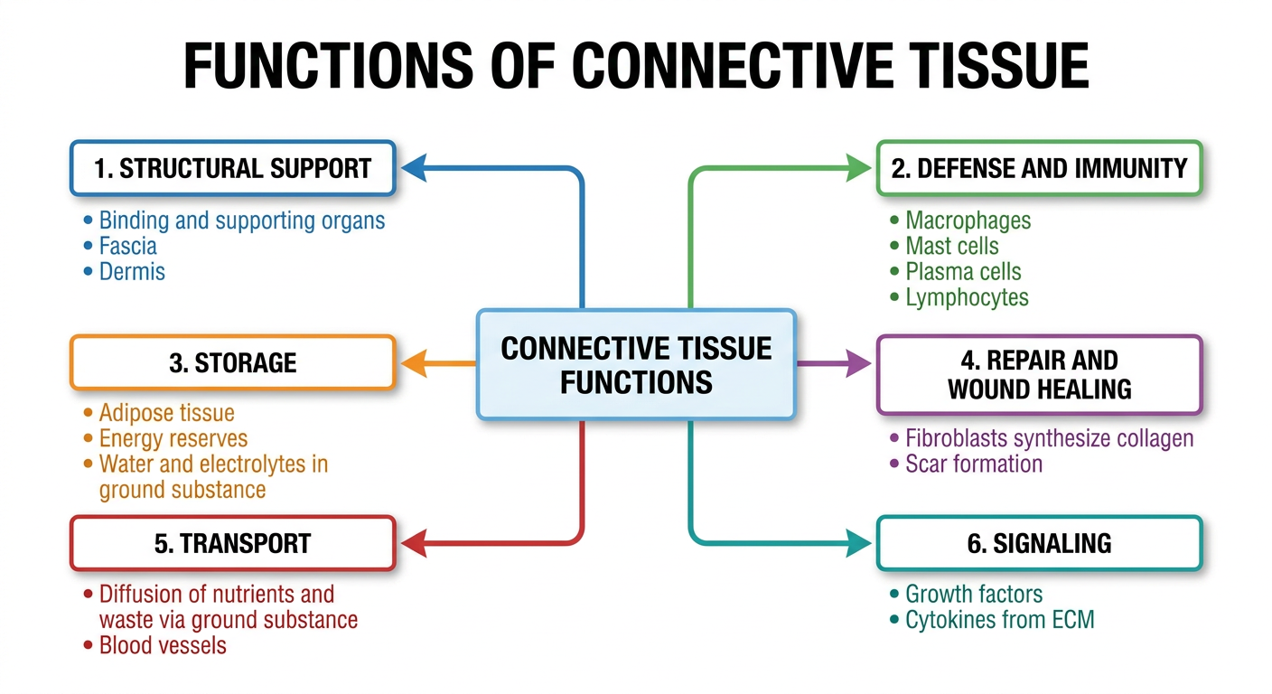
Functions of Connective Tissue - Flow Diagram

(This is the diagram Ten Cate's chapters do not include)
Functions of Connective Tissue flow diagram

1. Fibroblasts - The Principal Cell of Connective Tissue

The fibroblast is the master producer of the connective tissue ECM. A single fibroblast is capable of synthesizing all major ECM components:
  • Collagen fibers (types I, III - the structural backbone)
  • Elastic fibers (elastin + fibrillin microfibrils)
  • Reticular fibers (type III collagen - scaffolding for lymphoid organs)
  • Ground substance - glycosaminoglycans (GAGs), proteoglycans, multiadhesive glycoproteins (fibronectin, laminin)

Fibroblast Morphology - What You See on Slides

StateLight Microscopy Appearance
Resting (fibrocyte)Only the elongated/disc-like nucleus is visible; thin pale cytoplasm blends with collagen
Active (wound repair)More extensive, basophilic cytoplasm due to increased rER for protein synthesis

Fibroblast TEM - Fig. 6.21 (Pawlina)

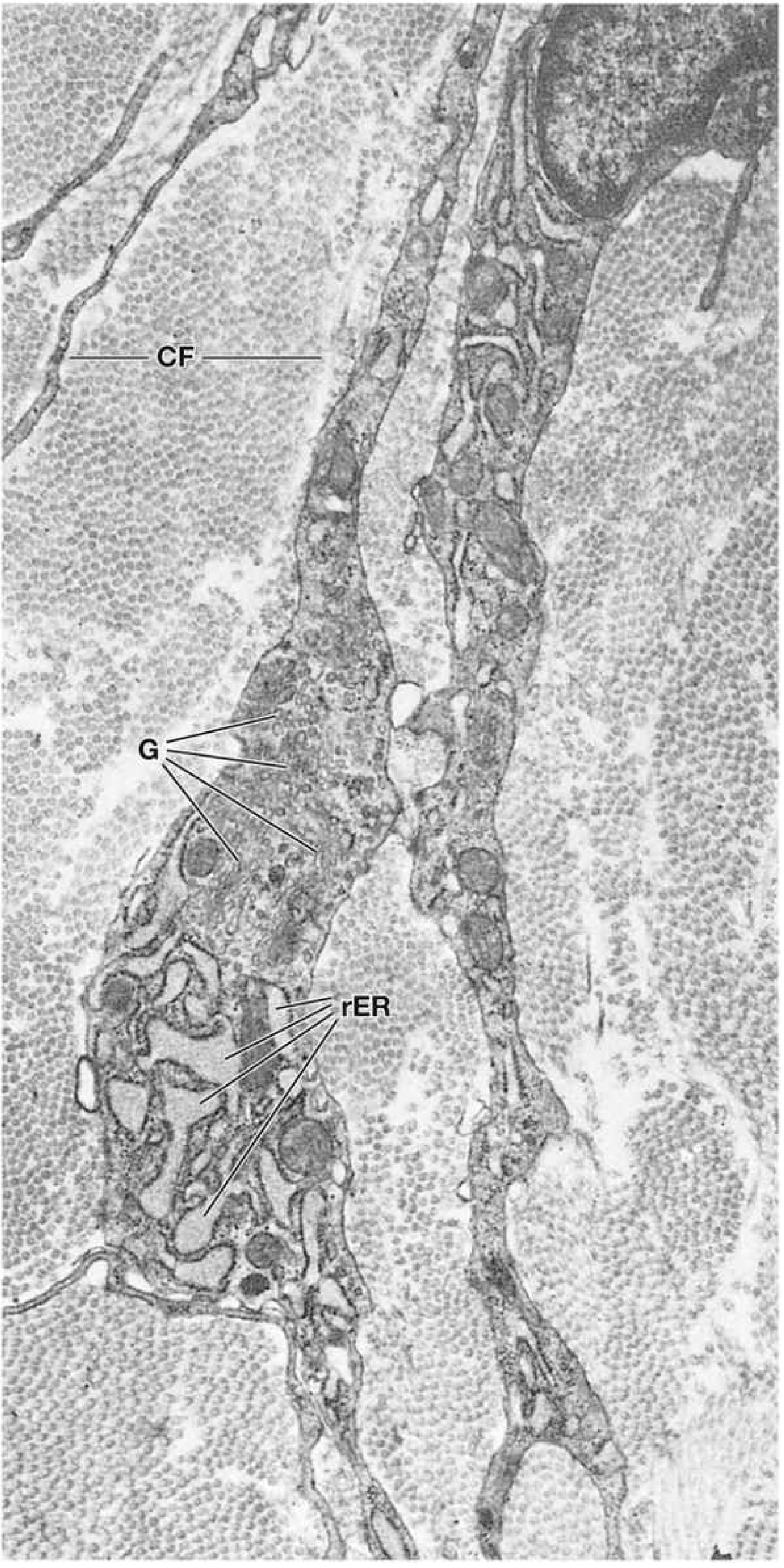
TEM of fibroblast showing rER, Golgi (G), and collagen fibers (CF)
At the ultrastructural level, the active fibroblast shows:
  • Abundant rough endoplasmic reticulum (rER) - site of procollagen synthesis
  • Prominent Golgi apparatus (G) - glycosylation and packaging of collagen and proteoglycans
  • Thin cytoplasmic processes lying between collagen fibers (CF)

Myofibroblast

During wound healing, fibroblasts differentiate into myofibroblasts - hybrid cells with contractile ability (actin stress fibers) used to close wounds. They are a key feature of healing oral wounds and the periodontal ligament.

2. Extracellular Matrix (ECM) Components

The ECM has two compartments: fibrous proteins and ground substance.

Fibrous Proteins

FiberCompositionKey PropertiesOral Location
CollagenFibrillar collagens (type I dominant)High tensile strength, 68 nm bandingPDL principal fibers, gingival CT, cementum, bone
ReticularType III collagenFine scaffolding, argyrophilic (silver stain)Around blood vessels, salivary gland acini
ElasticElastin core + fibrillin microfibrilsStretch and recoilElastic cartilage, large vessel walls, some ligaments
Collagen fiber formation (two-stage):
  1. Intracellular - procollagen synthesized in rER → glycosylated → assembled as triple helix → secreted as procollagen
  2. Extracellular - procollagen peptidase cleaves propeptides → tropocollagen → self-assembles into fibrils → cross-linked by lysyl oxidase → fibrils aggregate into fibers

Ground Substance - ECM and Proteoglycan Diagram

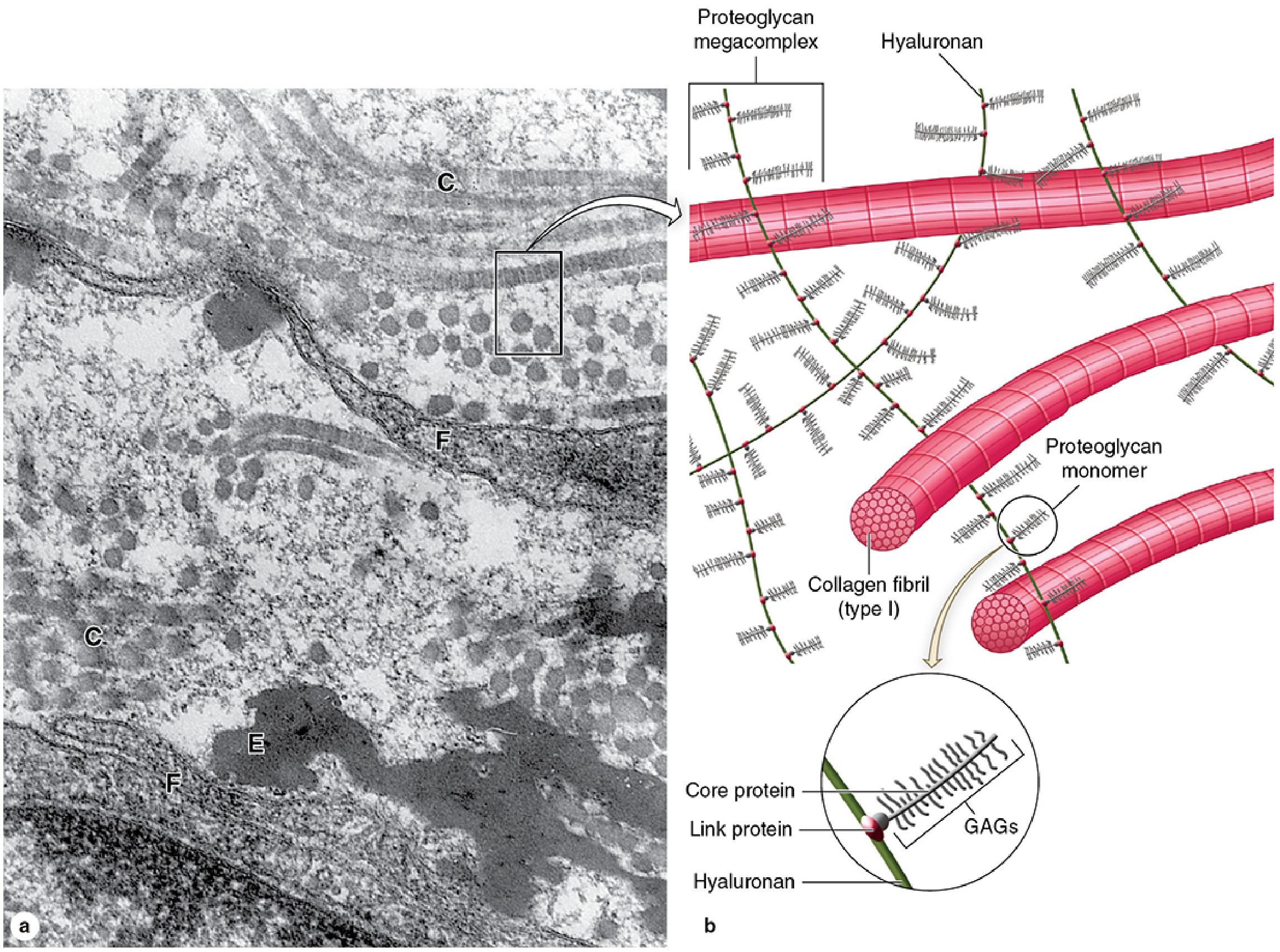
Ground substance ECM: TEM showing collagen (C), elastic (E), fibroblast processes (F), and schematic of proteoglycan megacomplex with hyaluronan, GAG chains, and collagen fibrils
Ground substance is a highly hydrated, transparent gel occupying the spaces between cells and fibers. It contains:
Glycosaminoglycans (GAGs) - long unbranched polysaccharide chains, highly negatively charged:
GAGLocationNotes
Hyaluronic acidUmbilical cord, synovial fluid, vitreous humor, cartilageNon-sulfated; forms backbone of proteoglycan megacomplex
Chondroitin 4- & 6-sulfateCartilage, bone, cornea, skinHigh collagen interaction (type II)
Heparan sulfateBasement membranesRegulates cell signaling
Dermatan sulfateSkin, tendons, heart valvesBinds collagen type I
Keratan sulfateCartilage, corneaCorneal transparency
Proteoglycans - core protein + covalently bound GAG side chains. Synthesized in rER and Golgi (like glycoproteins but more heavily glycosylated). The megacomplex - multiple proteoglycan monomers attached via link proteins to a central hyaluronan backbone - creates a sponge-like structure that:
  • Traps large volumes of water (hydration, turgor)
  • Acts as a lubricant between moving parts
  • Acts as a barrier to pathogen penetration
  • Enables diffusion of small molecules (O2, nutrients, waste)
Multiadhesive glycoproteins (fibronectin, laminin, tenascin) - link cells to ECM components via integrins, regulate cell migration, proliferation, and differentiation.

3. Functions of Connective Tissue - Detailed Breakdown

Since Ten Cate's chapters cover illustrations of structure without a dedicated functions diagram, here is the functional framework organized to map onto each structural component:

Structural / Mechanical Support

  • Dense CT of the gingival lamina propria maintains the gingival architecture and resists masticatory forces
  • Periodontal ligament (dense regular CT) transmits and absorbs occlusal loads via organized principal fiber groups
  • Submucosa of palatal mucosa = dense irregular CT providing rigidity for food processing

Defense and Immune Surveillance

Loose CT contains the resident immune cell population:
  • Macrophages - phagocytose pathogens, present antigen, secrete cytokines
  • Mast cells - release histamine and heparin in immediate hypersensitivity
  • Plasma cells - antibody secretion (especially IgA in oral submucosa)
  • Lymphocytes - adaptive immunity
  • Eosinophils - parasite defense, allergy
  • In gingivitis/periodontitis, neutrophils and monocytes invade from vessels into the loose CT of the lamina propria

Storage

  • Adipose tissue (specialized CT) - energy storage (triglycerides in adipocytes), insulation, cushioning; found in the buccal fat pad and some palatal regions
  • Ground substance - stores water, electrolytes, and growth factors

Transport / Metabolic Exchange

Ground substance acts as the medium through which oxygen, nutrients, hormones, and waste products diffuse between blood vessels and tissue cells. The PDL is especially vascular, allowing rapid metabolite exchange to support high fibroblast turnover.

Repair and Regeneration

  • Following oral injury, fibroblasts activate (→ myofibroblasts), upregulate collagen synthesis, and proliferate
  • Sequential stages: inflammation → granulation tissue (loose CT with new capillaries) → collagen remodeling → scar maturation
  • The high fibroblast turnover rate in oral tissues (especially PDL) means repair is faster than in skin

Signaling and Regulation

  • ECM-bound growth factors (TGF-β, FGF, PDGF) are released during remodeling and direct cell behavior
  • Proteoglycans modulate signaling gradients (e.g., heparan sulfate binds FGF)
  • Fibronectin and laminin provide positional cues for migrating cells during development and wound healing

4. Cytoskeleton - Relevance to Connective Tissue Cells

Ten Cate's cytoskeleton chapter, as it relates to connective tissue cells:
Cytoskeletal ComponentRole in Fibroblasts / Connective Tissue
Actin filamentsCell motility, wound contraction (myofibroblasts have prominent actin stress fibers)
Intermediate filaments (vimentin)Structural support of fibroblast; vimentin is the marker used to identify cells of mesenchymal origin
MicrotubulesIntracellular transport of procollagen vesicles from Golgi to cell surface

Oral Histology Integration Summary

Ten Cate Chapter ThemeStructural BasisFunction
Structure of oral tissuesEpithelium rests on loose CT lamina propriaDiffusion, defense, epithelial support
FibroblastsSpindle cells with rER + Golgi; activated in repairCollagen, elastic, reticular fiber + ground substance synthesis
ECM / CollagenType I dominant; 68 nm banding; organized in PDL as principal fibersTensile strength, load transmission, scaffolding
Ground substanceGAGs + proteoglycans + glycoproteins; gel-likeHydration, diffusion, barrier, lubrication, signaling
CytoskeletonActin, vimentin, microtubules in fibroblastsMotility, structural integrity, vesicle transport
  • Histology: A Text and Atlas (Pawlina), pp. 500-502 (Fibroblasts), pp. 440-443 (Connective Tissue Proper)
  • Junqueira's Basic Histology 17e, pp. 289-290 (Ground Substance, GAGs, Proteoglycans)

Ten Cate's Oral Histology: Development, Structure, and Function – Chapters Structure of the Oral Tissues and Cytoskeleton, Fibroblasts & Extracellular Matrix contain connective tissue illustrations, but not a dedicated "functions of connective tissue" flow diagram

Asking for Preferences
Reading File
Finding Sources
Finding Sources
Finding Sources
Reading File
Reading File
Reading File
Reading File
Reading File
Reading File
Loading Image
Loading Image
Loading Image
Reading File
Reading File
Loading Image
Here are the full notes for all three chapters, structured as Ten Cate's covers them, with illustrations sourced from the library's histology textbooks.
Library note: Ten Cate's Oral Histology is not in the collection. Notes below are compiled from Junqueira's Basic Histology (17e), Histology: A Text and Atlas (Pawlina), and Robbins & Cotran Pathologic Basis of Disease, which cover exactly the same material as Ten Cate's three chapters on Structure of Oral Tissues, Cytoskeleton, and Fibroblasts & ECM.

Chapter Notes: Cytoskeleton | Fibroblasts | Extracellular Matrix

(Equivalent to Ten Cate's Oral Histology - Structure of Oral Tissues chapters)

PART 1 — THE CYTOSKELETON

The cytoplasmic cytoskeleton is a complex array of three protein polymer systems that determine cell shape, enable movement, and direct intracellular transport.

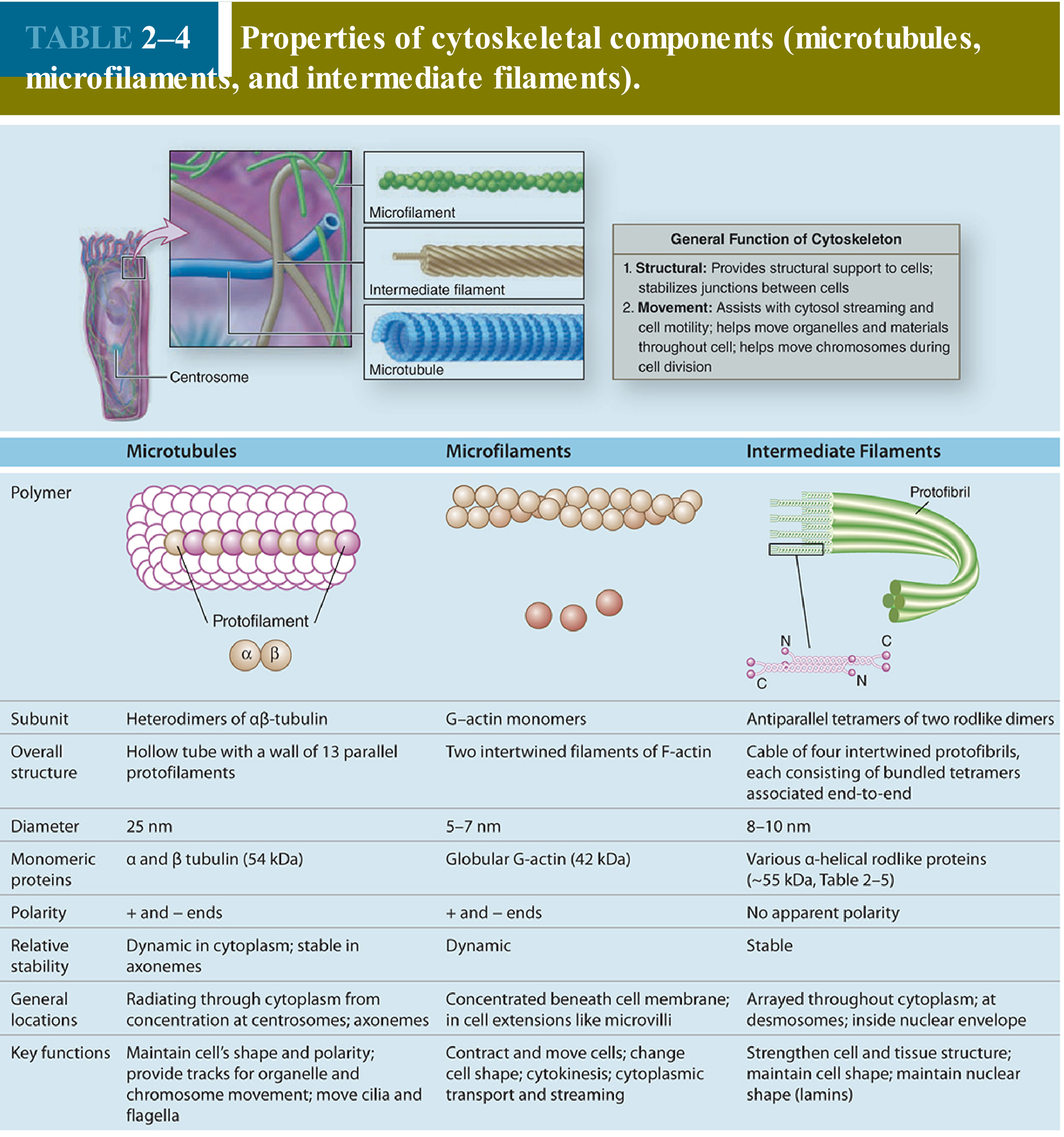
Table 2-4: Properties of Cytoskeletal Components

Table 2-4: Properties of cytoskeletal components - microtubules, microfilaments, and intermediate filaments with diagrams and key functions

1. Microtubules

  • Subunit: Heterodimers of α- and β-tubulin (54 kDa each)
  • Structure: Hollow tube; wall of 13 parallel protofilaments; outer diameter 25 nm; 5-nm-thick wall
  • Polarity: + and - ends; dynamic in cytoplasm, stable in axonemes
  • Location: Radiate through cytoplasm from the centrosome (microtubule-organizing center) near the nucleus; form axonemes of cilia and flagella
  • Key functions:
    • Maintain cell shape and polarity
    • Provide tracks for kinesin (anterograde, toward + end) and dynein (retrograde, toward - end) motor proteins carrying vesicles and organelles
    • Move chromosomes during cell division (mitotic spindle)
    • Transport procollagen secretory vesicles from Golgi to cell surface in fibroblasts

2. Microfilaments (Actin Filaments)

  • Subunit: Globular G-actin monomers (42 kDa) → polymerize to filamentous F-actin
  • Structure: Two intertwined filaments of F-actin; diameter 5-7 nm
  • Polarity: + (barbed) and - (pointed) ends; highly dynamic
  • Location: Concentrated beneath the cell membrane; in microvilli; throughout cytoplasm in stress fibers
  • Key functions:
    • Maintain and change cell shape
    • Cell locomotion and migration (critical for fibroblast migration in wound healing)
    • Cytokinesis (contractile ring at cell division)
    • Cytoplasmic streaming; endocytosis
Actin-binding proteins regulate filament length and cross-linking - examples include filamin (cross-linking), cofilin (severing), profilin (promotes polymerization), and formins (nucleation). Cross-linking increases cytoplasmic viscosity; severing decreases it.

3. Intermediate Filaments

  • Diameter: 8-10 nm (between the other two - hence "intermediate")
  • Structure: Stable cables of antiparallel tetramers of two rodlike dimers; four protofibrils per cable; no polarity
  • Stability: Stable (unlike the other two, which are dynamic)
  • Location: Throughout cytoplasm; at desmosomes; inside nuclear envelope (lamins)
  • Key functions: Strengthen cell and tissue structure; maintain cell and nuclear shape
Cell-type-specific intermediate filament proteins (Table 2-5):
ClassProteinSize (kDa)Cell TypeOral Relevance
I/IICytokeratins (acidic/basic)40-65Epithelial cellsOral mucosal epithelium, enamel organ
IIIVimentin54Fibroblasts, endothelium, leukocytesMarker of mesenchymal cells in CT
IIIDesmin53Muscle cellsPDL smooth muscle-like cells
IIIGFAP51Glial cells (astrocytes)Peripheral nerve sheaths
IVNeurofilament proteins57-200NeuronsDental pulp nerve fibers
VLamins A, B, C60-70All nucleated cellsNuclear envelope scaffold
Key exam point: Vimentin is the diagnostic intermediate filament marker for cells of mesenchymal origin - fibroblasts, odontoblasts, pulpal cells. Cytokeratins mark epithelial cells. This distinction is used in histopathology to characterize oral tumors.

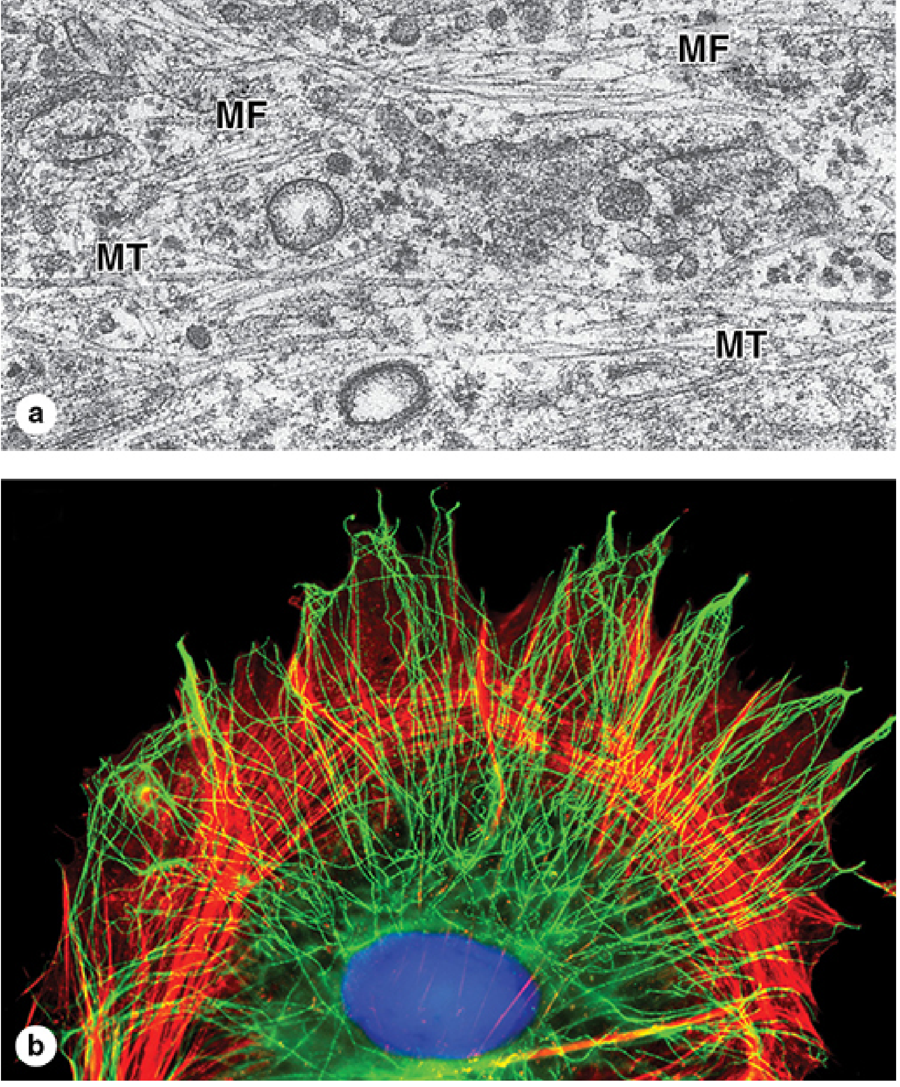
Cytoskeleton TEM and Immunofluorescence - Figure 2-22

Figure 2-22: (a) TEM of fibroblast cytoplasm showing microtubules (MT) and actin microfilaments (MF) - compare relative diameters (×60,000). (b) Immunofluorescence of cultured cell: actin filaments (red) concentrated peripherally; microtubules (green) radiate from centrosome
(a) TEM showing MT (microtubules, ~25 nm, darker thick profiles) and MF (microfilaments, 5-7 nm, finer profiles) side by side in fibroblast cytoplasm.
(b) Immunofluorescence: actin (red) forms circumferential bundles at cell periphery and projects into lamellipodia; microtubules (green) radiate centrifugally from centrosome toward cell periphery.

PART 2 — FIBROBLASTS

The Fibroblast as the Principal Cell of Connective Tissue

The fibroblast is the primary resident cell of all connective tissues. A single fibroblast can synthesize all ECM components:
  • Collagen fibers (type I, III, V...)
  • Elastic fibers (elastin + fibrillin microfibrils)
  • Reticular fibers (type III collagen)
  • All ground substance components: GAGs, proteoglycans, multiadhesive glycoproteins

Morphology

In routine H&E:
  • Only the elongated or disc-like nucleus is typically visible
  • Thin, pale-staining cytoplasmic processes blend invisibly with surrounding collagen fibers
  • The resting cell (fibrocyte) has a condensed, darkly-staining nucleus
In active/wound repair state:
  • More extensive cytoplasm
  • Basophilic cytoplasm due to expanded rER (increased protein synthesis)
  • Distinguishable as a cell body separate from the fibrous matrix
By TEM - Figure 6.21:
TEM of active fibroblast showing prominent rER cisternae (distended = active synthesis), Golgi apparatus (G), and surrounding collagen fibrils (CF) cut in cross section appearing as dots (×11,000)
The active fibroblast TEM shows:
  • rER - distended cisternae indicating active protein synthesis (procollagen)
  • Golgi apparatus (G) - packaging and glycosylation of procollagen
  • Collagen fibrils (CF) in cross-section appearing as small dots surrounding the cell

Fibroblast Activation - Resting vs. Active States

FeatureFibrocyte (resting)Active Fibroblast
TriggerNormal homeostasisGrowth, wound repair, inflammation
NucleusElongated, dark, condensedLarger, with visible nucleolus
CytoplasmBarely visible (pale, blends with collagen)Basophilic, extensive
rERSparseAbundant, distended
GolgiSmallProminent
FunctionMaintenance of existing ECMActive synthesis of new collagen + ECM

Myofibroblast

When tissue is injured, fibroblasts can differentiate into myofibroblasts - cells with hybrid properties of fibroblasts and smooth muscle cells.
Key features of myofibroblasts:
  • Express α-smooth muscle actin (α-SMA) - the key identifying marker
  • Contain prominent stress fibers of actin filaments visible on LM
  • Capable of wound contraction - physically pulling wound edges together
  • Produce large amounts of collagen type I and type III during granulation tissue formation
  • After wound healing, most undergo apoptosis; persistence leads to fibrosis
Oral relevance: Myofibroblasts are important in:
  • Healing of oral mucosal wounds
  • PDL remodeling under orthodontic forces
  • Formation of oral submucosal fibrosis (OSF) - a precancerous condition where myofibroblast persistence drives progressive fibrosis

Other Connective Tissue Cells

CellOriginKey FunctionOral Location
MacrophageMonocyte (bone marrow)Phagocytosis, antigen presentation, cytokine secretionLamina propria, around blood vessels
Mast cellHematopoietic stem cellIgE-mediated degranulation (histamine, heparin, tryptase)Perivascular in loose CT
Plasma cellB lymphocyteAntibody secretion (IgA, IgG)Gingival CT, lamina propria
LymphocyteLymphoid precursorAdaptive immunity (T and B cells)Throughout loose CT, increased in inflammation
EosinophilBone marrowParasite defense; allergyAllergic/parasitic lesions
AdipocyteMesenchymal stem cellLipid storage, energy reserve, signalingBuccal fat pad, palatal submucosa

PART 3 — EXTRACELLULAR MATRIX (ECM)

Overview

The ECM is a complex, dynamic, interactive structural network surrounding and supporting connective tissue cells. It is far more than passive scaffolding - it actively regulates cell behavior.
ECM functions (Robbins):
  1. Mechanical support - cell anchorage, migration pathways, maintenance of cell polarity
  2. Regulator of proliferation - binds and displays growth factors; provides depot of latent growth factors released at sites of injury
  3. Scaffold for tissue renewal - basement membrane integrity is required for organized tissue regeneration; ECM disruption prevents effective repair
  4. Foundation for tissue microenvironments - basement membranes create tissue boundaries (e.g., epithelium-connective tissue interface in oral mucosa)

ECM-Cell Signaling Diagram (Robbins Fig. 1.14)

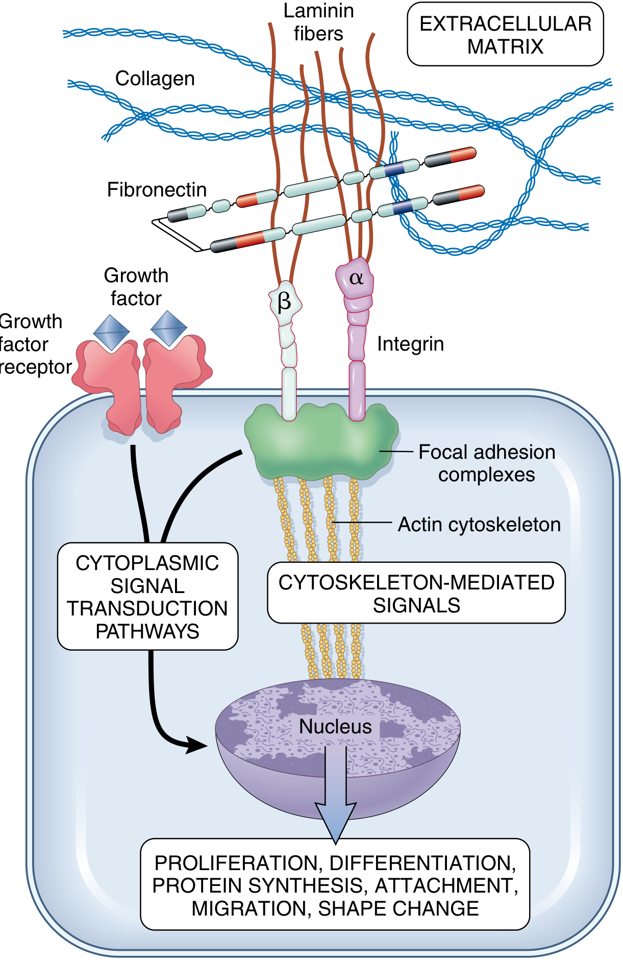
ECM-integrin-cytoskeleton signaling diagram: collagen, laminin, fibronectin in ECM; α and β integrin subunits span membrane; connect to actin cytoskeleton via focal adhesion complexes (vinculin, α-actinin, talin); signal to nucleus → proliferation, differentiation, migration, shape change
This diagram is the key to understanding how the cytoskeleton and ECM are functionally linked:
  • ECM components (collagen, fibronectin, laminin) bind integrin receptors (α and β subunits) on the cell surface
  • Integrins connect extracellularly to ECM and intracellularly to the actin cytoskeleton via focal adhesion complexes (containing vinculin, α-actinin, talin)
  • This connection transmits both mechanical signals (mechanosensing) and biochemical signals to the nucleus
  • Outcome: changes in proliferation, differentiation, protein synthesis, cell attachment, migration, and shape

ECM Forms

Interstitial matrix:
  • Fills spaces between stromal cells in connective tissue
  • Synthesized by mesenchymal cells (fibroblasts)
  • Semi-fluid, amorphous gel
  • Components: fibrillar collagens, fibronectin, elastin, proteoglycans, hyaluronate
  • Functions as cushion against compression (e.g., in peristalsis, pulsatile arterial flow)
Basement membrane:
  • Highly organized ECM around epithelial cells, endothelial cells, and smooth muscle cells
  • Bilaminar structure: lamina lucida (electron-lucent, contains laminin) + lamina densa (electron-dense, type IV collagen network)
  • Components: type IV collagen, laminin, nidogen, perlecan (heparan sulfate proteoglycan)
  • In the oral cavity, the basement membrane separates oral epithelium from lamina propria and anchors epithelial cells via hemidesmosomes

ECM Components in Detail

A. Collagen Fibers

Collagen is the most abundant structural protein in the body (~30% of total protein mass). Over 28 types exist; types I, II, III, IV, and V are most relevant.
Collagen biosynthesis - Figure 6.8:
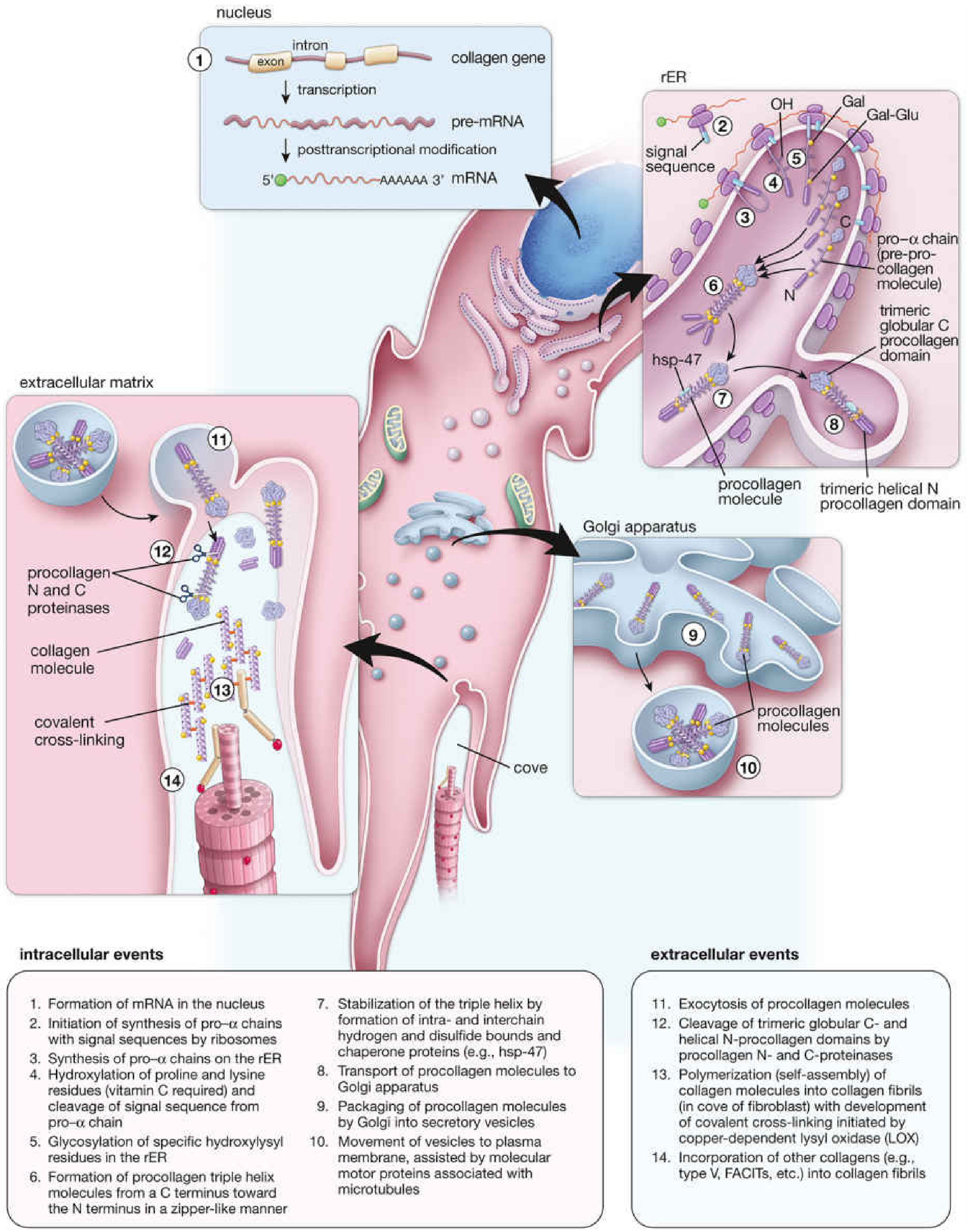
Collagen biosynthesis diagram showing intracellular events (nucleus → rER → Golgi → secretory vesicles) and extracellular events (fibril assembly, cross-linking by lysyl oxidase)
Intracellular steps:
  1. Collagen gene transcription in nucleus → mRNA
  2. Pro-α-chains synthesized on rER ribosomes with signal sequence
  3. Signal sequence cleaved in rER cisternae
  4. Hydroxylation of proline and lysine residues - requires vitamin C as cofactor (prolyl and lysyl hydroxylase enzymes)
    • Without hydroxylation → no stable triple helix → defective collagen → scurvy (wounds fail to heal, bleeding gums, bone formation impaired)
  5. Glycosylation of hydroxylysine residues (O-linked sugars in rER; N-linked sugars at terminals)
  6. Triple helix formation starting from C-terminus toward N-terminus ("zipper-like")
  7. hsp-47 chaperone stabilizes the triple helix and prevents premature aggregation
  8. Procollagen transported to Golgi apparatus → packaged into secretory vesicles → exocytosis
Extracellular steps: 11. Exocytosis of procollagen molecules into ECM 12. Procollagen N- and C-proteinases cleave the globular propeptide domains → tropocollagen 13. Self-assembly of tropocollagen into collagen fibrils (in "cove" of fibroblast cell surface indentations); covalent cross-linking initiated by lysyl oxidase (LOX) - copper-dependent enzyme 14. Fibrils aggregate into collagen fibers; other collagen types (type V, FACITs) incorporated
Key collagen types in oral tissues:
TypeFiber FormLocation in Oral Cavity
IFibrillar (large fibers)PDL principal fibers, gingival CT, alveolar bone matrix, dentinal tubules, cementum
IIFibrillar (thinner fibers)Cartilage (TMJ disc)
IIIFibrillar (reticular fibers, thin)Loose CT, around blood vessels, early wound repair
IVNon-fibrillar (network)Basement membranes (oral epithelium-CT junction)
VFibrillar (associated with I)PDL, cornea, interstitial tissues

B. Elastic Fibers

  • Core of cross-linked elastin molecules surrounded by a sheath of fibrillin microfibrils
  • Elastic fibers allow tissues to stretch and recoil (unlike collagen, which resists stretch)
  • Produced by fibroblasts, chondrocytes, endothelial cells, smooth muscle cells
  • Special stains: orcein, resorcin-fuchsin (stain elastic fibers selectively)
  • Oral relevance: present in elastic cartilage of epiglottis; large vessels; some ligaments

C. Ground Substance

Ground substance is a viscous, clear, highly hydrated gel that fills the space between cells and fibers. It is invisible in routine H&E (extracted during fixation/dehydration) - the apparent "empty" space between cells and fibers is actually ground substance.
Three molecular groups:
1. Glycosaminoglycans (GAGs)
  • Long-chain, unbranched polysaccharides of repeating disaccharide units
  • Highly negatively charged (sulfate and carboxylate groups) → bind enormous quantities of water → responsible for gel-like physical properties
GAGSulfationKey LocationsOral Relevance
Hyaluronic acid (hyaluronan)NoneUmbilical cord, synovial fluid, vitreous humor, cartilage, loose CTGround substance of dental pulp; forms backbone of proteoglycan aggregates
Chondroitin 4- & 6-sulfateYesCartilage, bone, cornea, skinTMJ disc, bone matrix
Dermatan sulfateYesSkin, tendons, heart valvesGingival CT, PDL
Heparan sulfateYesBasement membranesOral epithelial BM
Keratan sulfateYesCartilage, corneaTMJ disc
HeparinYesMast cell granulesReleased in allergic/inflammatory reactions in gingival CT
2. Proteoglycans
  • Core protein + covalently bound GAG side chains
  • Synthesized in rER and Golgi (more heavily glycosylated than glycoproteins)
  • Examples: aggrecan (cartilage), syndecan-1 (epithelial cell surface), perlecan (basement membranes), decorin, biglycan (bind collagen fibers in PDL)
  • Form proteoglycan megacomplex: multiple monomers linked via link proteins to a hyaluronan backbone
3. Multiadhesive Glycoproteins
  • Bridge cells to ECM components via integrin receptors
GlycoproteinKey Binding PartnersFunction
FibronectinCollagen, integrins, heparan sulfateCell attachment to matrix; guides cell migration during wound repair and development
LamininType IV collagen, integrins, perlecanCell attachment in basement membranes; specific to epithelial cells
TenascinFibronectin, proteoglycansModulates cell adhesion; expressed during development and wound healing; anti-adhesive
OsteopontinIntegrins, CD44Bone/cementum matrix; cell attachment in mineralized tissues

Cytoskeleton-ECM Integration: The Full Picture

This is the connection Ten Cate's chapters build toward - how cytoskeleton and ECM are functionally inseparable:
ECM (collagen + fibronectin + laminin)
         ↓  [binds]
    Integrin receptors (α/β heterodimers, transmembrane)
         ↓  [intracellular tail]
    Focal adhesion complexes (vinculin, talin, α-actinin)
         ↓  [connect to]
    Actin cytoskeleton (microfilaments + stress fibers)
         ↓  [relay signals via]
    Signal transduction (FAK, Src kinases, Rho GTPases)
         ↓
    Nucleus → gene expression changes
         ↓
    Cell response: proliferation / differentiation / migration / shape change / ECM synthesis
In the oral context:
  • Odontoblast processes are maintained by microtubules (transport of vesicles) and actin (process shape)
  • Fibroblasts of the PDL use actin-based motility to migrate and remodel matrix under mechanical load
  • Oral epithelial cells are anchored to the basement membrane via laminin-5/integrin-α6β4 at hemidesmosomes - disruption leads to oral mucosal blistering diseases (pemphigoid)
  • Vimentin (intermediate filament) marks all CT cells of mesenchymal origin; cytokeratins mark epithelial cells - used routinely in diagnostic histopathology of oral lesions

Sources: Junqueira's Basic Histology 17e, pp. 118-131 (Cytoskeleton); Pawlina Histology pp. 488-502 (ECM, Fibroblasts, Collagen biosynthesis); Robbins & Cotran Pathologic Basis of Disease pp. 35-38 (ECM functions and signaling)
This is a shared conversation. Sign in to Orris to start your own chat.