
How to put injection in hips
intramuscular injection hip dorsogluteal ventrogluteal technique steps
https://www.webmd.com/a-to-z-guides/what-is-the-ventroglutea…
Intramuscular (IM) Injection in the Hip
- Ventrogluteal site (preferred - gluteus medius/minimus)
- Dorsogluteal site (buttock - now discouraged)
Ventrogluteal Site (Recommended)
Why it's preferred over the dorsogluteal site
- No major nerves or vessels nearby (the dorsogluteal site is close to the sciatic nerve and can cause leg paralysis if the nerve is hit)
- Thinner skin means the needle reliably reaches muscle
- Less painful
- Works for thin, average, and overweight patients
Step-by-Step: Finding the Ventrogluteal Site
Landmarking - V Method (standard BMI)
- Use the opposite hand - if injecting the right hip, use your left hand
- Place your palm over the greater trochanter (the bony bump on the side of the hip)
- Point your index finger toward the anterior superior iliac spine (ASIS) - the bony point at the front of the hip
- Spread your middle finger back toward the iliac crest (top ridge of the pelvis)
- This creates a V shape - inject into the center of the V
Landmarking - G Method (geometric, works for all BMIs)
Performing the Injection
| Step | Action |
|---|---|
| 1 | Wash hands, gather equipment (syringe, needle, alcohol swab, gloves, medication) |
| 2 | Select correct needle length - typically 38 mm (1.5 inch) for most adults; longer for obese patients |
| 3 | Draw up medication, remove air bubbles |
| 4 | Clean the site with an alcohol swab and let it dry completely |
| 5 | Spread or bunch the skin depending on the patient (spread for most adults) |
| 6 | Insert the needle at a 90-degree angle in one smooth motion |
| 7 | Aspirate (pull back the plunger slightly) - check for blood; if blood appears, withdraw and restart |
| 8 | Inject the medication slowly (about 1 mL per 10 seconds) |
| 9 | Withdraw the needle smoothly at the same angle |
| 10 | Apply gentle pressure with a cotton ball or gauze - do not rub (rubbing can cause irritation or force medication into subcutaneous tissue) |
| 11 | Dispose of needle safely in a sharps container - never recap |
Needle Size Guidelines
| Patient | Needle Gauge | Needle Length |
|---|---|---|
| Average adult | 21-23 G | 38 mm (1.5 in) |
| Thin adult | 23-25 G | 25 mm (1 in) |
| Obese adult | 21-23 G | 50-75 mm (2-3 in) |
Z-Track Technique (for irritating medications)
- Pull the skin and subcutaneous tissue 2-3 cm to the side before inserting the needle
- Insert and inject normally
- Wait 10 seconds after injecting
- Withdraw the needle, then release the skin - this seals the track and prevents leakage upward
Key Safety Points
- Never inject into the dorsogluteal (upper outer buttock) unless specifically directed - risk of sciatic nerve injury
- Always aspirate before injecting (especially for the gluteal area)
- Rotate sites if giving frequent injections
- Maximum volume per ventrogluteal site: 3-5 mL for an adult
- The ventrogluteal site should not be used in children under 7 months
Note: This is a clinical skill. If you are a patient giving injections at home (e.g., for self-administered medications), your healthcare provider should train you in person before you attempt this independently.
Make a photo of the bone showing at hips and forming a v shaped
Medical anatomical diagram of the hip bones showing the ventrogluteal injection site landmarks. Clear illustration of the pelvis and upper femur from the lateral/side view, highlighting the iliac crest, anterior superior iliac spine (ASIS), and greater trochanter of the femur. Show the V-shaped hand landmarking technique with index finger pointing to the ASIS and middle finger spread toward the iliac crest, forming a V shape. Label the bones clearly: iliac crest, anterior superior iliac spine, greater trochanter, and mark the injection point at the center of the V. Medical textbook style, clean white background, labeled diagram.
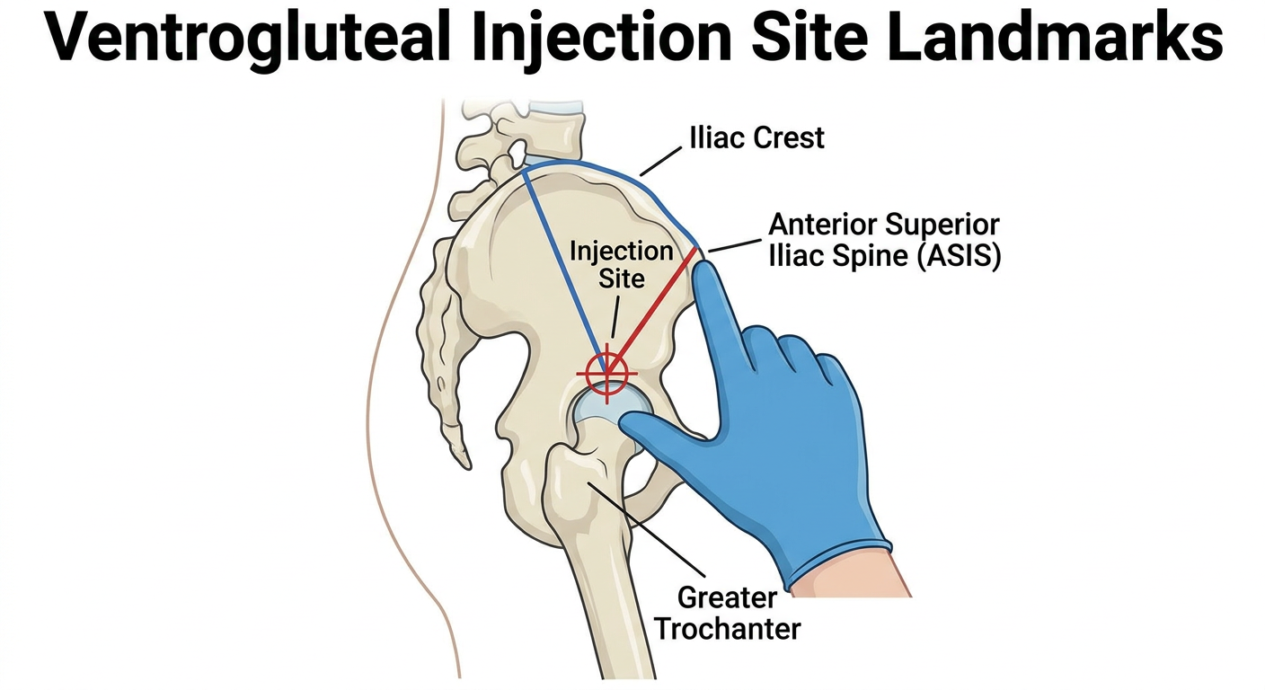
- Iliac Crest - the top ridge of the pelvis (your middle finger follows this)
- Anterior Superior Iliac Spine (ASIS) - the bony point at the front of the hip (your index finger points here)
- Greater Trochanter of the Femur - the large bony bump on the outer side of the upper thigh (your palm rests on this)