
PAS positivity in nodular hidradenoma
nodular hidradenoma clear cell histology PAS microscopy
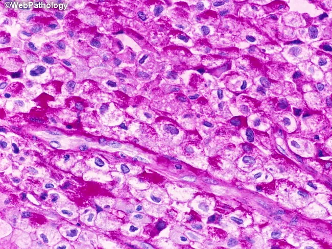
High-fidelity histopathology image captured with brightfield microscopy of a bladder mucosa biopsy stained with Periodic acid–Schiff (PAS). The tumor displays urothelial cells with abundant clear cytoplasm due to glycogen accumulation, characteristic of the clear cell variant of urothelial carcinoma. Cells are arranged in solid sheets and irregular nests within the urothelial/lamina propria region, with nuclear atypia including enlarged, hyperchromatic nuclei and conspicuous nucleoli. The PAS positivity highlights cytoplasmic glycogen; this reaction is typically diastase-labile (glycogen) and may require diastase treatment to confirm glycogen as the staining source (not shown). The tumor cells often border around a fibrovascular stroma, with occasional mitotic figures and occasional inflammatory infiltrates. The differential includes other glycogen-rich clear cell neoplasms and metastasis; immunohistochemical workup (e.g., GATA3, uroplakin, p63) supports urothelial origin while ruling out clear cell renal cell carcinoma. Clinically, this histology correlates with bladder mass lesions in adults and informs prognosis and treatment decisions, including cystectomy or intravesical therapy depending on invasion. This image demonstrates typical features of clear cell urothelial carcinoma, including glycogen-rich cytoplasm, clear cell morphology, and urothelial differentiation markers in adjunct studies. Correlation with imaging and flow cytometry may further refine staging; pathologist interpretation relies on morphological context and corroborative IHC profiles.
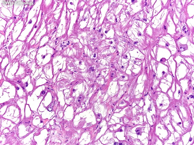
Light microscopy histology image of ventricular myocardial tissue stained with Periodic Acid-Schiff (PAS) highlighting glycogen-rich, vacuolated cardiac myocytes with spider cell morphology. Centrally placed nuclei with radiating, slender myofibrils produce the characteristic spider-like appearance and a distinctive periphery of cytoplasmic processes. PAS stain yields strong magenta coloration because of abundant glycogen, while conventional hematoxylin provides nuclear contrast. Electron microscopy described in the referenced material shows abundant glycogen, few irregular myofibrils, and poorly developed sarcoplasmic reticulum, supporting a benign, hamartomatous rhabdomyoma rather than a malignant sarcoma. Morphologically, tumor cells are large, polygonal, with clear cytoplasm and delicate, sparse myofilaments; cell borders may appear interwoven within the myocardium, reflecting a proliferative but well-differentiated muscular lineage. This constellation—spider cells, glycogen-rich cytoplasm, central nuclei, radiating myofibrils—constitutes a classic histopathologic signature for cardiac rhabdomyoma, especially in pediatric patients and in the context of tuberous sclerosis complex. Clinically, the presence of such tumors can yield arrhythmias or obstruction depending on size and location; prognosis is often favorable with potential spontaneous regression. Diagnostic relevance excludes most malignant cardiac neoplasms; differential diagnoses include rhabdomyosarcoma and myxoma, but immunophenotype and ultrastructure typically resolve the diagnosis. Correlation with clinical imaging and genetics further strengthens the definitive assessment in pediatric care.
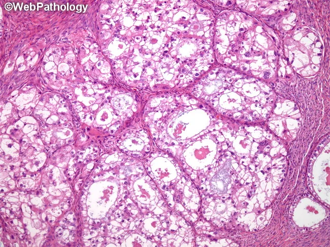
This is a bright-field light microscopy image of an ovarian tumor tissue section stained with Hematoxylin and Eosin. The specimen shows classic histology of ovarian clear cell carcinoma with abundant clear cytoplasm and distinct cell borders. Cells are polygonal and arranged in glandular, tubulocystic, and occasionally solid architectural patterns; hobnail cells with apical nuclei line some gland lumina. The cytoplasm is clear due to glycogen content; PAS-positive and diastase-sensitive. Nuclei are generally central to eccentric, with mild to moderate nuclear atypia and prominent nucleoli in a subset of cells. The tumor demonstrates tubulocystic structures embedded in a fibrous stromal background with delicate microvasculature. Overall distribution is infiltrative, lacking well-defined capsules in this field. There may be areas of microcystic change and focal hobnailing that help distinguish clear cell histology from other ovarian epithelial tumors. The image emphasizes characteristic features: mucin-free clear cytoplasm, glycogen-rich cytoplasm, increased cytoplasmic clearing, and distinct cell borders. Diagnostic significance lies in recognizing the cell morphology and architectural pattern that differentiate clear cell carcinoma from serous or endometrioid subtypes and from metastatic renal cell carcinoma. This morphology has clinical relevance for prognosis and guides platinum-based therapy and surgical management; endometriosis association may be noted in the clinical history.
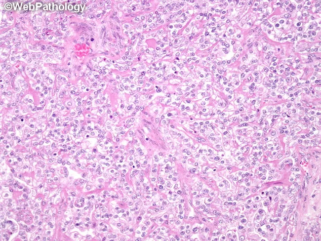
Histology: ovarian clear cell carcinoma, hematoxylin-eosin stained, evaluated at light microscopy. The tissue shows nests and sheets of polygonal cells with distinct cell borders and abundant clear cytoplasm, due to glycogen. Cells are arranged in glandular, tubular or tubulocystic structures; hobnail cells with apically placed nuclei are present, imparting a characteristic appearance. Nuclear enlargement is mild, with minimal pleomorphism; mitotic activity is not appreciably increased. Background stroma is often fibrous; occasional small cystic spaces may be seen. The cytoplasmic clearing is PAS-positive and diastase-sensitive, consistent with glycogen content. Nucleoli are inconspicuous in many cells; cytoplasmic borders crisp. The overall pattern demonstrates a combination of tubulocystic, papillary, and solid growth, which is typical for ovarian CCC. The slide shows features that help distinguish CCC from other ovarian epithelial tumors, such as endometrioid, serous, and mucinous carcinomas. Clinically, ovarian CCC tends to be associated with endometriosis and can show chemoresistance; prognostic implications depend on stage and grade. The presence of hobnail cells is a supportive diagnostic feature. This image likely represents typical example of clear cell histology and may be used for educational purposes to illustrate cellular morphology, glycogen-rich cytoplasm, and glandular architecture in CCC.
PAS Positivity in Nodular Hidradenoma
Background
PAS-Positive Structures
| Feature | PAS Result | Diastase Treatment | Significance |
|---|---|---|---|
| Clear cell cytoplasm (glycogen) | Strongly positive | Diastase-labile (digested) | Confirms glycogen as the source of clearing |
| Intraluminal/intracystic secretory material | Positive | Partially resistant | Mucopolysaccharides/neutral mucins |
| Hyalinized eosinophilic stroma | Weakly positive | Resistant | Basement membrane material |
Key Points
- The "clear cells" in nodular/clear cell hidradenoma have optically clear cytoplasm on H&E due to abundant intracytoplasmic glycogen.
- These cells stain strongly PAS-positive with a magenta/purple color.
- After treatment with diastase (amylase), the PAS positivity is abolished - confirming the material is glycogen (not mucin).
- This diastase-labile PAS positivity is one of the classic histochemical hallmarks of this tumor.
- The second population (non-clear, "dark" or polygonal cells with eosinophilic cytoplasm) shows less prominent or negative PAS staining - these contain fewer glycogen stores.
- Areas of duct differentiation within the tumor may contain secretory material that is PAS-positive and diastase-resistant, indicating neutral glycoproteins/mucins rather than pure glycogen.
- A distinctive feature of nodular hidradenoma is areas of eosinophilic hyalinized stroma (mentioned in Andrews'). This may show faint PAS positivity due to basement membrane-type glycoproteins, and is diastase-resistant.
Histological Context
- Solid lobules and cystic spaces in the dermis (not connected to epidermis in most cases)
- Two cell types: clear cells (PAS+, diastase-labile) and dark polyhedral cells
- Focal ductal differentiation
- Areas of cystic degeneration with eosinophilic hyalinized stroma
- Distinguishes it from sebaceous tumors (which show lipid rather than glycogen)
- Helps confirm eccrine/sweat gland differentiation
- Is shared with clear cell acanthoma (also glycogen-rich, PAS+/diastase-labile) but the two are distinguished by other morphological and clinical features
Summary
- Andrews' Diseases of the Skin, p. 766 - Acrospiromas section
Diastase labile means diastase sensitive or resistant??
| Term | Meaning | What happens to PAS staining after diastase? |
|---|---|---|
| Diastase-labile | Sensitive to diastase | PAS positivity is lost/abolished |
| Diastase-resistant | Not broken down by diastase | PAS positivity remains |
The logic:
- Diastase (alpha-amylase) digests glycogen.
- If the PAS-positive material disappears after diastase treatment → it was glycogen → diastase-labile.
- If the PAS positivity persists after diastase → it is not glycogen (it's mucin, basement membrane glycoprotein, etc.) → diastase-resistant.
In nodular hidradenoma:
- Clear cell cytoplasm → PAS+ and diastase-labile → confirms the material is glycogen (gets digested, staining disappears).
- Ductal secretions → PAS+ and diastase-resistant → confirms mucins/glycoproteins (staining persists).
Does hidradenoma have connection with the epidermis
Hidradenoma and Epidermal Connection
Nodular Hidradenoma (Clear Cell Hidradenoma)
- Sits entirely within the dermis as an encapsulated nodule
- Does NOT arise from or connect to the overlying epidermis
- This is in contrast to its acrospiroma relatives (poroma, hidroacanthoma simplex) which DO connect to the epidermis
Comparison within the Acrospiroma Group
| Tumor | Epidermal Connection |
|---|---|
| Hidroacanthoma simplex | Entirely intraepidermal (within epidermis) |
| Poroma | Connects to epidermis - arises from it, extends downward |
| Dermal duct tumor | Small dermal islands - no epidermal connection |
| Nodular hidradenoma | No epidermal connection - purely dermal nodule |
| Hidradenoma papilliferum | No connection with epidermis - encapsulated in dermis |
The Key Point
- Andrews' Diseases of the Skin, p. 766
What if the tumor has all the features of nodular hidradenoma but there is an epidermal connection
Nodular Hidradenoma Features + Epidermal Connection
Answer: It is still within the Acrospiroma spectrum - termed a "Poroma with hidradenoma differentiation" or "Hybrid Acrospiroma"
Why This Makes Sense
"Hybrid combinations of different patterns of acrospiroma are very common."
What the Epidermal Connection Tells You
| Scenario | Interpretation |
|---|---|
| Purely dermal nodule, no epidermal attachment | Classic nodular hidradenoma |
| Dermal nodule with clear cells + epidermal connection | Poroma with hidradenoma (clear cell) differentiation |
| Entire tumor within epidermis | Hidroacanthoma simplex |
| Connects to epidermis, extends into dermis as cords | Classic poroma |
Practical Diagnostic Approach
- Do NOT forcibly call it nodular hidradenoma - epidermal connection is not a feature of classic nodular hidradenoma
- Classify it as a hybrid acrospiroma or "poroma with clear cell/hidradenoma differentiation"
- The presence of epidermal origin actually pushes it toward the poroma end of the spectrum
- Behaviour and treatment remain the same - benign, treated by complete excision
Key Concept
Hidroacanthoma → Poroma → Dermal Duct Tumor → Nodular Hidradenoma
(intraepidermal)  (epidermal    (pure dermis,     (pure dermis,
                   + dermis)     small nodule)     large nodule)
- Andrews' Diseases of the Skin, p. 766 - "Hybrid combinations of different patterns of acrospiroma are very common"
What are the immunohistochemical markers (K5/14, K1/10, K77) that distinguish acrospiromas?
"All the cells in a poroma, except entrapped ducts, stain with K5/14. Focally, they are K1/10 positive and uniformly K77 negative. This is the staining pattern of the sweat duct ridge and acrosyringium (intraepidermal portions of sweat duct)."
Keratin IHC in Acrospiromas
The Three Keratins Explained
- Expressed in basal cells of the epidermis and outer root sheath
- Mark undifferentiated / progenitor epithelial cells
- Present in the acrosyringium (intraepidermal sweat duct)
- Expressed in suprabasal keratinocytes undergoing terminal differentiation
- Mark mature, differentiated epidermal cells
- Specifically expressed in the secretory coil and deeper portions of the eccrine sweat gland
- It is the marker of secretory differentiation in eccrine glands
- Absent from the acrosyringium/intraepidermal duct
IHC Profile of Acrospiromas
| Marker | Acrospiroma cells | Interpretation |
|---|---|---|
| K5/14 | Positive (all tumor cells except entrapped ducts) | Confirms origin from acrosyringeal progenitor cells |
| K1/10 | Focally positive | Shows partial/focal suprabasal differentiation |
| K77 | Uniformly NEGATIVE | Rules out secretory coil origin; confirms acrosyringeal (not secretory) differentiation |
What This Tells Us About Cell of Origin
- It confirms acrospiromas do NOT arise from the secretory coil (which would be K77+)
- They arise from the acrosyringium - the most superficial, intraepidermal segment of the duct
- This is why poroma classically connects to the epidermis - it mirrors the normal anatomy of its cell of origin
Practical Diagnostic Use
-
K77 negativity is the most useful discriminator - it separates acrospiromas from tumors with true secretory coil differentiation (e.g., spiradenoma, cylindroma)
-
K5/14 positivity confirms epithelial/sweat duct origin
-
Entrapped normal ducts within the tumor will be K77+ (since they represent normal deeper duct), helping distinguish them from the tumor cells themselves
-
Andrews' Diseases of the Skin, p. 766
Compare keratin profiles of spiradenoma vs acrospiroma
Keratin Profile: Spiradenoma vs Acrospiroma
Core Difference in Cell of Origin
| Feature | Acrospiroma | Spiradenoma |
|---|---|---|
| Cell of origin | Acrosyringium (intraepidermal sweat duct) | Uncertain - originally thought intradermal duct; now evidence for folliculosebaceous-apocrine origin (follicular stem cells) |
| Location | Epidermis → dermis continuum | Purely dermal, no epidermal connection |
Keratin Profile Comparison
| Keratin | Acrospiroma | Spiradenoma | Significance |
|---|---|---|---|
| K5/14 | Positive (all tumor cells) | Positive | Basal/progenitor marker - both express it |
| K1/10 | Focally positive | Negative | Terminal epidermal differentiation - absent in spiradenoma |
| K77 | Uniformly NEGATIVE | Positive (reported) | K77 marks secretory coil/deeper duct differentiation - key differentiator |
| Follicular stem cell markers (e.g., CD200, PHLDA1) | Negative | Positive | Supports folliculosebaceous origin of spiradenoma |
The Critical Differentiator: K77
- Acrospiromas are K77 NEGATIVE - confirming they do NOT differentiate toward the secretory coil
- Spiradenoma is K77 POSITIVE - reflecting deeper eccrine/secretory differentiation (or at minimum, a different progenitor pool)
Acrosyringium → Poroma/Hidradenoma → K77(-)
Secretory coil/follicular stem cell → Spiradenoma → K77(+)
Additional IHC Differences
| Marker | Acrospiroma | Spiradenoma |
|---|---|---|
| CD34 | Negative | Positive in stromal cells |
| Bcl-2 | Variable | Strongly positive (peripheral cells) |
| Scattered lymphocytes | Absent | Characteristic - scattered T-lymphocytes throughout tumor |
| PAS-positive hyaline material | In stroma of hidradenoma (eosinophilic hyalinized stroma) | PAS-positive basement membrane deposits within tumor lobules - classic finding |
Histological Correlate of the PAS Difference
- Hidradenoma: PAS+ is mainly glycogen in clear cells (diastase-labile)
- Spiradenoma: PAS+ is hyaline basement membrane material within tumor lobules (diastase-resistant) - this is a classic and distinctive histological feature of spiradenoma
Summary Mnemonic
Acrospiromas = Acrosyringium = Above (superficial) = K77 Absent Spiradenoma = Secretory/follicular = Subepidermal deep = K77 Stained
- Andrews' Diseases of the Skin, p. 766 (acrospiroma keratins)
- Fitzpatrick's Dermatology, p. 1856 (spiradenoma histology and follicular stem cell origin)