
Histology of spleen
Reading File
This is a Visual + Broad query - histology involves both images and comprehensive coverage. I'll run parallel searches now.
Finding Sources
Finding Sources
Finding Sources
Reading File
I found Junqueira's Basic Histology and Histology A Text and Atlas - the two primary histology textbooks. Let me read those in parallel along with Robbins for pathological context.
Reading File
Reading File
Reading File
Excellent content found. Now I need to read the dedicated Junqueira's spleen section and get histology images.
Reading File
Loading Image
Loading Image
Now let me get the spleen microscopy images from Junqueira's and the Histology text & atlas, plus the remaining content.
Loading Image
Reading File
Loading Image
Reading File
Excellent - I now have rich textbook content and multiple high-quality images. Let me fetch a couple more key images to show the sinusoids and red pulp.
Loading Image
Loading Image
I now have comprehensive content from Junqueira's, Robbins, and Cellular Immunology textbooks along with multiple excellent histology images. I have enough to write a thorough, well-illustrated answer.
Histology of the Spleen
The spleen is the largest secondary lymphoid organ and the only one dedicated to filtering blood (not lymph). It weighs ~150 g in adults and sits in the left upper quadrant. Its parenchyma - the splenic pulp - is divided into red pulp and white pulp, embedded in a reticular framework.
1. Capsule and Trabeculae
The spleen is covered by a dense connective tissue capsule (smooth muscle + collagen fibers, stained red in Picro-Sirius-hematoxylin). From the capsule, trabeculae extend inward carrying branches of the splenic artery, vein, lymphatics, and nerves. The trabeculae form the internal scaffolding of the organ.
Low-power micrograph - Capsule (C), Trabeculae (T), White Pulp (W) and Red Pulp (R):
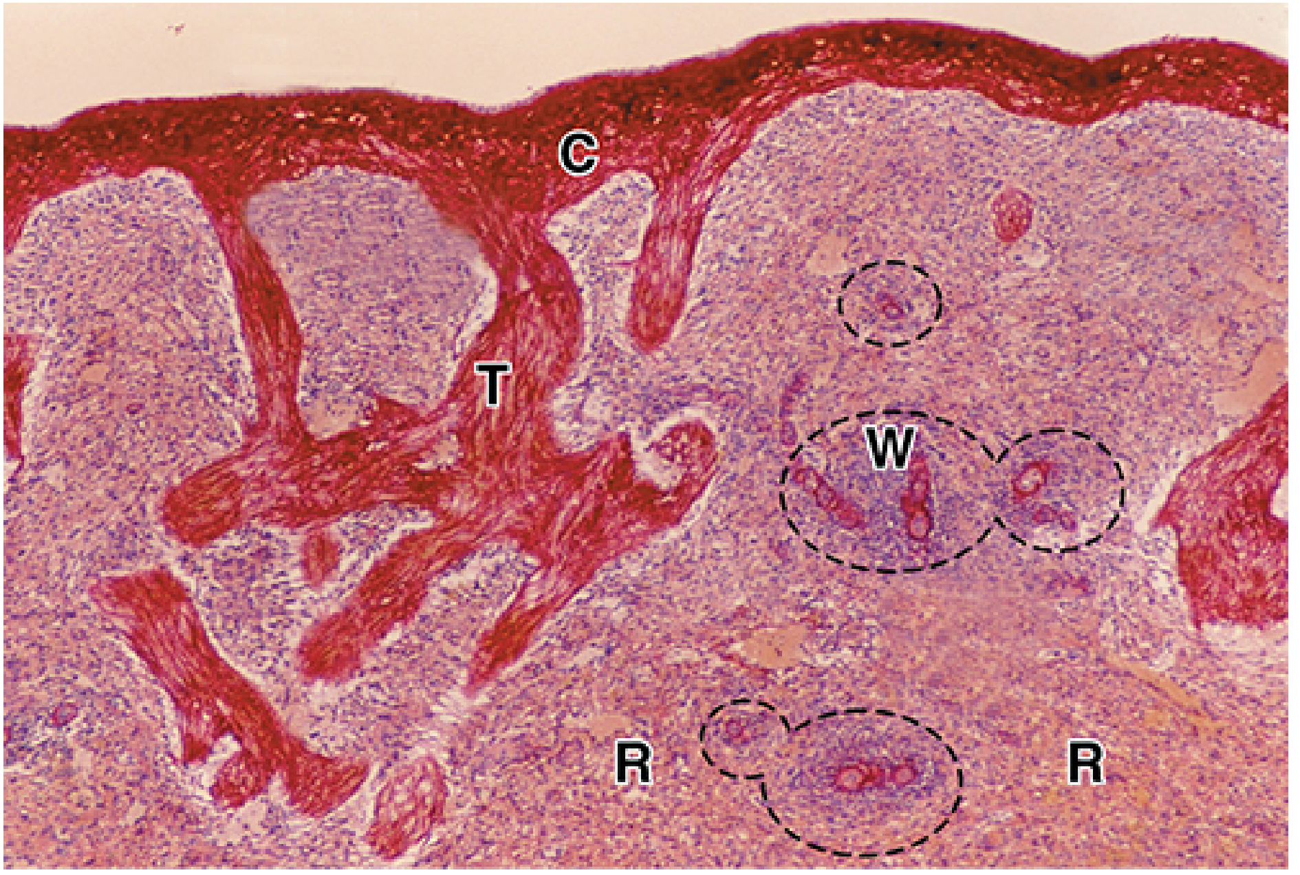
Capsule (C), trabeculae (T), white pulp (W, dashed circles), and red pulp (R). x20; Picro-Sirius-hematoxylin. - Junqueira's Basic Histology 17e, Fig. 14-21
2. White Pulp (~20% of splenic mass)
The white pulp appears as pale gray-white nodules on fresh cut surface and as darker blue-purple islands on H&E (densely packed lymphocytes). It has two main components:
a. Periarteriolar Lymphoid Sheath (PALS)
- Surrounds the central arteriole (a small muscular arteriole that has left the trabecular connective tissue)
- Composed predominantly of T lymphocytes (T cell zone), with scattered macrophages, dendritic cells, and plasma cells
- This is analogous to the paracortex of lymph nodes
b. Lymphoid Nodules (Follicles)
- B cells are located in follicles within and adjacent to the PALS
- On antigenic stimulation, B cells proliferate and form germinal centers - pale central areas with activated blasts, identical to those in lymph nodes
- When a nodule enlarges, the arteriole is pushed to an eccentric position but is still called the central arteriole
- Nodules include primary follicles (quiescent, small dark B cells) and secondary follicles (with pale germinal centers)
White pulp (W) with central arteriole (arrowhead) in PALS, surrounded by red pulp (R):
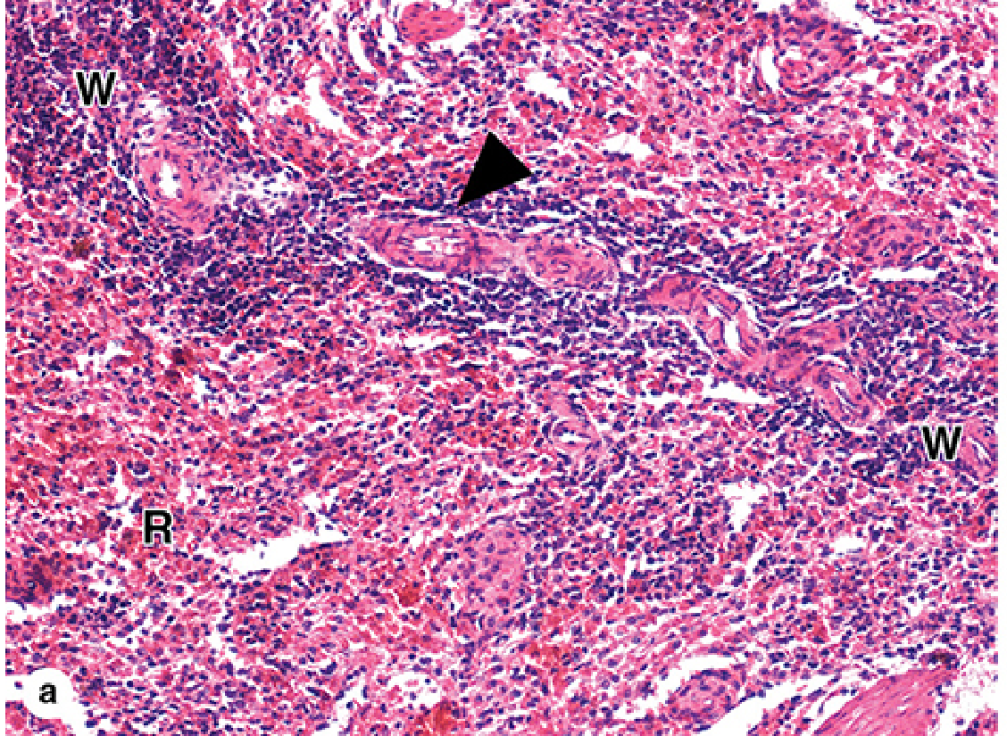
White pulp (W) - PALS with central arteriole (arrowhead), surrounded by red pulp (R). - Junqueira's Basic Histology 17e, Fig. 14-23a
c. Marginal Zone
- A ring of specialized B cells and macrophages encircling each lymphoid nodule at the outer edge of the white pulp
- Receives blood from marginal zone sinuses (branches of the central arteriole)
- Marginal zone B cells have a limited repertoire and respond quickly to blood-borne antigens (esp. bacterial polysaccharides)
- In humans there is an inner and outer marginal zone plus a perifollicular zone
- Antigens are delivered here by circulating dendritic cells or sampled by macrophages
3. Red Pulp (~80% of splenic mass)
The red pulp is filled with blood and consists of two components:
a. Splenic Cords (Cords of Billroth)
- Irregular masses of reticular tissue (reticular cells + reticular fibers) between the sinusoids
- Contain T and B lymphocytes, macrophages, other leukocytes, and red blood cells
- Macrophages in the cords form a "labyrinth" connected by long dendritic processes
- Key function: macrophages phagocytose aging/damaged RBCs that squeeze through the sinusoidal wall; they also "pit" inclusions such as Heinz bodies and Howell-Jolly bodies from RBCs
b. Splenic Sinusoids
- Wide, irregular, blood-filled channels lined by specialized stave cells (endothelial cells)
- Stave cells are elongated, oriented parallel to blood flow, with large nuclei bulging into the lumen
- The basement membrane beneath them is highly discontinuous - blood cells must squeeze through gaps between stave cells to re-enter the sinusoid from the cords
- This acts as a mechanical filter: healthy, deformable RBCs pass through; rigid or abnormal RBCs are trapped and phagocytosed
H&E micrograph of red pulp - sinusoids (S) and cords (C):
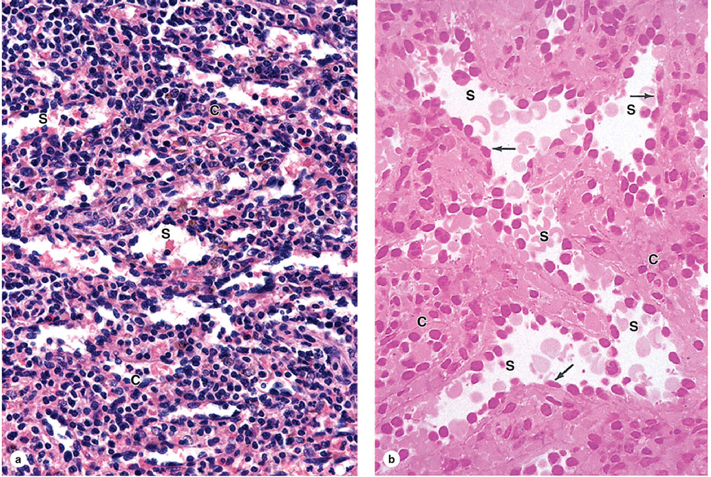
(a) Red pulp with sinusoids (S) and cords (C), both containing blood cells. x140. (b) Higher magnification - stave cell nuclei (arrows) bulging into sinusoidal lumens (S); splenic cords (C) lateral to sinusoids. x200. H&E. - Junqueira's Basic Histology 17e, Fig. 14-25
4. Blood Circulation in the Spleen
The splenic microvasculature is uniquely organized to facilitate immune surveillance. Blood flow:
Splenic artery → Trabecular artery → Central arteriole (in PALS) → Penicillar arterioles (in red pulp) → Sheathed capillaries → Two routes:
| Route | Path | Speed | Function |
|---|---|---|---|
| Closed (fast) circulation | Capillaries → directly into sinusoids → trabecular vein | Rapid | Most blood takes this route |
| Open (slow) circulation | Capillaries dumped into splenic cords → blood cells squeeze through sinusoidal walls | Slow | Blood cells examined by macrophages; effete cells trapped |
Only a small fraction takes the open route at any moment, but over a day the entire blood volume is screened by this system.
Schematic of splenic blood flow (PALS, white pulp, red pulp circulation):
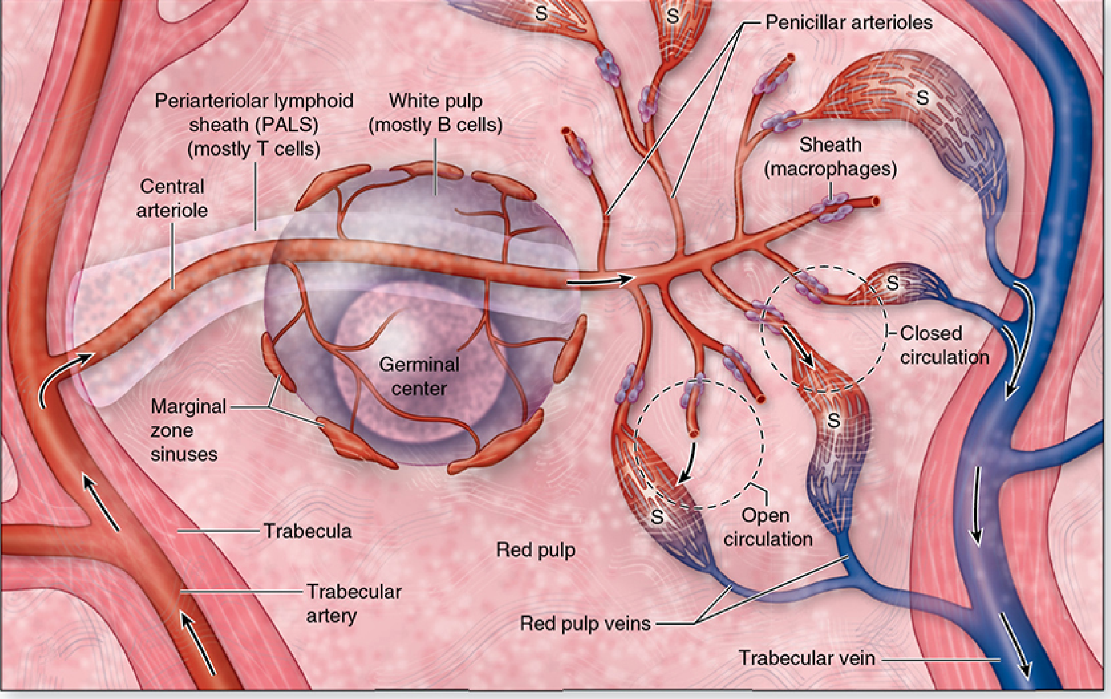
PALS (T cells), white pulp B cell nodule with germinal center, penicillar arterioles with macrophage sheaths (S), open and closed circulation routes. - Junqueira's Basic Histology 17e, Fig. 14-22
5. Normal Splenic Architecture Diagram (Robbins)
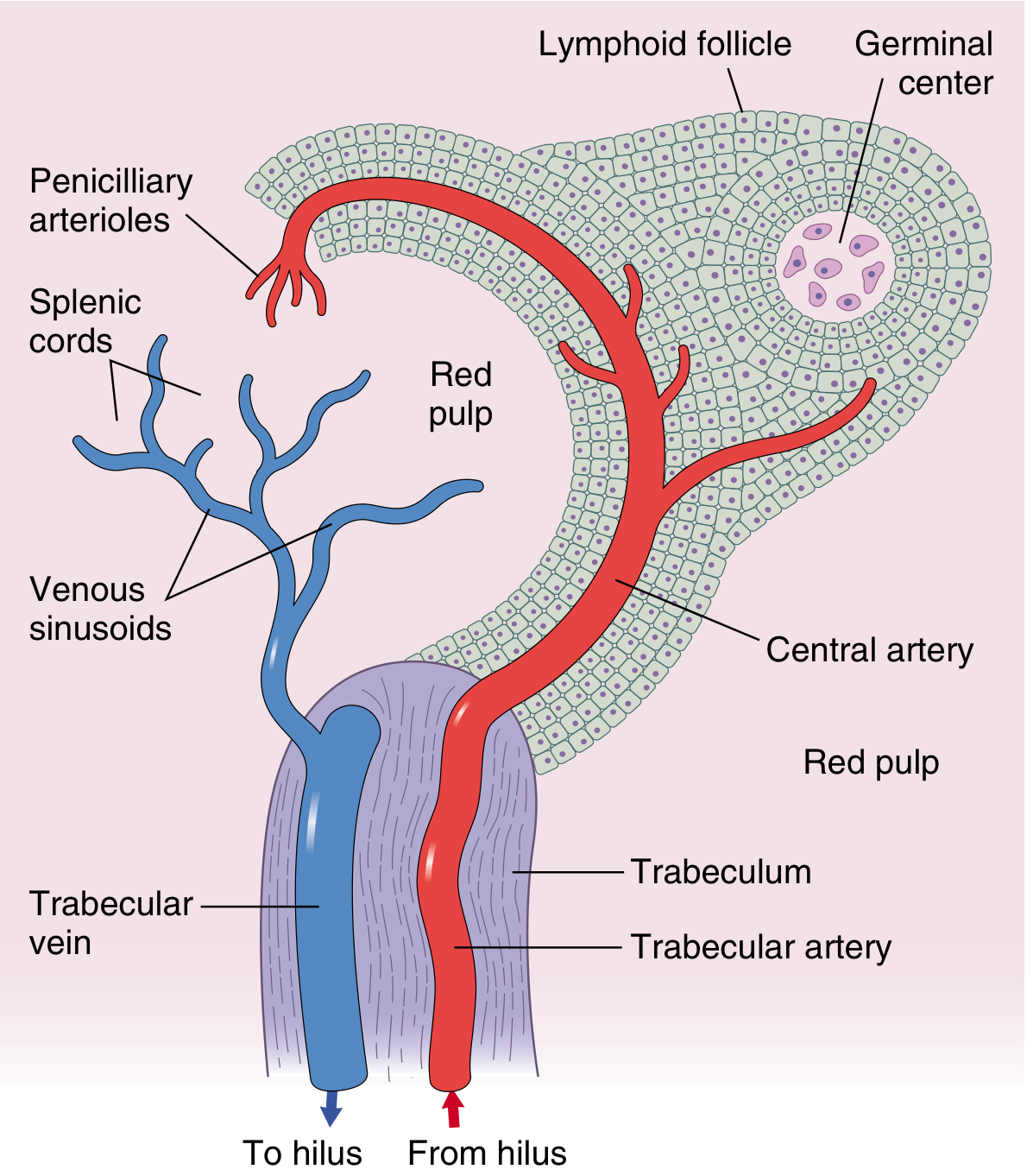
Normal splenic architecture showing trabecular artery/vein, central artery, lymphoid follicle with germinal center, penicilliary arterioles, venous sinusoids, and splenic cords. - Robbins Cotran & Kumar, Pathologic Basis of Disease, Fig. 13.41
6. Framework and Supporting Stroma
- The entire parenchyma rests on a reticular fiber framework (Type III collagen, demonstrated by silver stain)
- Reticular fibers wrap around the sinusoids and fill the cords
- Reticular cells are the stromal cells that produce these fibers and form channels guiding lymphocyte traffic
- A small amount of smooth muscle in the capsule and trabeculae allows minor contraction to release stored blood
7. Functions of Each Histological Compartment
| Compartment | Cell Population | Main Functions |
|---|---|---|
| White pulp - PALS | T lymphocytes, DCs | T-cell activation; antigen presentation |
| White pulp - Follicles | B lymphocytes | Antibody production; germinal center reactions |
| Marginal zone | Marginal zone B cells, macrophages, DCs | First-line response to blood-borne antigens, esp. encapsulated bacteria |
| Red pulp - Cords | Macrophages, lymphocytes, plasma cells, all blood cells | Phagocytosis of old/damaged RBCs; platelet sequestration; extramedullary hematopoiesis (fetal) |
| Red pulp - Sinusoids | Stave cells (endothelium) | Blood filtration; discrimination of healthy vs. rigid RBCs |
Key Histological Landmarks to Identify on a Slide
- Capsule - thick pink dense CT layer at periphery
- Trabeculae - pink CT bands projecting inward
- White pulp - darker blue-purple islands (dense lymphocytes) containing a central arteriole
- Germinal center - pale zone within a secondary follicle
- PALS - T cell sleeve around central arteriole
- Red pulp - lighter pink area with alternating pale sinusoids and darker cords
- Stave cells - elongated endothelial nuclei protruding into sinusoidal lumen
Sources:
- Junqueira's Basic Histology, 17e - Figs. 14-21 to 14-26 (primary source)
- Robbins, Cotran & Kumar - Pathologic Basis of Disease - Spleen section, Fig. 13.41
- Cellular and Molecular Immunology (Abbas) - Spleen microarchitecture
This is a shared conversation. Sign in to Orris to start your own chat.