
Glycolysis
Reading File
Glycolysis is the 10-step metabolic pathway that converts one glucose (6C) into two pyruvate (3C) molecules, yielding a net gain of 2 ATP and 2 NADH. It occurs in the cytoplasm and does not require oxygen.
Searching sources...
Finding Sources
Searching the Web
glycolysis steps enzymes ATP yield biochemistry overview
I have great sources. Let me pull content from the best biochemistry textbooks - Lippincott and Harper's, plus Basic Medical Biochemistry - simultaneously.
Reading File
Reading File
Reading File
Excellent content. Now let me read the detailed 10-step reactions from Lippincott and fetch the key diagrams.
Reading File
Loading Image
Loading Image
Now let me read the detailed 10 steps from Lippincott and the regulation section.
Reading File
Reading File
Now let me get the regulation content and fetch one more key diagram showing the full 10-step pathway.
Finding Sources
Reading File
Reading File
Loading Image
I now have comprehensive content from multiple authoritative biochemistry textbooks. Let me compose the full answer.
Glycolysis
Glycolysis is the universal 10-step pathway that converts one molecule of glucose (6 carbons) into two molecules of pyruvate (3 carbons each). It takes place entirely in the cytosol, requires no oxygen, and is the central hub of carbohydrate metabolism because nearly all sugars from the diet can be fed into it after conversion to glucose intermediates.
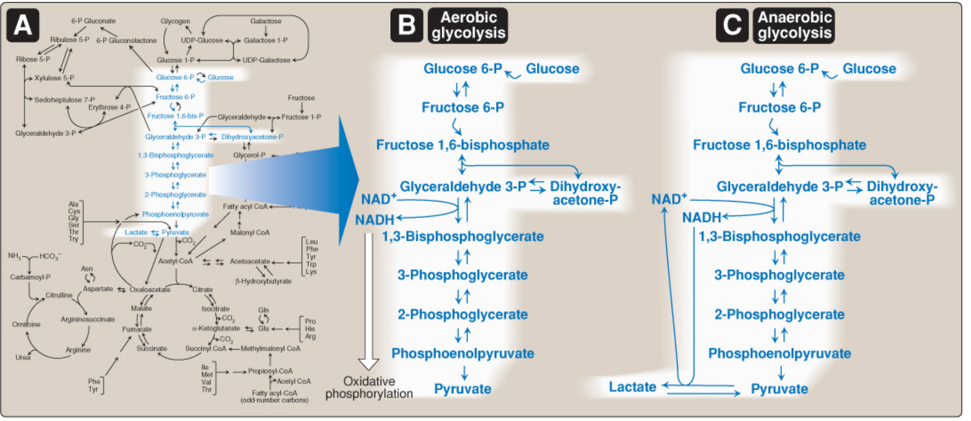
Overall equation (aerobic):
Glucose + 2 NAD⁺ + 2 ADP + 2 Pi → 2 Pyruvate + 2 NADH + 2 H⁺ + 2 ATP + 2 H₂O
Overall equation (anaerobic - to lactate):
Glucose + 2 ADP + 2 Pi → 2 Lactate + 2 ATP + 2 H₂O
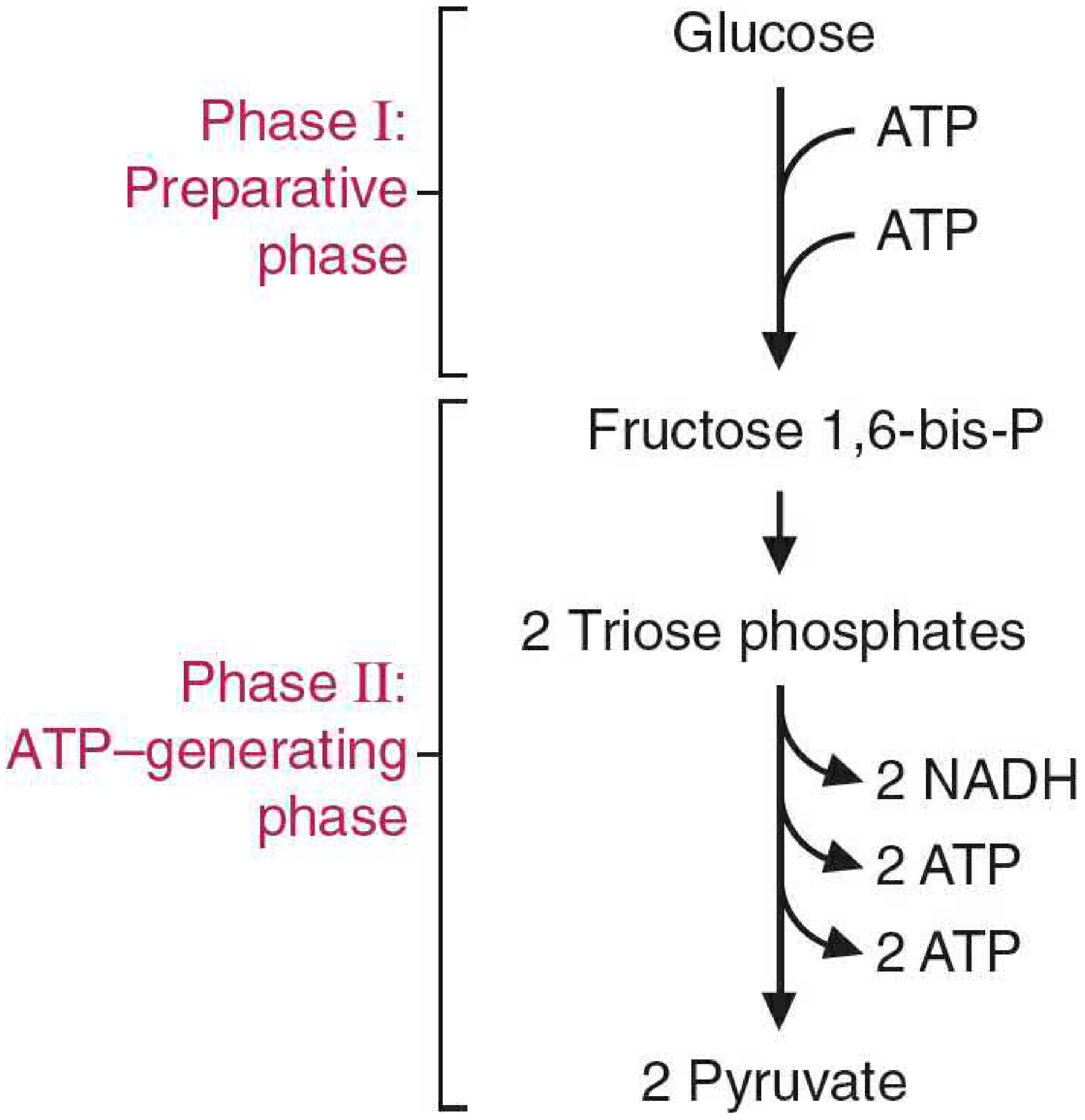
Two Phases
| Phase | Steps | ATP Status | Key Product |
|---|---|---|---|
| Phase I: Preparatory (Investment) | 1-5 | Consumes 2 ATP | Fructose 1,6-bisphosphate → 2 triose phosphates |
| Phase II: Payoff (ATP-generating) | 6-10 | Produces 4 ATP + 2 NADH | 2 Pyruvate |
| Net | - | +2 ATP, +2 NADH | 2 Pyruvate |
The 10 Steps
Phase I - Energy Investment (Steps 1-5)
Step 1 - Glucose → Glucose 6-phosphate
- Enzyme: Hexokinase (HK I-III in most tissues) / Glucokinase (HK IV in liver and β-cells)
- Costs 1 ATP; irreversible under physiological conditions
- Traps glucose inside the cell (charged molecule cannot cross membranes)
- Hexokinase I-III: low Km (~0.1 mM) - high affinity, inhibited by product G6P
- Glucokinase: high Km (~10 mM) - acts as a glucose sensor in liver/pancreas; not inhibited by G6P; induced by insulin
Step 2 - Glucose 6-phosphate → Fructose 6-phosphate
- Enzyme: Phosphoglucose isomerase (PGI)
- Reversible aldose-to-ketose isomerization
- Positions the keto group next to C3, preparing for later cleavage
Step 3 - Fructose 6-phosphate → Fructose 1,6-bisphosphate
- Enzyme: Phosphofructokinase-1 (PFK-1) - the major rate-limiting step
- Costs 1 ATP; highly irreversible
- Key regulatory enzyme: inhibited by ATP, citrate, and H⁺; activated by AMP, ADP, and fructose 2,6-bisphosphate (F-2,6-BP)
- F-2,6-BP is the most potent allosteric activator and is made by PFK-2 (regulated by insulin/glucagon)
Step 4 - Fructose 1,6-bisphosphate → DHAP + Glyceraldehyde 3-phosphate (G3P)
- Enzyme: Aldolase
- Cleaves the 6C sugar into two 3C molecules:
- Dihydroxyacetone phosphate (DHAP)
- Glyceraldehyde 3-phosphate (G3P)
Step 5 - DHAP → Glyceraldehyde 3-phosphate
- Enzyme: Triose phosphate isomerase (TPI)
- Converts DHAP to G3P, effectively doubling the substrate entering Phase II
- From this point on, all steps count ×2 per glucose
Phase II - ATP Generation (Steps 6-10)
Step 6 - G3P → 1,3-Bisphosphoglycerate (1,3-BPG)
- Enzyme: Glyceraldehyde 3-phosphate dehydrogenase (GAPDH)
- Oxidation by NAD⁺ → 2 NADH produced (×2 per glucose)
- Uses inorganic phosphate (Pi), not ATP - this is where the "high-energy" bond is made
- Inhibited by arsenate (pentavalent arsenic), which substitutes for Pi and prevents ATP synthesis
Step 7 - 1,3-BPG → 3-Phosphoglycerate + ATP
- Enzyme: Phosphoglycerate kinase (PGK)
- First ATP-generating step by substrate-level phosphorylation
- Replaces the 2 ATP consumed in Phase I (×2 per glucose = 2 ATP made)
- Physiologically reversible (unusual for a kinase)
- Note: Some 1,3-BPG in RBCs is diverted by bisphosphoglycerate mutase to form 2,3-BPG, which reduces hemoglobin's O₂ affinity (right-shifts the O₂-dissociation curve), aiding O₂ delivery to tissues
Step 8 - 3-Phosphoglycerate → 2-Phosphoglycerate
- Enzyme: Phosphoglycerate mutase
- Shifts the phosphate from carbon 3 to carbon 2; freely reversible
Step 9 - 2-Phosphoglycerate → Phosphoenolpyruvate (PEP) + H₂O
- Enzyme: Enolase
- Dehydration reaction; creates a high-energy enol-phosphate bond in PEP
- Inhibited by fluoride (used in clinical fluoride-oxalate blood tubes to inhibit glycolysis)
Step 10 - PEP → Pyruvate + ATP
- Enzyme: Pyruvate kinase (PK)
- Second ATP-generating step by substrate-level phosphorylation (×2 per glucose = 2 more ATP)
- Irreversible; strongly exergonic
- Activated by fructose 1,6-bisphosphate (feedforward activation)
- Inhibited by ATP, alanine; phosphorylated (inactivated) by glucagon-mediated cAMP/PKA in liver
Energy Yield Summary
| Event | ATP/NADH |
|---|---|
| Step 1 (Hexokinase) | -1 ATP |
| Step 3 (PFK-1) | -1 ATP |
| Step 6 (GAPDH) ×2 | +2 NADH |
| Step 7 (PGK) ×2 | +2 ATP |
| Step 10 (PK) ×2 | +2 ATP |
| Net per glucose | +2 ATP, +2 NADH |
The 2 NADH can yield additional ATP via oxidative phosphorylation in aerobic conditions (~5 ATP in mitochondria), making aerobic glycolysis far more productive overall.
Regulation - Three Key Control Points
Glycolysis is controlled primarily at its three irreversible steps:
| Enzyme | Activators | Inhibitors |
|---|---|---|
| Hexokinase (I-III) | Glucose | Glucose 6-phosphate (product inhibition) |
| PFK-1 | AMP, ADP, F-2,6-BP, Pi | ATP, citrate, H⁺ |
| Pyruvate kinase | Fructose 1,6-bisphosphate | ATP, alanine; glucagon (phosphorylation in liver) |
Fructose 2,6-bisphosphate (F-2,6-BP) is the master regulator:
- Made by PFK-2 when insulin is high (fed state) → stimulates glycolysis
- Broken down by FBPase-2 when glucagon is high (fasted state) → inhibits glycolysis
Fate of Pyruvate
| Condition | Fate | Enzyme |
|---|---|---|
| Aerobic, mitochondria present | → Acetyl-CoA + CO₂ | Pyruvate dehydrogenase complex |
| Anaerobic (or no mitochondria) | → Lactate + NAD⁺ regenerated | Lactate dehydrogenase (LDH) |
| Yeast/anaerobic microbes | → Ethanol + CO₂ | Pyruvate decarboxylase + alcohol dehydrogenase |
| Gluconeogenesis (liver, kidney) | → Glucose | Pyruvate carboxylase → OAA pathway |
The conversion of pyruvate to lactate is essential for regenerating NAD⁺ to sustain glycolysis when oxygen is limited - without NAD⁺, GAPDH (Step 6) cannot function and the pathway halts.
Clinically Relevant Points
- Pyruvate kinase deficiency: most common enzyme defect in the glycolytic pathway; causes hemolytic anemia because RBCs depend entirely on glycolysis for ATP (no mitochondria). Heterozygotes show resistance to severe malaria.
- Arsenic poisoning: arsenite inhibits pyruvate dehydrogenase; arsenate (pentavalent) substitutes for Pi in Step 6, preventing ATP synthesis from 1,3-BPG.
- Warburg effect: cancer cells preferentially use aerobic glycolysis (glucose → lactate even in the presence of O₂) to generate biosynthetic precursors - the basis for FDG-PET scanning.
- RBC 2,3-BPG: the Luebering-Rapoport shunt in RBCs diverts 1,3-BPG to 2,3-BPG, which allosterically reduces hemoglobin's O₂ affinity, promoting O₂ release to tissues.
- Fluoride: inhibits enolase (Step 9) and is used in grey-top blood collection tubes to prevent glycolysis from falsely lowering measured blood glucose.
Sources: Lippincott Illustrated Reviews: Biochemistry, 8th ed., pp. 293-316 | Basic Medical Biochemistry: A Clinical Approach, 6th ed., pp. 790-800 | Harper's Illustrated Biochemistry, 32nd ed., p. 175
This is a shared conversation. Sign in to Orris to start your own chat.