
How to read hrctand cect chest
how to read HRCT CECT chest systematic approach radiology
https://radiologyassistant.nl/chest/hrct/basic-interpretatio…
CECT chest reading approach mediastinum pleura lung vessels systematic
How to Read HRCT and CECT Chest
PART 1 - HRCT CHEST (High-Resolution CT)
What is HRCT?
- Fishman's Pulmonary Diseases and Disorders, 2-Volume Set
- Murray & Nadel's Textbook of Respiratory Medicine, 2-Volume
- Supine, full inspiration - routine
- Prone - to distinguish true ILD from dependent atelectasis
- Expiratory - to detect air trapping (useful in obstructive/small airway disease)
Step 1 - Technical Assessment Before Interpretation
- Adequate inspiration (diaphragm at 9th-10th rib posteriorly)
- Correct window settings used (lung window: W: 1500, L: -700; mediastinal window: W: 350-400, L: 40)
- No motion artifact
Step 2 - Know the Secondary Pulmonary Lobule (SPL)
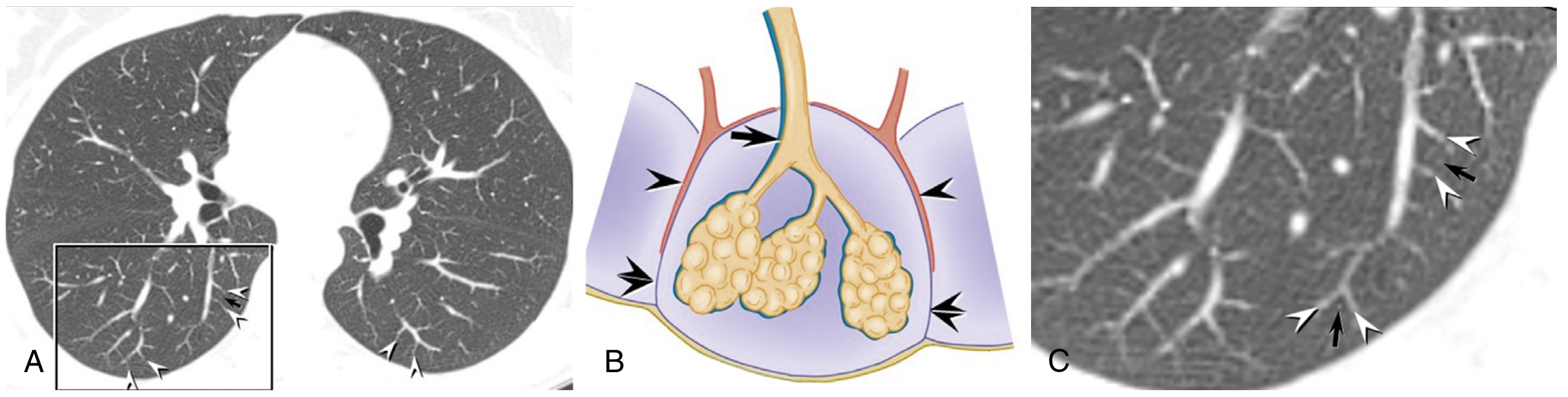
- Centrilobular region: bronchiole + centrilobular artery
- Interlobular septa: contain pulmonary veins + lymphatics
- Perilobular region: connective tissue septum
| Distribution | Pattern | Classic Disease |
|---|---|---|
| Centrilobular | Nodules near lobule center, spares pleura | Hypersensitivity pneumonitis, respiratory bronchiolitis, endobronchial spread of infection |
| Perilymphatic | Along septa, fissures, bronchovascular bundles | Sarcoidosis, lymphangitic carcinomatosis, pulmonary edema |
| Random | No lobular preference | Hematogenous metastases, miliary TB/fungal |
Step 3 - Identify the Dominant Pattern
A. Increased Attenuation Patterns
- Interlobular septal thickening (smooth): pulmonary edema, lymphangitic carcinomatosis
- Interlobular septal thickening (nodular/irregular): sarcoidosis, lymphangitic spread
- Intralobular lines (fine netlike): fibrotic ILDs (UIP, NSIP, asbestosis)
- Honeycombing: clustered cystic spaces 3-10 mm, subpleural - hallmark of end-stage fibrosis/UIP
- Size: micronodules (<3 mm) vs macronodules (3-10 mm) vs large nodules (>10 mm)
- Distribution: centrilobular, perilymphatic, or random (see table above)
- Borders: well-defined (metastases) vs ill-defined/ground glass halo (infection, eosinophilic)
- Calcification: old granuloma, hamartoma
- Active inflammation/infection (COVID-19 pneumonia, PCP)
- Pulmonary edema
- Early ILD (NSIP)
- Alveolar hemorrhage
B. Decreased Attenuation Patterns
- Centrilobular: smoking-related, upper lobe predominant, spares periphery
- Panlobular: alpha-1 antitrypsin deficiency, lower lobe predominant
- Paraseptal: along septa, subpleural, associated with bullae and spontaneous pneumothorax
- LAM (lymphangioleiomyomatosis): uniform thin-walled cysts throughout both lungs in young women
- LCH (Langerhans cell histiocytosis): bizarre-shaped thick-walled cysts + nodules, upper lobe, smoker
- LIP: cysts + GGO, usually immune disorder
Step 4 - Determine Distribution
| Location | Think of |
|---|---|
| Upper lobe predominant | Sarcoidosis, silicosis, LCH, PLCH, hypersensitivity pneumonitis (some) |
| Lower lobe predominant | UIP/IPF, NSIP, aspiration, pulmonary edema |
| Diffuse | NSIP, DIP, infections |
- Central (peribronchovascular): sarcoidosis, lymphangitic carcinomatosis, bronchitis
- Peripheral/subpleural: UIP, COP (organizing pneumonia), chronic eosinophilic pneumonia
- Bilateral symmetric: UIP, NSIP, edema, sarcoidosis
- Unilateral/asymmetric: lymphangitic carcinomatosis (~50% unilateral), post-obstructive changes, aspiration
Step 5 - UIP/ILD Assessment (Fleischner Society Criteria)
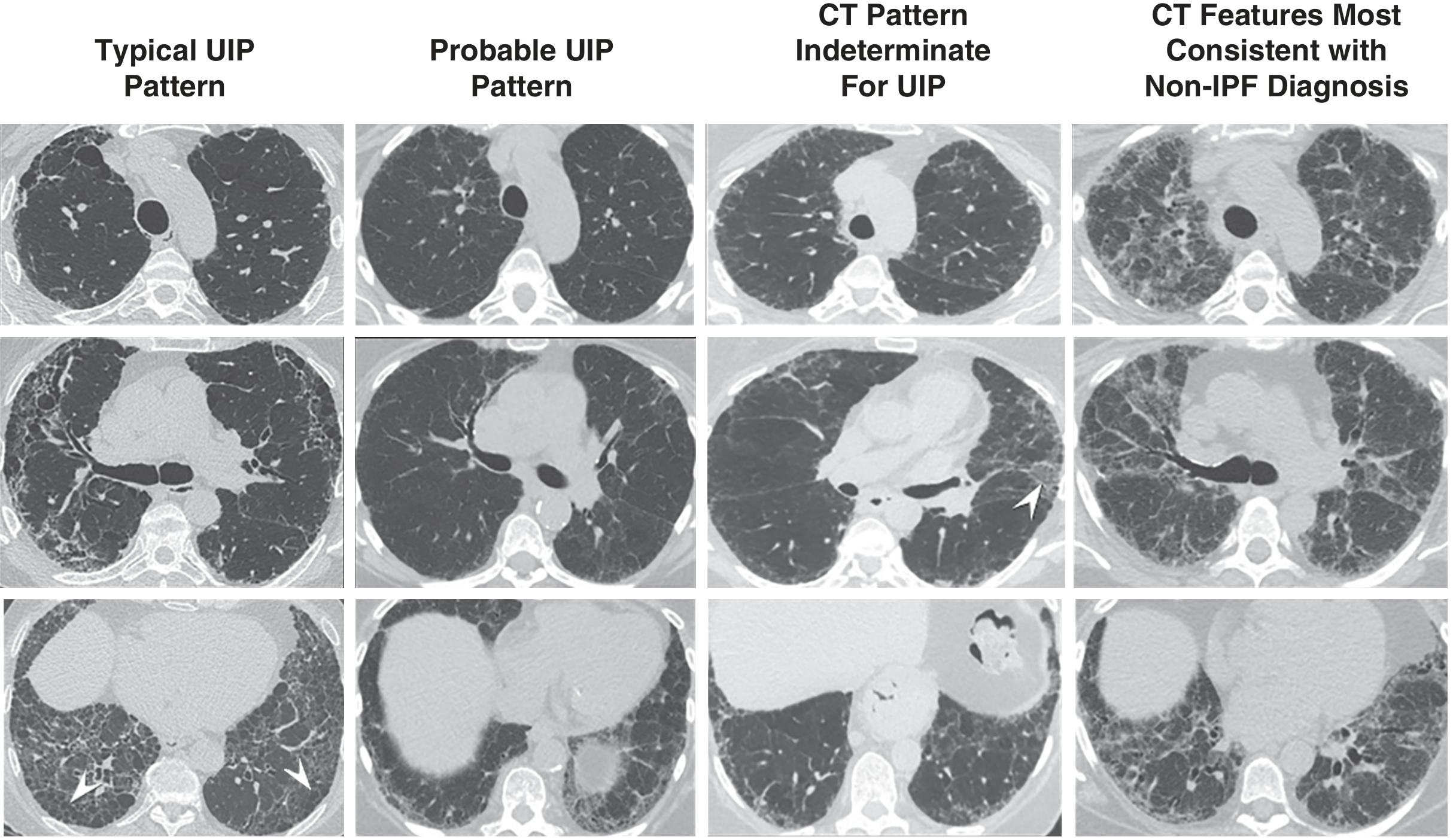
| Pattern | CT Features | Biopsy Needed? |
|---|---|---|
| Typical UIP | Subpleural + basal reticulation + honeycombing + traction bronchiectasis | No |
| Probable UIP | Subpleural + basal reticulation + traction bronchiectasis, NO honeycombing | Usually no |
| Indeterminate | Variable/diffuse distribution, some features of fibrosis but inconclusive | Yes |
| Non-IPF diagnosis | Upper/mid predominant, consolidation, diffuse GGO, extensive mosaic, diffuse nodules/cysts | Yes (alternate diagnosis) |
Step 6 - Additional Features (Don't Miss These)
- Pleural effusion: transudative (edema, heart failure) vs exudative (lymphangitic, mesothelioma, empyema)
- Lymphadenopathy: sarcoidosis shows classic "1-2-3 sign" (right paratracheal + bilateral hilar); lymphoma is infiltrative, encircles vessels; lung carcinoma usually unilateral
- Traction bronchiectasis: dilated bronchi within areas of fibrosis - sign of established fibrotic ILD
- Pleural plaques: asbestos exposure
- Pneumothorax: spontaneous (paraseptal emphysema, LAM, LCH)
PART 2 - CECT CHEST (Contrast-Enhanced CT)
When to Use CECT vs. HRCT
| Indication | HRCT (no contrast) | CECT (with contrast) |
|---|---|---|
| Interstitial lung disease | Yes | No |
| Pulmonary embolism (CTPA) | No | Yes (arterial phase) |
| Mediastinal masses | Less useful | Yes |
| Lung cancer staging | Less useful | Yes |
| Hilar/mediastinal nodes | Less useful | Yes |
| Pleural vs. parenchymal disease | Less useful | Yes |
| Aortic aneurysm/dissection | No | Yes |
Contrast Phases Used
| Phase | Timing after contrast | What it shows |
|---|---|---|
| Arterial (CT Pulmonary Angiography - CTPA) | 15-20 sec | Pulmonary arteries - PE, AVM |
| Venous/portal | 60-70 sec | Mediastinal structures, lymph nodes, liver |
| Delayed | 3-5 min | Vascular tumors, infection |
Systematic Approach to Reading CECT Chest
Use windows correctly:
- Lung window (W:1500, L:-700): parenchyma, airways, emphysema
- Mediastinal/soft tissue window (W:350-400, L:40): nodes, vessels, masses, pleura
- Bone window (W:2000, L:400): ribs, vertebrae, sternum
Go in a fixed order (prevents omission):
- Central position? Any displacement, stenosis, compression?
- Tracheal shape: saber sheath trachea (COPD), tracheomalacia
- Prevascular/Anterior: thymus, thymic tumors, germ cell tumors, thyroid
- Visceral/Middle: trachea, heart, great vessels, esophagus, lymph nodes
- Paravertebral/Posterior: neural tumors (schwannoma, neurofibroma), vertebral disease
- Normal pulmonary arteries vs. enlarged hila (lymph nodes, mass)
- Sarcoidosis: bilateral symmetric hilar enlargement
- Primary lung cancer: unilateral hilar mass
- Heart size, pericardial effusion (density: transudative vs hemorrhagic)
- Cardiac masses, thrombus
- Aneurysm (>4 cm thoracic), intimal flap (dissection), mural thrombus
- CTPA: filling defects in pulmonary arteries = PE (saddle embolus at bifurcation is classic)
- Nodules: size, density (solid, part-solid, GGO), margins (spiculated = malignant, smooth = benign)
- Masses (>3 cm), consolidation, GGO, atelectasis
- Fleischner 2017 guidelines for nodule follow-up (<6 mm = no routine follow-up in low-risk patients)
- Effusion: measure and note density (HU <20 = transudative; >35 = exudative/hemorrhagic)
- Pleural thickening: uniform (reactive) vs. nodular (malignant mesothelioma)
- Pneumothorax
- Rib fractures (acute = sharp margins; healing = callus formation)
- Lytic vs. sclerotic bone lesions
- Soft tissue masses
- Adrenal glands (common site of lung cancer metastasis)
- Liver, upper kidneys
CECT Chest: Common Clinical Applications
Pulmonary Embolism (CTPA)
- Scan in arterial phase (~15-20 seconds after contrast injection)
- Look for: intraluminal filling defect in opacified pulmonary arteries
- Saddle embolus straddles the main pulmonary artery bifurcation
- Right heart strain signs: RV:LV ratio >0.9, straightening of interventricular septum
- Peripheral wedge-shaped infarct (Hampton's hump)
Lung Cancer Staging (TNM)
- T: Tumor size, invasion of mediastinum/chest wall/diaphragm
- N: Mediastinal node involvement (short axis >1 cm considered enlarged)
- M: Distant metastases (liver, adrenals, brain with CT head)
Mediastinal Masses with CECT
- Thymoma: anterior mediastinal, heterogeneous enhancement, pleural nodules in invasive type
- Lymphoma: multiple nodes, infiltrative, soft tissue, "encases" vessels without invading
- Germ cell tumor: anterior, may contain fat/calcification (teratoma)
- Neurogenic tumor: posterior mediastinum, may extend through neural foramen (dumbbell)
Summary Mnemonic for Reading Chest CT
- T - Trachea & main bronchi
- A - Airspaces (lung parenchyma - lung window)
- L - Lymph nodes (hilar + mediastinal)
- E - Effusions (pleural and pericardial)
- P - Pleura and chest wall
- V - Vessels (aorta, pulmonary arteries, CTPA filling defects)
- B - Bones (ribs, vertebrae, sternum)
- Murray & Nadel's Textbook of Respiratory Medicine - HRCT patterns, secondary lobule anatomy
- Fishman's Pulmonary Diseases and Disorders - HRCT technique, ILD/UIP classification
- Radiology Assistant - HRCT Basic Interpretation for pattern-disease correlation
- Fleischner Society 2018 UIP diagnostic criteria
Create a visual reference guide with labeled HRCT patterns and pathology
What format would you like the visual reference guide in?
mkdir -p /home/daytona/workspace/hrct-guide && ls /home/daytona/workspace/hrct-guide/
~/hrct-guide/generate_hrct_guide.py
"""
HRCT & CECT Chest Visual Reference Guide
Multi-page PDF using reportlab with visual pattern diagrams drawn programmatically
"""
from reportlab.lib.pagesizes import A4
from reportlab.lib import colors
from reportlab.lib.units import cm, mm
from reportlab.platypus import (SimpleDocTemplate, Paragraph, Spacer, Table,
TableStyle, HRFlowable, KeepTogether, PageBreak)
from reportlab.lib.styles import getSampleStyleSheet, ParagraphStyle
from reportlab.lib.enums import TA_CENTER, TA_LEFT, TA_RIGHT
from reportlab.graphics.shapes import (Drawing, Rect, Circle, Line, String,
Ellipse, Polygon, PolyLine, Group)
from reportlab.graphics import renderPDF
from reportlab.platypus import Flowable
from reportlab.lib.colors import (HexColor, white, black, grey, lightgrey,
darkgrey, Color)
import math
# ── Colour palette ─────────────────────────────────────────────────────────────
NAVY = HexColor('#0D2B45')
TEAL = HexColor('#1B6CA8')
CYAN = HexColor('#3ABFD4')
GOLD = HexColor('#F5A623')
ORANGE = HexColor('#E07B3A')
RED = HexColor('#C0392B')
GREEN = HexColor('#27AE60')
PURPLE = HexColor('#8E44AD')
LBLUE = HexColor('#D6EAF8')
LGOLD = HexColor('#FEF9E7')
LGREEN = HexColor('#E9F7EF')
LRED = HexColor('#FDEDEC')
LPURPLE = HexColor('#F5EEF8')
LGREY = HexColor('#F2F3F4')
DARKGREY = HexColor('#2C3E50')
MIDGREY = HexColor('#7F8C8D')
PAGEBG = HexColor('#FAFBFC')
# ── Lung grey scale (CT appearance) ────────────────────────────────────────────
CT_AIRSPACE = HexColor('#0A0A0A') # almost black – normal aerated lung
CT_GGO = HexColor('#505050') # dark grey – ground glass
CT_CONSOL = HexColor('#C8C8C8') # light grey – consolidation / soft tissue
CT_VESSEL = HexColor('#FFFFFF') # white – vessels / enhanced structures
CT_HONEY = HexColor('#1A1A1A') # very dark – honeycombing cysts
CT_FIBROSIS = HexColor('#888888') # medium grey – reticular fibrosis
W, H = A4 # 595 x 842 pt
# ── Styles ─────────────────────────────────────────────────────────────────────
def make_styles():
base = getSampleStyleSheet()
def s(name, parent='Normal', **kw):
return ParagraphStyle(name, parent=base[parent], **kw)
styles = {
'cover_title': s('CoverTitle',
fontName='Helvetica-Bold', fontSize=28, textColor=white,
leading=34, alignment=TA_CENTER, spaceAfter=8),
'cover_sub': s('CoverSub',
fontName='Helvetica', fontSize=14, textColor=HexColor('#D6EAF8'),
leading=18, alignment=TA_CENTER, spaceAfter=4),
'cover_tag': s('CoverTag',
fontName='Helvetica-Oblique', fontSize=10, textColor=HexColor('#AED6F1'),
leading=14, alignment=TA_CENTER),
'section_h': s('SectionH',
fontName='Helvetica-Bold', fontSize=16, textColor=white,
leading=20, alignment=TA_LEFT, spaceAfter=0, spaceBefore=0),
'page_title': s('PageTitle',
fontName='Helvetica-Bold', fontSize=18, textColor=NAVY,
leading=22, alignment=TA_LEFT, spaceBefore=4, spaceAfter=6),
'sub_h': s('SubH',
fontName='Helvetica-Bold', fontSize=12, textColor=TEAL,
leading=15, spaceBefore=8, spaceAfter=3),
'body': s('Body',
fontName='Helvetica', fontSize=8.5, textColor=DARKGREY,
leading=13, spaceBefore=1, spaceAfter=1),
'small': s('Small',
fontName='Helvetica', fontSize=7.5, textColor=MIDGREY,
leading=11, spaceBefore=0, spaceAfter=0),
'label': s('Label',
fontName='Helvetica-Bold', fontSize=7.5, textColor=NAVY,
leading=10, alignment=TA_CENTER),
'caption': s('Caption',
fontName='Helvetica-Oblique', fontSize=7.5, textColor=MIDGREY,
leading=10, alignment=TA_CENTER, spaceAfter=4),
'table_h': s('TableH',
fontName='Helvetica-Bold', fontSize=8, textColor=white,
leading=11, alignment=TA_CENTER),
'table_cell': s('TableCell',
fontName='Helvetica', fontSize=8, textColor=DARKGREY,
leading=11, alignment=TA_LEFT),
'bullet': s('Bullet',
fontName='Helvetica', fontSize=8.5, textColor=DARKGREY,
leading=13, leftIndent=12, firstLineIndent=-8, spaceAfter=2),
'footer': s('Footer',
fontName='Helvetica', fontSize=7, textColor=MIDGREY,
alignment=TA_CENTER),
'tip_text': s('TipText',
fontName='Helvetica', fontSize=8, textColor=HexColor('#1A5276'),
leading=12),
}
return styles
ST = make_styles()
# ── Helper Flowables ────────────────────────────────────────────────────────────
class ColorBand(Flowable):
"""Full-width coloured header band"""
def __init__(self, text, bg=TEAL, fg=white, height=28, fs=13, bold=True):
super().__init__()
self.text = text
self.bg = bg
self.fg = fg
self.height = height
self.fs = fs
self.bold = bold
self.width = W - 3*cm
def wrap(self, aw, ah):
self.width = aw
return aw, self.height
def draw(self):
c = self.canv
c.setFillColor(self.bg)
c.roundRect(0, 0, self.width, self.height, 4, fill=1, stroke=0)
fn = 'Helvetica-Bold' if self.bold else 'Helvetica'
c.setFont(fn, self.fs)
c.setFillColor(self.fg)
c.drawString(10, self.height/2 - self.fs*0.35, self.text)
class TipBox(Flowable):
def __init__(self, text, bg=LBLUE, border=TEAL, label='KEY POINT', width=None):
super().__init__()
self.text = text
self.bg = bg
self.border = border
self.label = label
self._width = width or (W - 3*cm)
self.height = 40
def wrap(self, aw, ah):
self._width = aw
return aw, self.height
def draw(self):
c = self.canv
c.setFillColor(self.bg)
c.roundRect(0, 2, self._width, self.height-4, 5, fill=1, stroke=0)
c.setStrokeColor(self.border)
c.setLineWidth(1.5)
c.roundRect(0, 2, self._width, self.height-4, 5, fill=0, stroke=1)
# label
c.setFont('Helvetica-Bold', 7)
c.setFillColor(self.border)
c.drawString(8, self.height - 12, self.label)
c.setFont('Helvetica', 8)
c.setFillColor(DARKGREY)
c.drawString(8, 8, self.text)
# ── CT Pattern Drawing Functions ───────────────────────────────────────────────
def draw_lung_outline(d, x, y, w, h, fill_color=CT_AIRSPACE, label=None,
border_color=HexColor('#444444'), label_color=white):
"""Draw a simplified lung circle with label"""
d.add(Circle(x + w/2, y + h/2, min(w, h)/2 - 2,
fillColor=fill_color, strokeColor=border_color, strokeWidth=1))
if label:
d.add(String(x + w/2, y + h/2 - 4, label,
fontSize=7, fillColor=label_color,
textAnchor='middle', fontName='Helvetica-Bold'))
def draw_vessel(d, x1, y1, x2, y2, color=CT_VESSEL, width=2):
d.add(Line(x1, y1, x2, y2, strokeColor=color, strokeWidth=width))
def pattern_normal(w=120, h=100):
"""Normal HRCT - dark lung with thin branching vessels"""
d = Drawing(w, h)
# Background circle
d.add(Circle(w/2, h/2, min(w,h)/2-3,
fillColor=CT_AIRSPACE, strokeColor=HexColor('#333333'), strokeWidth=1))
# Main bronchus/artery
d.add(Line(w/2, h*0.9, w/2, h*0.55, strokeColor=CT_VESSEL, strokeWidth=2.5))
# Branching vessels
d.add(Line(w/2, h*0.75, w*0.3, h*0.5, strokeColor=CT_VESSEL, strokeWidth=1.5))
d.add(Line(w/2, h*0.75, w*0.7, h*0.5, strokeColor=CT_VESSEL, strokeWidth=1.5))
d.add(Line(w*0.3, h*0.5, w*0.2, h*0.3, strokeColor=CT_VESSEL, strokeWidth=1))
d.add(Line(w*0.3, h*0.5, w*0.4, h*0.3, strokeColor=CT_VESSEL, strokeWidth=1))
d.add(Line(w*0.7, h*0.5, w*0.6, h*0.3, strokeColor=CT_VESSEL, strokeWidth=1))
d.add(Line(w*0.7, h*0.5, w*0.8, h*0.3, strokeColor=CT_VESSEL, strokeWidth=1))
# Label
d.add(String(w/2, 6, 'NORMAL', fontSize=7, fillColor=CT_VESSEL,
textAnchor='middle', fontName='Helvetica-Bold'))
return d
def pattern_ggo(w=120, h=100):
"""Ground Glass Opacity - hazy grey overlay"""
d = Drawing(w, h)
d.add(Circle(w/2, h/2, min(w,h)/2-3,
fillColor=CT_AIRSPACE, strokeColor=HexColor('#333333'), strokeWidth=1))
# GGO patches
for cx, cy, r in [(w*0.45, h*0.6, 22), (w*0.55, h*0.45, 18), (w*0.35, h*0.4, 15)]:
d.add(Circle(cx, cy, r, fillColor=CT_GGO, strokeColor=None, strokeWidth=0))
# Vessels visible through GGO
d.add(Line(w/2, h*0.85, w/2, h*0.5, strokeColor=CT_VESSEL, strokeWidth=2))
d.add(Line(w/2, h*0.7, w*0.35, h*0.5, strokeColor=CT_VESSEL, strokeWidth=1.5))
d.add(String(w/2, 6, 'GROUND GLASS', fontSize=7, fillColor=CT_VESSEL,
textAnchor='middle', fontName='Helvetica-Bold'))
return d
def pattern_consolidation(w=120, h=100):
"""Consolidation - dense opacification"""
d = Drawing(w, h)
d.add(Circle(w/2, h/2, min(w,h)/2-3,
fillColor=CT_AIRSPACE, strokeColor=HexColor('#333333'), strokeWidth=1))
# Consolidated area (lower/peripheral)
pts = [w*0.2, h*0.25, w*0.8, h*0.25, w*0.85, h*0.65, w*0.15, h*0.65]
d.add(Polygon(pts, fillColor=CT_CONSOL, strokeColor=None))
# Air bronchogram - dark bronchi within consolidation
d.add(Line(w/2, h*0.6, w/2, h*0.3, strokeColor=CT_AIRSPACE, strokeWidth=2))
d.add(Line(w/2, h*0.45, w*0.35, h*0.3, strokeColor=CT_AIRSPACE, strokeWidth=1.5))
d.add(Line(w/2, h*0.45, w*0.65, h*0.3, strokeColor=CT_AIRSPACE, strokeWidth=1.5))
d.add(String(w/2, 6, 'CONSOLIDATION', fontSize=7, fillColor=CT_VESSEL,
textAnchor='middle', fontName='Helvetica-Bold'))
return d
def pattern_reticulation(w=120, h=100):
"""Reticular pattern - septal thickening / fibrosis"""
d = Drawing(w, h)
d.add(Circle(w/2, h/2, min(w,h)/2-3,
fillColor=CT_AIRSPACE, strokeColor=HexColor('#333333'), strokeWidth=1))
# Reticular lines (grid-like)
line_c = HexColor('#AAAAAA')
for i in range(5):
y = h*0.25 + i*h*0.12
d.add(Line(w*0.15, y, w*0.85, y, strokeColor=line_c, strokeWidth=0.8))
for i in range(5):
x = w*0.2 + i*w*0.14
d.add(Line(x, h*0.2, x, h*0.8, strokeColor=line_c, strokeWidth=0.8))
# Subpleural accentuation
d.add(Circle(w/2, h/2, min(w,h)/2-8,
fillColor=None, strokeColor=HexColor('#888888'), strokeWidth=1.5))
d.add(String(w/2, 6, 'RETICULATION', fontSize=7, fillColor=CT_VESSEL,
textAnchor='middle', fontName='Helvetica-Bold'))
return d
def pattern_honeycombing(w=120, h=100):
"""Honeycombing - clustered cystic spaces, subpleural"""
d = Drawing(w, h)
d.add(Circle(w/2, h/2, min(w,h)/2-3,
fillColor=CT_AIRSPACE, strokeColor=HexColor('#333333'), strokeWidth=1))
# Subpleural honeycomb cysts
cyst_c = CT_HONEY
wall_c = HexColor('#BBBBBB')
positions = [
(w*0.65, h*0.28), (w*0.75, h*0.35), (w*0.72, h*0.48),
(w*0.62, h*0.55), (w*0.52, h*0.50), (w*0.58, h*0.38),
(w*0.68, h*0.62), (w*0.78, h*0.55),
]
for cx, cy in positions:
r = 7
d.add(Circle(cx, cy, r, fillColor=cyst_c, strokeColor=wall_c, strokeWidth=1.5))
d.add(String(w/2, 6, 'HONEYCOMBING', fontSize=7, fillColor=CT_VESSEL,
textAnchor='middle', fontName='Helvetica-Bold'))
return d
def pattern_nodules(w=120, h=100, nodule_type='random'):
"""Nodular pattern"""
d = Drawing(w, h)
d.add(Circle(w/2, h/2, min(w,h)/2-3,
fillColor=CT_AIRSPACE, strokeColor=HexColor('#333333'), strokeWidth=1))
if nodule_type == 'centrilobular':
positions = [(w*0.3, h*0.65), (w*0.5, h*0.7), (w*0.7, h*0.65),
(w*0.25, h*0.45), (w*0.5, h*0.5), (w*0.75, h*0.45),
(w*0.35, h*0.3), (w*0.65, h*0.3)]
nc = HexColor('#DDDDDD')
elif nodule_type == 'perilymphatic':
positions = [(w*0.35, h*0.7), (w*0.5, h*0.75), (w*0.65, h*0.7),
(w*0.5, h*0.55), (w*0.3, h*0.4), (w*0.7, h*0.4)]
nc = HexColor('#EEEEEE')
else: # random
positions = [(w*0.25, h*0.7), (w*0.5, h*0.65), (w*0.72, h*0.7),
(w*0.3, h*0.45), (w*0.6, h*0.5), (w*0.75, h*0.35),
(w*0.4, h*0.28), (w*0.55, h*0.35)]
nc = HexColor('#FFFFFF')
for cx, cy in positions:
d.add(Circle(cx, cy, 5, fillColor=nc, strokeColor=None))
label = {'centrilobular': 'CENTRILOBULAR', 'perilymphatic': 'PERILYMPHATIC',
'random': 'RANDOM'}.get(nodule_type, 'NODULES')
d.add(String(w/2, 6, label, fontSize=7, fillColor=CT_VESSEL,
textAnchor='middle', fontName='Helvetica-Bold'))
return d
def pattern_emphysema(w=120, h=100):
"""Centrilobular emphysema - bullous low attenuation areas"""
d = Drawing(w, h)
d.add(Circle(w/2, h/2, min(w,h)/2-3,
fillColor=CT_AIRSPACE, strokeColor=HexColor('#333333'), strokeWidth=1))
# Emphysematous spaces (slightly darker than background, no walls)
emp_c = HexColor('#050505')
for cx, cy, r in [(w*0.35, h*0.65, 12), (w*0.55, h*0.6, 10),
(w*0.45, h*0.45, 14), (w*0.65, h*0.5, 11),
(w*0.3, h*0.45, 9)]:
d.add(Circle(cx, cy, r, fillColor=emp_c, strokeColor=HexColor('#1A1A1A'), strokeWidth=0.3))
# Centrilobular artery dot in center of some
for cx, cy in [(w*0.35, h*0.65), (w*0.45, h*0.45)]:
d.add(Circle(cx, cy, 2, fillColor=HexColor('#CCCCCC'), strokeColor=None))
d.add(String(w/2, 6, 'EMPHYSEMA', fontSize=7, fillColor=CT_VESSEL,
textAnchor='middle', fontName='Helvetica-Bold'))
return d
def pattern_bronchiectasis(w=120, h=100):
"""Bronchiectasis - signet ring sign"""
d = Drawing(w, h)
d.add(Circle(w/2, h/2, min(w,h)/2-3,
fillColor=CT_AIRSPACE, strokeColor=HexColor('#333333'), strokeWidth=1))
# Multiple signet ring signs
for cx, cy, br, ar in [(w*0.35, h*0.65, 10, 6),
(w*0.6, h*0.6, 9, 5),
(w*0.45, h*0.45, 11, 7)]:
# Dilated bronchus (ring)
d.add(Circle(cx, cy, br, fillColor=CT_AIRSPACE,
strokeColor=HexColor('#BBBBBB'), strokeWidth=2))
# Adjacent artery (solid)
d.add(Circle(cx + br + ar + 1, cy, ar,
fillColor=HexColor('#BBBBBB'), strokeColor=None))
d.add(String(w/2, 6, 'BRONCHIECTASIS', fontSize=7, fillColor=CT_VESSEL,
textAnchor='middle', fontName='Helvetica-Bold'))
return d
def pattern_crazy_paving(w=120, h=100):
"""Crazy paving - GGO + septal thickening"""
d = Drawing(w, h)
d.add(Circle(w/2, h/2, min(w,h)/2-3,
fillColor=CT_GGO, strokeColor=HexColor('#333333'), strokeWidth=1))
# Superimposed lines (septal thickening)
line_c = HexColor('#CCCCCC')
for i in range(6):
y = h*0.2 + i*h*0.11
d.add(Line(w*0.15, y, w*0.85, y, strokeColor=line_c, strokeWidth=1.2))
for i in range(5):
x = w*0.2 + i*w*0.15
d.add(Line(x, h*0.15, x, h*0.85, strokeColor=line_c, strokeWidth=1.2))
d.add(String(w/2, 6, 'CRAZY PAVING', fontSize=7, fillColor=CT_VESSEL,
textAnchor='middle', fontName='Helvetica-Bold'))
return d
def pattern_mosaic(w=120, h=100):
"""Mosaic attenuation - air trapping"""
d = Drawing(w, h)
d.add(Circle(w/2, h/2, min(w,h)/2-3,
fillColor=CT_AIRSPACE, strokeColor=HexColor('#333333'), strokeWidth=1))
# Alternating dark/grey lobules
lobule_positions = [
(w*0.35, h*0.65, HexColor('#050505')), # very dark = air trapped
(w*0.55, h*0.65, HexColor('#383838')), # slightly brighter
(w*0.25, h*0.50, HexColor('#383838')),
(w*0.45, h*0.50, HexColor('#050505')),
(w*0.65, h*0.50, HexColor('#383838')),
(w*0.35, h*0.35, HexColor('#050505')),
(w*0.55, h*0.35, HexColor('#383838')),
]
for cx, cy, fc in lobule_positions:
d.add(Rect(cx-12, cy-8, 22, 16, fillColor=fc, strokeColor=HexColor('#2A2A2A'), strokeWidth=0.5))
d.add(String(w/2, 6, 'MOSAIC/AIR TRAP', fontSize=7, fillColor=CT_VESSEL,
textAnchor='middle', fontName='Helvetica-Bold'))
return d
def pattern_mass(w=120, h=100):
"""Pulmonary mass with spiculation"""
d = Drawing(w, h)
d.add(Circle(w/2, h/2, min(w,h)/2-3,
fillColor=CT_AIRSPACE, strokeColor=HexColor('#333333'), strokeWidth=1))
cx, cy = w*0.5, h*0.55
r = 18
# Spicules
for angle_deg in range(0, 360, 30):
angle = math.radians(angle_deg)
d.add(Line(cx + r*math.cos(angle), cy + r*math.sin(angle),
cx + (r+8)*math.cos(angle), cy + (r+8)*math.sin(angle),
strokeColor=HexColor('#AAAAAA'), strokeWidth=0.8))
# Mass
d.add(Circle(cx, cy, r, fillColor=HexColor('#AAAAAA'), strokeColor=HexColor('#DDDDDD'), strokeWidth=0.5))
d.add(String(w/2, 6, 'SPICULATED MASS', fontSize=7, fillColor=CT_VESSEL,
textAnchor='middle', fontName='Helvetica-Bold'))
return d
# ── Secondary Lobule Diagram ────────────────────────────────────────────────────
def draw_secondary_lobule(w=300, h=220):
d = Drawing(w, h)
# Background
d.add(Rect(0, 0, w, h, fillColor=LGREY, strokeColor=None))
# Hexagonal lobule outline
cx, cy = w*0.4, h*0.52
side = 60
pts = []
for i in range(6):
angle = math.radians(60*i - 30)
pts.extend([cx + side*math.cos(angle), cy + side*math.sin(angle)])
d.add(Polygon(pts, fillColor=HexColor('#E8F4FD'), strokeColor=TEAL, strokeWidth=2))
# Centrilobular artery (center)
d.add(Circle(cx, cy, 5, fillColor=RED, strokeColor=None))
# Centrilobular bronchiole
d.add(Circle(cx+8, cy+3, 4, fillColor=CT_AIRSPACE, strokeColor=HexColor('#888888'), strokeWidth=1.5))
# Interlobular septa with veins
for i in range(6):
angle = math.radians(60*i - 30)
x1 = cx + 5*math.cos(angle)
y1 = cy + 5*math.sin(angle)
x2 = cx + side*math.cos(angle)
y2 = cy + side*math.sin(angle)
d.add(Line(x1, y1, x2, y2, strokeColor=PURPLE, strokeWidth=2))
# Vein along septa
d.add(Circle(cx + (side*0.6)*math.cos(angle),
cy + (side*0.6)*math.sin(angle),
3, fillColor=PURPLE, strokeColor=None))
# Alveolar clusters
for i in range(6):
angle = math.radians(60*i)
ax = cx + 30*math.cos(angle)
ay = cy + 30*math.sin(angle)
d.add(Circle(ax, ay, 7, fillColor=HexColor('#BDE3F0'),
strokeColor=HexColor('#7FB3C8'), strokeWidth=0.8))
# Labels
d.add(String(cx+12, cy+8, 'Centrilobular', fontSize=6.5, fillColor=RED,
fontName='Helvetica-Bold'))
d.add(String(cx+12, cy+1, 'artery + bronchiole', fontSize=6, fillColor=RED))
# Legend panel on right
lx = w*0.72
items = [
(RED, 'Centrilobular artery'),
(HexColor('#0A0A0A'), 'Centrilobular bronchiole'),
(PURPLE, 'Interlobular septum / vein'),
(HexColor('#7FB3C8'), 'Alveolar sacs'),
]
legend_y = h*0.85
for fc, lbl in items:
d.add(Circle(lx, legend_y, 5, fillColor=fc, strokeColor=MIDGREY, strokeWidth=0.5))
d.add(String(lx+10, legend_y-4, lbl, fontSize=6.5, fillColor=DARKGREY))
legend_y -= 16
d.add(String(lx-5, h*0.93, 'Legend:', fontSize=7, fillColor=DARKGREY, fontName='Helvetica-Bold'))
d.add(String(cx - 20, 8, 'Secondary Pulmonary Lobule', fontSize=8,
fillColor=TEAL, fontName='Helvetica-Bold', textAnchor='middle'))
return d
# ── UIP Pattern Diagram ────────────────────────────────────────────────────────
def draw_uip_comparison(w=500, h=130):
d = Drawing(w, h)
d.add(Rect(0, 0, w, h, fillColor=LGREY, strokeColor=None))
labels = ['Typical UIP', 'Probable UIP', 'Indeterminate', 'Non-UIP/Alt Dx']
colors_ = [GREEN, TEAL, GOLD, RED]
x_positions = [w*0.12, w*0.37, w*0.62, w*0.87]
findings = [
['Subpleural+basal', 'Reticulation', 'Traction bronchiect.', 'HONEYCOMBING'],
['Subpleural+basal', 'Reticulation', 'Traction bronchiect.', 'No honeycombing'],
['Variable/diffuse', 'Some fibrosis', 'Inconclusive', 'features'],
['Upper/mid lung', 'Consolidation', 'Diffuse GGO', 'Diffuse cysts'],
]
for i, (lbl, col, xc, flist) in enumerate(zip(labels, colors_, x_positions, findings)):
# Header circle
d.add(Circle(xc, h*0.82, 14, fillColor=col, strokeColor=None))
d.add(String(xc, h*0.78, str(i+1), fontSize=10, fillColor=white,
textAnchor='middle', fontName='Helvetica-Bold'))
d.add(String(xc, h*0.64, lbl, fontSize=7.5, fillColor=col,
textAnchor='middle', fontName='Helvetica-Bold'))
for j, f in enumerate(flist):
bullet = u'\u2022 '
d.add(String(xc - 38, h*0.50 - j*13, bullet + f, fontSize=6.5,
fillColor=DARKGREY))
# Biopsy indicator
d.add(String(w*0.62, 12, 'Biopsy needed', fontSize=7, fillColor=RED,
fontName='Helvetica-Bold', textAnchor='middle'))
d.add(String(w*0.87, 12, 'Biopsy needed', fontSize=7, fillColor=RED,
fontName='Helvetica-Bold', textAnchor='middle'))
d.add(String(w*0.12, 12, 'No biopsy', fontSize=7, fillColor=GREEN,
fontName='Helvetica-Bold', textAnchor='middle'))
d.add(String(w*0.37, 12, 'Usually no biopsy', fontSize=7, fillColor=GREEN,
fontName='Helvetica-Bold', textAnchor='middle'))
return d
# ── Window Settings Diagram ────────────────────────────────────────────────────
def draw_window_comparison(w=400, h=100):
d = Drawing(w, h)
d.add(Rect(0, 0, w, h, fillColor=LGREY, strokeColor=None))
windows = [
('Lung Window', 'W:1500 / L:-700',
[CT_AIRSPACE, HexColor('#505050'), HexColor('#888888'), HexColor('#CCCCCC')],
['Airspace', 'GGO', 'Vessels', 'Consol.']),
('Mediastinal', 'W:400 / L:40',
[HexColor('#888888'), HexColor('#AAAAAA'), HexColor('#FFFFFF'), HexColor('#DDDDDD')],
['Tissue', 'Fat', 'Bone', 'Vessels']),
('Bone Window', 'W:2000 / L:400',
[HexColor('#000000'), HexColor('#666666'), HexColor('#BBBBBB'), HexColor('#FFFFFF')],
['Soft Tiss.', 'Cortex', 'Medulla', 'Dense']),
]
wx = [w*0.18, w*0.5, w*0.82]
for (wname, wset, wcolors, wlabels), xc in zip(windows, wx):
d.add(String(xc, h*0.93, wname, fontSize=8, fillColor=TEAL,
textAnchor='middle', fontName='Helvetica-Bold'))
d.add(String(xc, h*0.82, wset, fontSize=7, fillColor=MIDGREY,
textAnchor='middle'))
for k, (wc, wl) in enumerate(zip(wcolors, wlabels)):
bx = xc - 30 + k*16
d.add(Rect(bx, h*0.45, 14, 22, fillColor=wc,
strokeColor=HexColor('#999999'), strokeWidth=0.5))
d.add(String(bx+7, h*0.35, wl, fontSize=5.5, fillColor=DARKGREY,
textAnchor='middle'))
return d
# ── CECT Applications Diagram ──────────────────────────────────────────────────
def draw_cect_phases(w=450, h=80):
d = Drawing(w, h)
d.add(Rect(0, 0, w, h, fillColor=LGREY, strokeColor=None))
phases = [
('Non-contrast', '0 sec', DARKGREY, 'Calcification\nHemorrhage'),
('Arterial/CTPA', '15-20 sec', RED, 'Pulm. arteries\nAorta\nEmbolism'),
('Venous', '60-70 sec', TEAL, 'Mediastinum\nLymph nodes\nMasses'),
('Delayed', '3-5 min', PURPLE, 'Fibrous tumors\nInfection'),
]
pw = w / len(phases)
for i, (name, timing, col, desc) in enumerate(phases):
xc = (i + 0.5) * pw
# Arrow connector
if i < len(phases) - 1:
d.add(Line((i+1)*pw - 5, h*0.65, (i+1)*pw + 5, h*0.65,
strokeColor=MIDGREY, strokeWidth=1))
d.add(Rect(xc-30, h*0.5, 60, 22, fillColor=col, strokeColor=None,
rx=4, ry=4))
d.add(String(xc, h*0.56, name, fontSize=7, fillColor=white,
textAnchor='middle', fontName='Helvetica-Bold'))
d.add(String(xc, h*0.44, timing, fontSize=6.5, fillColor=col,
textAnchor='middle', fontName='Helvetica-Bold'))
for j, line in enumerate(desc.split('\n')):
d.add(String(xc, h*0.28 - j*10, line, fontSize=6, fillColor=DARKGREY,
textAnchor='middle'))
return d
# ── Distribution Map ───────────────────────────────────────────────────────────
def draw_distribution_map(w=460, h=150):
d = Drawing(w, h)
d.add(Rect(0, 0, w, h, fillColor=LGREY, strokeColor=None))
def lung_shape(d, xc, yc, wr, hr, fill, label_text):
# Simplified lung as ellipse
d.add(Ellipse(xc, yc, wr, hr, fillColor=fill,
strokeColor=HexColor('#555555'), strokeWidth=1))
d.add(String(xc, yc - 4, label_text, fontSize=6, fillColor=white,
textAnchor='middle', fontName='Helvetica-Bold'))
# Coronal view pair
cx = w*0.25
# Right lung
lung_shape(d, cx - 22, h*0.5, 18, 50, HexColor('#1B6CA8'), 'R')
# Left lung
lung_shape(d, cx + 22, h*0.5, 16, 48, HexColor('#1B6CA8'), 'L')
# Upper zone highlight (e.g. sarcoidosis)
d.add(Ellipse(cx - 22, h*0.75, 16, 15,
fillColor=HexColor('#F5A623'), strokeColor=None))
d.add(Ellipse(cx + 22, h*0.75, 14, 14,
fillColor=HexColor('#F5A623'), strokeColor=None))
d.add(String(cx, h*0.93, 'Upper Zone', fontSize=7, fillColor=GOLD,
textAnchor='middle', fontName='Helvetica-Bold'))
d.add(String(cx, h*0.85, 'Sarcoid, Silicosis,', fontSize=6.5, fillColor=DARKGREY,
textAnchor='middle'))
d.add(String(cx, h*0.77, 'LCH, Smoking-ILD', fontSize=6.5, fillColor=DARKGREY,
textAnchor='middle'))
d.add(String(cx, h*0.10, 'UPPER ZONE', fontSize=7.5, fillColor=GOLD,
textAnchor='middle', fontName='Helvetica-Bold'))
# Lower zone
cx2 = w*0.5
lung_shape(d, cx2 - 22, h*0.5, 18, 50, HexColor('#1B6CA8'), 'R')
lung_shape(d, cx2 + 22, h*0.5, 16, 48, HexColor('#1B6CA8'), 'L')
d.add(Ellipse(cx2 - 22, h*0.28, 16, 18,
fillColor=HexColor('#E74C3C'), strokeColor=None))
d.add(Ellipse(cx2 + 22, h*0.28, 14, 17,
fillColor=HexColor('#E74C3C'), strokeColor=None))
d.add(String(cx2, h*0.93, 'Lower Zone', fontSize=7, fillColor=RED,
textAnchor='middle', fontName='Helvetica-Bold'))
d.add(String(cx2, h*0.85, 'IPF/UIP, NSIP,', fontSize=6.5, fillColor=DARKGREY,
textAnchor='middle'))
d.add(String(cx2, h*0.77, 'Aspiration, Edema', fontSize=6.5, fillColor=DARKGREY,
textAnchor='middle'))
d.add(String(cx2, h*0.10, 'LOWER ZONE', fontSize=7.5, fillColor=RED,
textAnchor='middle', fontName='Helvetica-Bold'))
# Peripheral / Central
cx3 = w*0.76
lung_shape(d, cx3 - 22, h*0.5, 18, 50, HexColor('#1B6CA8'), 'R')
lung_shape(d, cx3 + 22, h*0.5, 16, 48, HexColor('#1B6CA8'), 'L')
# Peripheral ring
d.add(Ellipse(cx3 - 22, h*0.5, 18, 50,
fillColor=None, strokeColor=GREEN, strokeWidth=3))
d.add(Ellipse(cx3 + 22, h*0.5, 16, 48,
fillColor=None, strokeColor=GREEN, strokeWidth=3))
d.add(String(cx3, h*0.93, 'Peripheral', fontSize=7, fillColor=GREEN,
textAnchor='middle', fontName='Helvetica-Bold'))
d.add(String(cx3, h*0.85, 'UIP, COP,', fontSize=6.5, fillColor=DARKGREY,
textAnchor='middle'))
d.add(String(cx3, h*0.77, 'Eosinophilic PNA', fontSize=6.5, fillColor=DARKGREY,
textAnchor='middle'))
d.add(String(cx3, h*0.10, 'PERIPH/CENTRAL', fontSize=7.5, fillColor=GREEN,
textAnchor='middle', fontName='Helvetica-Bold'))
return d
# ── Page header/footer callbacks ────────────────────────────────────────────────
def page_header_footer(canvas, doc):
canvas.saveState()
# Header bar
canvas.setFillColor(NAVY)
canvas.rect(0, H - 28, W, 28, fill=1, stroke=0)
canvas.setFont('Helvetica-Bold', 9)
canvas.setFillColor(white)
canvas.drawString(1.5*cm, H - 18, 'HRCT & CECT CHEST | Visual Reference Guide')
canvas.setFont('Helvetica', 8)
canvas.setFillColor(CYAN)
canvas.drawRightString(W - 1.5*cm, H - 18, 'Orris Medical 2026')
# Footer
canvas.setFillColor(LGREY)
canvas.rect(0, 0, W, 18, fill=1, stroke=0)
canvas.setFont('Helvetica', 7)
canvas.setFillColor(MIDGREY)
canvas.drawCentredString(W/2, 5, f'Page {doc.page} | For educational use only')
canvas.restoreState()
def cover_page(canvas, doc):
canvas.saveState()
# Full gradient-like background
canvas.setFillColor(NAVY)
canvas.rect(0, 0, W, H, fill=1, stroke=0)
# Accent band top
canvas.setFillColor(TEAL)
canvas.rect(0, H - 8, W, 8, fill=1, stroke=0)
# Bottom accent
canvas.setFillColor(TEAL)
canvas.rect(0, 0, W, 8, fill=1, stroke=0)
# Decorative circles
canvas.setFillColor(HexColor('#1A3A5C'))
canvas.circle(W*0.85, H*0.75, 120, fill=1, stroke=0)
canvas.setFillColor(HexColor('#153250'))
canvas.circle(W*0.1, H*0.2, 80, fill=1, stroke=0)
canvas.restoreState()
# ── Build PDF ──────────────────────────────────────────────────────────────────
def build_pdf(path):
doc = SimpleDocTemplate(
path,
pagesize=A4,
leftMargin=1.5*cm,
rightMargin=1.5*cm,
topMargin=1.8*cm,
bottomMargin=1.2*cm,
)
story = []
# ── COVER PAGE ───────────────────────────────────────────────────────────
story.append(Spacer(1, 4*cm))
story.append(Paragraph('HRCT & CECT CHEST', ST['cover_title']))
story.append(Paragraph('Visual Reference Guide', ST['cover_sub']))
story.append(Spacer(1, 0.4*cm))
story.append(Paragraph('Patterns · Pathology · Systematic Approach', ST['cover_tag']))
story.append(Spacer(1, 0.6*cm))
story.append(Paragraph('Fleischner Society Classification · CT Windows · CECT Phases',
ST['cover_tag']))
story.append(Spacer(1, 2*cm))
# Mini contents list
toc_data = [
[Paragraph('Section', ST['table_h']), Paragraph('Page', ST['table_h'])],
[Paragraph('1. Technical Essentials & CT Windows', ST['table_cell']), Paragraph('2', ST['table_cell'])],
[Paragraph('2. Secondary Pulmonary Lobule Anatomy', ST['table_cell']), Paragraph('3', ST['table_cell'])],
[Paragraph('3. HRCT Patterns Gallery', ST['table_cell']), Paragraph('4', ST['table_cell'])],
[Paragraph('4. Distribution & Zonal Patterns', ST['table_cell']), Paragraph('5', ST['table_cell'])],
[Paragraph('5. Nodule Distribution: Centrilobular vs Perilymphatic vs Random', ST['table_cell']), Paragraph('6', ST['table_cell'])],
[Paragraph('6. Fleischner UIP Classification', ST['table_cell']), Paragraph('7', ST['table_cell'])],
[Paragraph('7. CECT Chest: Phases & Applications', ST['table_cell']), Paragraph('8', ST['table_cell'])],
[Paragraph('8. Systematic Reading Checklist', ST['table_cell']), Paragraph('9', ST['table_cell'])],
[Paragraph('9. Quick Pattern-Disease Lookup Table', ST['table_cell']), Paragraph('10', ST['table_cell'])],
]
toc_style = TableStyle([
('BACKGROUND', (0, 0), (-1, 0), TEAL),
('TEXTCOLOR', (0, 0), (-1, 0), white),
('FONTNAME', (0, 0), (-1, 0), 'Helvetica-Bold'),
('ROWBACKGROUNDS', (0, 1), (-1, -1), [HexColor('#D6EAF8'), HexColor('#EBF5FB')]),
('GRID', (0, 0), (-1, -1), 0.5, HexColor('#AED6F1')),
('FONTSIZE', (0, 1), (-1, -1), 9),
('LEFTPADDING', (0, 0), (-1, -1), 8),
('TOPPADDING', (0, 0), (-1, -1), 5),
('BOTTOMPADDING', (0, 0), (-1, -1), 5),
])
toc = Table(toc_data, colWidths=[12*cm, 2*cm])
toc.setStyle(toc_style)
story.append(toc)
story.append(Spacer(1, 1.5*cm))
story.append(Paragraph('Based on Murray & Nadel\'s Respiratory Medicine, Fishman\'s Pulmonary Diseases, '
'Fleischner Society 2018 UIP Criteria, and Radiological Society guidelines.',
ST['cover_tag']))
story.append(PageBreak())
# ── PAGE 2 – Technical Essentials ────────────────────────────────────────
story.append(ColorBand(' SECTION 1 – TECHNICAL ESSENTIALS & CT WINDOWS', bg=NAVY, height=30))
story.append(Spacer(1, 6))
# Window settings
story.append(Paragraph('CT Window Settings', ST['sub_h']))
win_data = [
[Paragraph('Window', ST['table_h']),
Paragraph('Width (W)', ST['table_h']),
Paragraph('Level (L)', ST['table_h']),
Paragraph('Best For', ST['table_h'])],
[Paragraph('Lung', ST['table_cell']),
Paragraph('1500 HU', ST['table_cell']),
Paragraph('-700 HU', ST['table_cell']),
Paragraph('Parenchyma, airways, emphysema, ILD', ST['table_cell'])],
[Paragraph('Mediastinal / Soft Tissue', ST['table_cell']),
Paragraph('350-400 HU', ST['table_cell']),
Paragraph('40 HU', ST['table_cell']),
Paragraph('Lymph nodes, masses, vessels, pleura', ST['table_cell'])],
[Paragraph('Bone', ST['table_cell']),
Paragraph('2000 HU', ST['table_cell']),
Paragraph('400 HU', ST['table_cell']),
Paragraph('Ribs, vertebrae, sternum, cortex', ST['table_cell'])],
[Paragraph('High-Res Lung (HRCT)', ST['table_cell']),
Paragraph('1500-2000 HU', ST['table_cell']),
Paragraph('-650 HU', ST['table_cell']),
Paragraph('Fine interstitial detail, GGO, fibrosis', ST['table_cell'])],
]
win_ts = TableStyle([
('BACKGROUND', (0, 0), (-1, 0), TEAL),
('ROWBACKGROUNDS', (0, 1), (-1, -1), [white, LGREEN]),
('GRID', (0, 0), (-1, -1), 0.5, HexColor('#BDC3C7')),
('FONTNAME', (0, 0), (-1, 0), 'Helvetica-Bold'),
('FONTSIZE', (0, 0), (-1, -1), 8),
('LEFTPADDING', (0, 0), (-1, -1), 6),
('TOPPADDING', (0, 0), (-1, -1), 5),
('BOTTOMPADDING', (0, 0), (-1, -1), 5),
])
win_t = Table(win_data, colWidths=[4.5*cm, 2.8*cm, 2.5*cm, 6.2*cm])
win_t.setStyle(win_ts)
story.append(win_t)
story.append(Spacer(1, 6))
story.append(Paragraph('Window Grayscale Visual', ST['sub_h']))
story.append(draw_window_comparison(460, 90))
story.append(Paragraph('Each window reveals different structures. Always view chest CT in at least lung AND mediastinal windows.',
ST['caption']))
story.append(Spacer(1, 6))
# HRCT vs Standard CT
story.append(Paragraph('HRCT vs. Standard (Non-contrast) CT Chest', ST['sub_h']))
diff_data = [
[Paragraph('Feature', ST['table_h']),
Paragraph('Standard CT', ST['table_h']),
Paragraph('HRCT', ST['table_h'])],
[Paragraph('Slice thickness', ST['table_cell']),
Paragraph('3-5 mm', ST['table_cell']),
Paragraph('\u22641.5 mm (ideally 0.625-1 mm)', ST['table_cell'])],
[Paragraph('Algorithm', ST['table_cell']),
Paragraph('Standard / smooth', ST['table_cell']),
Paragraph('High spatial-frequency (sharp)', ST['table_cell'])],
[Paragraph('Contrast used?', ST['table_cell']),
Paragraph('Optional', ST['table_cell']),
Paragraph('NO (not needed for ILD)', ST['table_cell'])],
[Paragraph('Best for', ST['table_cell']),
Paragraph('Mediastinum, staging, PE', ST['table_cell']),
Paragraph('ILD, emphysema, bronchiectasis, fine parenchyma', ST['table_cell'])],
[Paragraph('Extra sequences', ST['table_cell']),
Paragraph('None routine', ST['table_cell']),
Paragraph('Prone (atelectasis vs ILD), Expiratory (air trapping)', ST['table_cell'])],
]
diff_ts = TableStyle([
('BACKGROUND', (0, 0), (-1, 0), NAVY),
('ROWBACKGROUNDS', (0, 1), (-1, -1), [white, LBLUE]),
('GRID', (0, 0), (-1, -1), 0.5, HexColor('#BDC3C7')),
('FONTNAME', (0, 0), (-1, 0), 'Helvetica-Bold'),
('FONTSIZE', (0, 0), (-1, -1), 8),
('LEFTPADDING', (0, 0), (-1, -1), 6),
('TOPPADDING', (0, 0), (-1, -1), 5),
('BOTTOMPADDING', (0, 0), (-1, -1), 5),
])
diff_t = Table(diff_data, colWidths=[4.5*cm, 4.5*cm, 7*cm])
diff_t.setStyle(diff_ts)
story.append(diff_t)
story.append(Spacer(1, 8))
story.append(TipBox(
'Tip: Prone images distinguish true lower-lobe ILD from dependent atelectasis. '
'Expiratory scans unmask air trapping (lobular hyperlucency) in small airway disease.',
bg=LGREEN, border=GREEN, label='CLINICAL PEARL'))
story.append(PageBreak())
# ── PAGE 3 – Secondary Lobule ─────────────────────────────────────────────
story.append(ColorBand(' SECTION 2 – SECONDARY PULMONARY LOBULE ANATOMY', bg=NAVY, height=30))
story.append(Spacer(1, 6))
story.append(Paragraph(
'The secondary pulmonary lobule (SPL) is the basic anatomical unit of HRCT interpretation. '
'It is a polyhedral unit (~1-2.5 cm diameter) bounded by interlobular septa containing pulmonary veins and lymphatics. '
'The centrilobular region in the center contains the pulmonary arteriole and bronchiole. '
'Disease localization within the SPL forms the basis of HRCT differential diagnosis.',
ST['body']))
story.append(Spacer(1, 6))
story.append(draw_secondary_lobule(480, 220))
story.append(Spacer(1, 6))
# Distribution within SPL
story.append(Paragraph('Disease Distribution Within the Secondary Lobule', ST['sub_h']))
spl_data = [
[Paragraph('Distribution', ST['table_h']),
Paragraph('Anatomic Site', ST['table_h']),
Paragraph('HRCT Appearance', ST['table_h']),
Paragraph('Classic Diseases', ST['table_h'])],
[Paragraph('Centrilobular', ST['table_cell']),
Paragraph('Center of lobule\n(artery + bronchiole)', ST['table_cell']),
Paragraph('Nodules/GGO sparing\nthe pleural surface\n(2-3 mm from pleura)', ST['table_cell']),
Paragraph('Hypersensitivity pneumonitis,\nRespiratory bronchiolitis,\nEndobronchial TB/infection,\nSubacute HP', ST['table_cell'])],
[Paragraph('Perilymphatic', ST['table_cell']),
Paragraph('Along septa, fissures,\nbronchovascular bundles', ST['table_cell']),
Paragraph('Nodules at pleura,\nfissures and along\nbronchi', ST['table_cell']),
Paragraph('Sarcoidosis (classic),\nLymphangitic carcinomatosis,\nSilicosis, Coal workers PD', ST['table_cell'])],
[Paragraph('Random', ST['table_cell']),
Paragraph('No lobular predilection\n(hematogenous spread)', ST['table_cell']),
Paragraph('Uniform distribution,\nno zonal preference,\npleura involved', ST['table_cell']),
Paragraph('Miliary TB, Fungal\n(histoplasma), Haematogenous\nmetastases, Varicella PNA', ST['table_cell'])],
]
spl_ts = TableStyle([
('BACKGROUND', (0, 0), (-1, 0), PURPLE),
('ROWBACKGROUNDS', (0, 1), (-1, -1), [white, LPURPLE]),
('GRID', (0, 0), (-1, -1), 0.5, HexColor('#BDC3C7')),
('FONTNAME', (0, 0), (-1, 0), 'Helvetica-Bold'),
('FONTSIZE', (0, 0), (-1, -1), 8),
('LEFTPADDING', (0, 0), (-1, -1), 6),
('TOPPADDING', (0, 0), (-1, -1), 5),
('BOTTOMPADDING', (0, 0), (-1, -1), 5),
('VALIGN', (0, 0), (-1, -1), 'TOP'),
])
spl_t = Table(spl_data, colWidths=[3*cm, 3.5*cm, 4*cm, 5.5*cm])
spl_t.setStyle(spl_ts)
story.append(spl_t)
story.append(PageBreak())
# ── PAGE 4 – HRCT Patterns Gallery ───────────────────────────────────────
story.append(ColorBand(' SECTION 3 – HRCT PATTERNS GALLERY', bg=NAVY, height=30))
story.append(Spacer(1, 6))
story.append(Paragraph(
'Four dominant HRCT patterns are recognized. Each circle below simulates an axial CT view of the lung '
'(black = normal air density). Identify the dominant pattern first, then refine by distribution and additional features.',
ST['body']))
story.append(Spacer(1, 8))
# Row 1: Normal, GGO, Consolidation, Reticulation
pw = 120
ph = 100
patterns_row1 = [
(pattern_normal(pw, ph), 'NORMAL', 'Fine branching vessels on dark background', HexColor('#D6EAF8')),
(pattern_ggo(pw, ph), 'GROUND GLASS (GGO)', 'Hazy opacity; vessels still visible through it', HexColor('#EAF7EA')),
(pattern_consolidation(pw, ph), 'CONSOLIDATION', 'Dense opacity obscuring vessels; air bronchogram', HexColor('#FDEDEC')),
(pattern_reticulation(pw, ph), 'RETICULATION', 'Intersecting lines; subpleural accentuation', HexColor('#FEF9E7')),
]
patterns_row2 = [
(pattern_honeycombing(pw, ph), 'HONEYCOMBING', 'Clustered subpleural cysts 3-10 mm; fibrosis', LRED),
(pattern_emphysema(pw, ph), 'EMPHYSEMA', 'Lucent areas WITHOUT walls; centrilobular artery dot', LGREY),
(pattern_bronchiectasis(pw, ph), 'BRONCHIECTASIS', 'Signet ring sign: dilated bronchus > adjacent artery', HexColor('#EAF7EA')),
(pattern_crazy_paving(pw, ph), 'CRAZY PAVING', 'GGO + superimposed septal lines; classic: alveolar proteinosis', LPURPLE),
]
for row_patterns in [patterns_row1, patterns_row2]:
row_cells = []
for drw, title, desc, bg in row_patterns:
cell_content = [
Spacer(1, 3),
drw,
Spacer(1, 3),
Paragraph(f'<b>{title}</b>', ParagraphStyle('pc', fontName='Helvetica-Bold',
fontSize=8, textColor=NAVY, alignment=TA_CENTER)),
Paragraph(desc, ParagraphStyle('pd', fontName='Helvetica', fontSize=7,
textColor=DARKGREY, alignment=TA_CENTER, leading=10)),
Spacer(1, 4),
]
row_cells.append(cell_content)
pat_t = Table([row_cells],
colWidths=[3.8*cm, 3.8*cm, 3.8*cm, 3.8*cm])
pat_t.setStyle(TableStyle([
('BACKGROUND', (0, 0), (0, 0), patterns_row1[0][3] if row_patterns == patterns_row1 else patterns_row2[0][3]),
('BACKGROUND', (1, 0), (1, 0), patterns_row1[1][3] if row_patterns == patterns_row1 else patterns_row2[1][3]),
('BACKGROUND', (2, 0), (2, 0), patterns_row1[2][3] if row_patterns == patterns_row1 else patterns_row2[2][3]),
('BACKGROUND', (3, 0), (3, 0), patterns_row1[3][3] if row_patterns == patterns_row1 else patterns_row2[3][3]),
('ALIGN', (0, 0), (-1, -1), 'CENTER'),
('VALIGN', (0, 0), (-1, -1), 'TOP'),
('BOX', (0, 0), (-1, -1), 0.5, HexColor('#AEB6BF')),
('INNERGRID', (0, 0), (-1, -1), 0.5, HexColor('#AEB6BF')),
('TOPPADDING', (0, 0), (-1, -1), 4),
('BOTTOMPADDING', (0, 0), (-1, -1), 4),
]))
story.append(pat_t)
story.append(Spacer(1, 6))
# Additional pattern: Mosaic & Mass
story.append(Paragraph('Additional Important Patterns', ST['sub_h']))
extra_row = [
(pattern_mosaic(pw, ph), 'MOSAIC ATTENUATION', 'Lobular patchwork; darker lobules = air trapping', LBLUE),
(pattern_mass(pw, ph), 'SPICULATED MASS', 'Radiating spicules = malignancy until proven otherwise', LRED),
]
extra_cells = []
for drw, title, desc, bg in extra_row:
extra_cells.append([
Spacer(1, 3), drw, Spacer(1, 3),
Paragraph(f'<b>{title}</b>', ParagraphStyle('pc2', fontName='Helvetica-Bold',
fontSize=8, textColor=NAVY, alignment=TA_CENTER)),
Paragraph(desc, ParagraphStyle('pd2', fontName='Helvetica', fontSize=7,
textColor=DARKGREY, alignment=TA_CENTER, leading=10)),
Spacer(1, 4),
])
extra_t = Table([extra_cells + [[''] * 6, [''] * 6]],
colWidths=[3.8*cm, 3.8*cm, 3.8*cm, 3.8*cm])
extra_t2 = Table([extra_cells],
colWidths=[7.6*cm, 7.6*cm])
extra_t2.setStyle(TableStyle([
('BACKGROUND', (0, 0), (0, 0), LBLUE),
('BACKGROUND', (1, 0), (1, 0), LRED),
('ALIGN', (0, 0), (-1, -1), 'CENTER'),
('VALIGN', (0, 0), (-1, -1), 'TOP'),
('BOX', (0, 0), (-1, -1), 0.5, HexColor('#AEB6BF')),
('INNERGRID', (0, 0), (-1, -1), 0.5, HexColor('#AEB6BF')),
('TOPPADDING', (0, 0), (-1, -1), 4),
('BOTTOMPADDING', (0, 0), (-1, -1), 4),
]))
story.append(extra_t2)
story.append(PageBreak())
# ── PAGE 5 – Distribution ────────────────────────────────────────────────
story.append(ColorBand(' SECTION 4 – DISTRIBUTION & ZONAL PATTERNS', bg=NAVY, height=30))
story.append(Spacer(1, 6))
story.append(Paragraph(
'After identifying the dominant pattern, determine WHERE it is located. '
'Zonal and central-peripheral distribution narrows the differential significantly.',
ST['body']))
story.append(Spacer(1, 8))
story.append(draw_distribution_map(480, 155))
story.append(Spacer(1, 8))
dist_data = [
[Paragraph('Distribution', ST['table_h']),
Paragraph('Diseases to Consider', ST['table_h']),
Paragraph('Key Differentiating Feature', ST['table_h'])],
[Paragraph('Upper zone\n(upper lobes)', ST['table_cell']),
Paragraph('Sarcoidosis, Silicosis, Coal workers PD, LCH,\nHypersensitivity pneumonitis (some), Old TB', ST['table_cell']),
Paragraph('Sarcoid: perilymphatic nodules + hilar nodes\nLCH: bizarre cysts + history of smoking', ST['table_cell'])],
[Paragraph('Lower zone\n(basal)', ST['table_cell']),
Paragraph('UIP/IPF, NSIP, Aspiration pneumonia,\nPulmonary edema, BOOP/COP', ST['table_cell']),
Paragraph('UIP: subpleural honeycombing\nNSIP: diffuse GGO, less honeycombing\nEdema: septal lines, effusion', ST['table_cell'])],
[Paragraph('Perihilar / Central', ST['table_cell']),
Paragraph('Sarcoidosis, Lymphangitic carcinomatosis,\nBronchitis, Cardiogenic pulmonary edema', ST['table_cell']),
Paragraph('Sarcoid: peribronchial thickening\nLymphangitic: irregular septal lines + nodes', ST['table_cell'])],
[Paragraph('Peripheral / Subpleural', ST['table_cell']),
Paragraph('UIP/IPF (classic), COP,\nChronic eosinophilic pneumonia, NSIP', ST['table_cell']),
Paragraph('COP: peripheral consolidation, "reverse halo"\nIPF: honeycombing + traction bronchiectasis', ST['table_cell'])],
[Paragraph('Diffuse / Random', ST['table_cell']),
Paragraph('Pulmonary edema (bilateral), Miliary TB,\nHematogenous metastases, NSIP', ST['table_cell']),
Paragraph('Miliary: uniform tiny nodules in all zones\nEdema: basal GGO + bilateral effusions', ST['table_cell'])],
]
dist_ts = TableStyle([
('BACKGROUND', (0, 0), (-1, 0), TEAL),
('ROWBACKGROUNDS', (0, 1), (-1, -1), [white, LBLUE]),
('GRID', (0, 0), (-1, -1), 0.5, HexColor('#BDC3C7')),
('FONTNAME', (0, 0), (-1, 0), 'Helvetica-Bold'),
('FONTSIZE', (0, 0), (-1, -1), 8),
('LEFTPADDING', (0, 0), (-1, -1), 6),
('TOPPADDING', (0, 0), (-1, -1), 5),
('BOTTOMPADDING', (0, 0), (-1, -1), 5),
('VALIGN', (0, 0), (-1, -1), 'TOP'),
])
dist_t = Table(dist_data, colWidths=[3*cm, 6*cm, 7*cm])
dist_t.setStyle(dist_ts)
story.append(dist_t)
story.append(PageBreak())
# ── PAGE 6 – Nodule Types ────────────────────────────────────────────────
story.append(ColorBand(' SECTION 5 – NODULE DISTRIBUTION: CENTRILOBULAR vs PERILYMPHATIC vs RANDOM',
bg=NAVY, height=30))
story.append(Spacer(1, 6))
story.append(Paragraph(
'Nodule distribution within the secondary pulmonary lobule is one of the most powerful differentiators in HRCT. '
'The three patterns have fundamentally different pathological mechanisms and disease associations.',
ST['body']))
story.append(Spacer(1, 8))
# Three nodule pattern circles side by side
nod_cells = [
[Spacer(1, 3),
pattern_nodules(120, 100, 'centrilobular'),
Spacer(1, 3),
Paragraph('<b>CENTRILOBULAR</b>', ParagraphStyle('nc', fontName='Helvetica-Bold',
fontSize=9, textColor=TEAL, alignment=TA_CENTER)),
Paragraph('Spares the pleura (2-3 mm gap)\nCentrilobular artery at center', ParagraphStyle('nd',
fontName='Helvetica', fontSize=7.5, textColor=DARKGREY, alignment=TA_CENTER, leading=11)),
Spacer(1, 4)],
[Spacer(1, 3),
pattern_nodules(120, 100, 'perilymphatic'),
Spacer(1, 3),
Paragraph('<b>PERILYMPHATIC</b>', ParagraphStyle('nc2', fontName='Helvetica-Bold',
fontSize=9, textColor=PURPLE, alignment=TA_CENTER)),
Paragraph('Along pleura, fissures &\nbronchovascular bundles', ParagraphStyle('nd2',
fontName='Helvetica', fontSize=7.5, textColor=DARKGREY, alignment=TA_CENTER, leading=11)),
Spacer(1, 4)],
[Spacer(1, 3),
pattern_nodules(120, 100, 'random'),
Spacer(1, 3),
Paragraph('<b>RANDOM</b>', ParagraphStyle('nc3', fontName='Helvetica-Bold',
fontSize=9, textColor=RED, alignment=TA_CENTER)),
Paragraph('No lobular preference\nPleural surface also involved', ParagraphStyle('nd3',
fontName='Helvetica', fontSize=7.5, textColor=DARKGREY, alignment=TA_CENTER, leading=11)),
Spacer(1, 4)],
]
nod_t = Table([nod_cells], colWidths=[5.3*cm, 5.3*cm, 5.3*cm])
nod_t.setStyle(TableStyle([
('BACKGROUND', (0, 0), (0, 0), LBLUE),
('BACKGROUND', (1, 0), (1, 0), LPURPLE),
('BACKGROUND', (2, 0), (2, 0), LRED),
('ALIGN', (0, 0), (-1, -1), 'CENTER'),
('VALIGN', (0, 0), (-1, -1), 'TOP'),
('BOX', (0, 0), (-1, -1), 1, TEAL),
('INNERGRID', (0, 0), (-1, -1), 0.5, HexColor('#BDC3C7')),
('TOPPADDING', (0, 0), (-1, -1), 6),
('BOTTOMPADDING', (0, 0), (-1, -1), 6),
]))
story.append(nod_t)
story.append(Spacer(1, 8))
nod2_data = [
[Paragraph('Feature', ST['table_h']),
Paragraph('Centrilobular', ST['table_h']),
Paragraph('Perilymphatic', ST['table_h']),
Paragraph('Random', ST['table_h'])],
[Paragraph('Pleura spared?', ST['table_cell']),
Paragraph('YES (classic)', ST['table_cell']),
Paragraph('NO (nodules at pleura)', ST['table_cell']),
Paragraph('NO (random)', ST['table_cell'])],
[Paragraph('Fissure nodules?', ST['table_cell']),
Paragraph('No', ST['table_cell']),
Paragraph('YES (classic)', ST['table_cell']),
Paragraph('Sometimes', ST['table_cell'])],
[Paragraph('Zonal predominance', ST['table_cell']),
Paragraph('Upper/mid (HP)\nUpper (smoking-RB)', ST['table_cell']),
Paragraph('Upper (sarcoid)\nBases (lymphangitic)', ST['table_cell']),
Paragraph('None (uniform)', ST['table_cell'])],
[Paragraph('Key diseases', ST['table_cell']),
Paragraph('Hypersensitivity pneumonitis\nRespiratory bronchiolitis\nEndobronchial TB spread\nSubacute HP', ST['table_cell']),
Paragraph('Sarcoidosis (1-2-3 sign)\nLymphangitic carcinomatosis\nSilicosis / Coal workers PD', ST['table_cell']),
Paragraph('Miliary TB / Fungal\nHematogenous metastases\nVaricella pneumonia', ST['table_cell'])],
[Paragraph('Associated features', ST['table_cell']),
Paragraph('"Tree-in-bud" = endobronchial\ninfection', ST['table_cell']),
Paragraph('Bilateral hilar nodes (sarcoid)\nKnown malignancy (lymphangitic)', ST['table_cell']),
Paragraph('Very uniform, tiny (<3 mm)\nNo lymphadenopathy', ST['table_cell'])],
]
nod2_ts = TableStyle([
('BACKGROUND', (0, 0), (-1, 0), PURPLE),
('ROWBACKGROUNDS', (0, 1), (-1, -1), [white, LPURPLE]),
('GRID', (0, 0), (-1, -1), 0.5, HexColor('#BDC3C7')),
('FONTNAME', (0, 0), (-1, 0), 'Helvetica-Bold'),
('FONTSIZE', (0, 0), (-1, -1), 8),
('LEFTPADDING', (0, 0), (-1, -1), 6),
('TOPPADDING', (0, 0), (-1, -1), 5),
('BOTTOMPADDING', (0, 0), (-1, -1), 5),
('VALIGN', (0, 0), (-1, -1), 'TOP'),
])
nod2_t = Table(nod2_data, colWidths=[3.5*cm, 4.5*cm, 4.5*cm, 4.5*cm])
nod2_t.setStyle(nod2_ts)
story.append(nod2_t)
story.append(PageBreak())
# ── PAGE 7 – UIP / Fleischner ────────────────────────────────────────────
story.append(ColorBand(' SECTION 6 – FLEISCHNER SOCIETY UIP/IPF CLASSIFICATION', bg=NAVY, height=30))
story.append(Spacer(1, 6))
story.append(Paragraph(
'The Fleischner Society 2018 classification divides HRCT findings for suspected UIP/IPF into four categories '
'with direct implications for biopsy decisions and management.',
ST['body']))
story.append(Spacer(1, 8))
story.append(draw_uip_comparison(480, 135))
story.append(Spacer(1, 8))
uip_data = [
[Paragraph('Pattern', ST['table_h']),
Paragraph('Distribution', ST['table_h']),
Paragraph('CT Features', ST['table_h']),
Paragraph('Biopsy?', ST['table_h']),
Paragraph('PPV for UIP histology', ST['table_h'])],
[Paragraph('Typical UIP', ParagraphStyle('u1', fontName='Helvetica-Bold',
fontSize=8, textColor=GREEN)),
Paragraph('Subpleural + basal\npredominant', ST['table_cell']),
Paragraph('Reticulation + traction bronchiectasis\n+ HONEYCOMBING\n(no alternative diagnosis features)', ST['table_cell']),
Paragraph('NO', ParagraphStyle('nb', fontName='Helvetica-Bold',
fontSize=9, textColor=GREEN)),
Paragraph('>90%', ST['table_cell'])],
[Paragraph('Probable UIP', ParagraphStyle('u2', fontName='Helvetica-Bold',
fontSize=8, textColor=TEAL)),
Paragraph('Subpleural + basal\npredominant', ST['table_cell']),
Paragraph('Reticulation + traction bronchiectasis\nNO honeycombing', ST['table_cell']),
Paragraph('Usually NO', ParagraphStyle('unb', fontName='Helvetica-Bold',
fontSize=9, textColor=TEAL)),
Paragraph('>70%', ST['table_cell'])],
[Paragraph('Indeterminate', ParagraphStyle('u3', fontName='Helvetica-Bold',
fontSize=8, textColor=GOLD)),
Paragraph('Variable/diffuse\nor inconclusive', ST['table_cell']),
Paragraph('Fibrotic features but inconclusive;\nsome non-UIP features present', ST['table_cell']),
Paragraph('YES', ParagraphStyle('yb', fontName='Helvetica-Bold',
fontSize=9, textColor=GOLD)),
Paragraph('~50%', ST['table_cell'])],
[Paragraph('Non-IPF / Alt Dx', ParagraphStyle('u4', fontName='Helvetica-Bold',
fontSize=8, textColor=RED)),
Paragraph('Upper/mid lung OR\ncentral/peribronchovasc.', ST['table_cell']),
Paragraph('Consolidation, diffuse pure GGO,\nextensive mosaic, diffuse cysts/nodules', ST['table_cell']),
Paragraph('YES\n(different Dx)', ParagraphStyle('yb2', fontName='Helvetica-Bold',
fontSize=9, textColor=RED)),
Paragraph('Low', ST['table_cell'])],
]
uip_ts = TableStyle([
('BACKGROUND', (0, 0), (-1, 0), NAVY),
('ROWBACKGROUNDS', (0, 1), (-1, -1), [LGREEN, HexColor('#EBF5FB'), LGOLD, LRED]),
('GRID', (0, 0), (-1, -1), 0.5, HexColor('#BDC3C7')),
('FONTNAME', (0, 0), (-1, 0), 'Helvetica-Bold'),
('FONTSIZE', (0, 0), (-1, -1), 8),
('LEFTPADDING', (0, 0), (-1, -1), 6),
('TOPPADDING', (0, 0), (-1, -1), 5),
('BOTTOMPADDING', (0, 0), (-1, -1), 5),
('VALIGN', (0, 0), (-1, -1), 'TOP'),
])
uip_t = Table(uip_data, colWidths=[2.8*cm, 3*cm, 5.5*cm, 2.2*cm, 2.5*cm])
uip_t.setStyle(uip_ts)
story.append(uip_t)
story.append(Spacer(1, 8))
# Additional ILD patterns
story.append(Paragraph('Common ILD Patterns on HRCT', ST['sub_h']))
ild_data = [
[Paragraph('ILD Type', ST['table_h']),
Paragraph('Predominant Zone', ST['table_h']),
Paragraph('Key HRCT Features', ST['table_h']),
Paragraph('Distinguishing Feature', ST['table_h'])],
[Paragraph('UIP (IPF)', ST['table_cell']),
Paragraph('Lower + subpleural', ST['table_cell']),
Paragraph('Reticulation, honeycombing, traction bronchiectasis', ST['table_cell']),
Paragraph('Honeycombing is hallmark', ST['table_cell'])],
[Paragraph('NSIP', ST['table_cell']),
Paragraph('Lower + diffuse', ST['table_cell']),
Paragraph('Diffuse GGO, reticulation, NO honeycombing (or rare)', ST['table_cell']),
Paragraph('Subpleural sparing in 50%', ST['table_cell'])],
[Paragraph('COP (Organizing PNA)', ST['table_cell']),
Paragraph('Peripheral, lower', ST['table_cell']),
Paragraph('Peripheral consolidation, "reverse halo sign"', ST['table_cell']),
Paragraph('Migrates over time', ST['table_cell'])],
[Paragraph('HP (Hypersensitivity P.)', ST['table_cell']),
Paragraph('Upper/mid, centrilobular', ST['table_cell']),
Paragraph('Centrilobular nodules, GGO, air trapping', ST['table_cell']),
Paragraph('Spares bases; exposure history', ST['table_cell'])],
[Paragraph('DIP', ST['table_cell']),
Paragraph('Lower, diffuse', ST['table_cell']),
Paragraph('Diffuse GGO (macrophage filling)', ST['table_cell']),
Paragraph('Heavy smoker, under 40', ST['table_cell'])],
[Paragraph('Sarcoidosis', ST['table_cell']),
Paragraph('Upper + perilymphatic', ST['table_cell']),
Paragraph('Perilymphatic nodules, bilateral hilar adenopathy', ST['table_cell']),
Paragraph('"1-2-3 sign"; perihilar', ST['table_cell'])],
]
ild_ts = TableStyle([
('BACKGROUND', (0, 0), (-1, 0), TEAL),
('ROWBACKGROUNDS', (0, 1), (-1, -1), [white, LBLUE]),
('GRID', (0, 0), (-1, -1), 0.5, HexColor('#BDC3C7')),
('FONTNAME', (0, 0), (-1, 0), 'Helvetica-Bold'),
('FONTSIZE', (0, 0), (-1, -1), 7.5),
('LEFTPADDING', (0, 0), (-1, -1), 5),
('TOPPADDING', (0, 0), (-1, -1), 4),
('BOTTOMPADDING', (0, 0), (-1, -1), 4),
('VALIGN', (0, 0), (-1, -1), 'TOP'),
])
ild_t = Table(ild_data, colWidths=[3*cm, 3*cm, 5.5*cm, 4.5*cm])
ild_t.setStyle(ild_ts)
story.append(ild_t)
story.append(PageBreak())
# ── PAGE 8 – CECT Chest ──────────────────────────────────────────────────
story.append(ColorBand(' SECTION 7 – CECT CHEST: PHASES & APPLICATIONS', bg=NAVY, height=30))
story.append(Spacer(1, 6))
story.append(Paragraph(
'Contrast-enhanced CT (CECT) uses IV iodinated contrast to evaluate vessels, mediastinum, lymph nodes, '
'and enhancement patterns of masses. Contrast is NOT used for standard ILD/HRCT evaluation.',
ST['body']))
story.append(Spacer(1, 8))
story.append(Paragraph('CT Contrast Phases', ST['sub_h']))
story.append(draw_cect_phases(480, 85))
story.append(Spacer(1, 8))
phase_data = [
[Paragraph('Phase', ST['table_h']),
Paragraph('Timing', ST['table_h']),
Paragraph('Indication / Use', ST['table_h']),
Paragraph('Key Findings', ST['table_h'])],
[Paragraph('Non-contrast', ST['table_cell']),
Paragraph('Before injection', ST['table_cell']),
Paragraph('Calcification, hemorrhage, baseline HU', ST['table_cell']),
Paragraph('Dense = calcium/hemorrhage\nFat = lipoid lesion', ST['table_cell'])],
[Paragraph('Arterial / CTPA', ST['table_cell']),
Paragraph('15-20 seconds', ST['table_cell']),
Paragraph('Pulmonary embolism (CTPA), aortic dissection, AVM', ST['table_cell']),
Paragraph('Filling defect in pulmonary artery = PE\nIntimal flap = dissection', ST['table_cell'])],
[Paragraph('Venous / Portal', ST['table_cell']),
Paragraph('60-70 seconds', ST['table_cell']),
Paragraph('Mediastinal masses, lymph nodes, lung cancer staging', ST['table_cell']),
Paragraph('Enhancing nodes, mass characterization\nPleural enhancement', ST['table_cell'])],
[Paragraph('Delayed', ST['table_cell']),
Paragraph('3-5 minutes', ST['table_cell']),
Paragraph('Fibrous tumors, infection, vascular anomalies', ST['table_cell']),
Paragraph('Progressive enhancement = carcinoid\nWash-out = HCC', ST['table_cell'])],
]
phase_ts = TableStyle([
('BACKGROUND', (0, 0), (-1, 0), DARKGREY),
('ROWBACKGROUNDS', (0, 1), (-1, -1), [white, LBLUE]),
('GRID', (0, 0), (-1, -1), 0.5, HexColor('#BDC3C7')),
('FONTNAME', (0, 0), (-1, 0), 'Helvetica-Bold'),
('FONTSIZE', (0, 0), (-1, -1), 8),
('LEFTPADDING', (0, 0), (-1, -1), 6),
('TOPPADDING', (0, 0), (-1, -1), 5),
('BOTTOMPADDING', (0, 0), (-1, -1), 5),
('VALIGN', (0, 0), (-1, -1), 'TOP'),
])
phase_t = Table(phase_data, colWidths=[2.8*cm, 2.5*cm, 5.5*cm, 5.2*cm])
phase_t.setStyle(phase_ts)
story.append(phase_t)
story.append(Spacer(1, 8))
# Mediastinal compartments
story.append(Paragraph('Mediastinal Compartments & Masses (ITMIG Classification)', ST['sub_h']))
med_data = [
[Paragraph('Compartment', ST['table_h']),
Paragraph('Contents', ST['table_h']),
Paragraph('Masses (4 T\'s)', ST['table_h']),
Paragraph('CT Features', ST['table_h'])],
[Paragraph('Prevascular\n(Anterior)', ParagraphStyle('ac', fontName='Helvetica-Bold',
fontSize=8, textColor=RED)),
Paragraph('Thymus, lymph nodes\nthyroid extension', ST['table_cell']),
Paragraph('Thymoma, Teratoma,\nTerrible (lymphoma),\nThyroid mass', ST['table_cell']),
Paragraph('Thymoma: heterogeneous, may invade pleura\nLymphoma: infiltrates vessels', ST['table_cell'])],
[Paragraph('Visceral\n(Middle)', ParagraphStyle('vc', fontName='Helvetica-Bold',
fontSize=8, textColor=TEAL)),
Paragraph('Trachea, heart, great vessels,\nesophagus, lymph nodes', ST['table_cell']),
Paragraph('Lymphoma, mediastinal\ncysts, vascular tumors', ST['table_cell']),
Paragraph('Pericardial cyst: homogeneous, no enhancement\nLymph node >1 cm SAX = abnormal', ST['table_cell'])],
[Paragraph('Paravertebral\n(Posterior)', ParagraphStyle('pc3', fontName='Helvetica-Bold',
fontSize=8, textColor=PURPLE)),
Paragraph('Sympathetic chain,\nneural structures\nalong spine', ST['table_cell']),
Paragraph('Neurogenic tumors\n(Schwannoma, NF,\nNeuroblastoma)', ST['table_cell']),
Paragraph('Dumbbell sign: foraminal extension\nSmooth margins, well-defined', ST['table_cell'])],
]
med_ts = TableStyle([
('BACKGROUND', (0, 0), (-1, 0), NAVY),
('ROWBACKGROUNDS', (0, 1), (-1, -1), [LRED, HexColor('#EBF5FB'), LPURPLE]),
('GRID', (0, 0), (-1, -1), 0.5, HexColor('#BDC3C7')),
('FONTNAME', (0, 0), (-1, 0), 'Helvetica-Bold'),
('FONTSIZE', (0, 0), (-1, -1), 8),
('LEFTPADDING', (0, 0), (-1, -1), 6),
('TOPPADDING', (0, 0), (-1, -1), 5),
('BOTTOMPADDING', (0, 0), (-1, -1), 5),
('VALIGN', (0, 0), (-1, -1), 'TOP'),
])
med_t = Table(med_data, colWidths=[2.8*cm, 3.5*cm, 4*cm, 5.7*cm])
med_t.setStyle(med_ts)
story.append(med_t)
story.append(PageBreak())
# ── PAGE 9 – Systematic Checklist ────────────────────────────────────────
story.append(ColorBand(' SECTION 8 – SYSTEMATIC READING CHECKLIST', bg=NAVY, height=30))
story.append(Spacer(1, 6))
story.append(Paragraph(
'Use a fixed order every time. A systematic approach prevents errors of omission. '
'Read each CT in BOTH lung AND mediastinal windows before reporting.',
ST['body']))
story.append(Spacer(1, 8))
checklist = [
('1', 'TRACHEA & MAIN BRONCHI', TEAL,
['Position: central? Deviated?',
'Calibre: normal / narrowed / widened?',
'Wall thickening, intraluminal lesion?',
'Tracheal shape: saber-sheath (COPD)',
'Carina angle (normal <70 degrees)']),
('2', 'LUNG PARENCHYMA', GREEN,
['Use LUNG WINDOW (W:1500, L:-700)',
'Identify dominant pattern: GGO / Consolidation / Reticulation / Nodules / Low attenuation',
'Determine distribution: upper vs lower, central vs peripheral',
'Note: honeycombing, traction bronchiectasis, air trapping (expiratory scan)',
'Nodule: size, density, margins, distribution (centrilobular/perilymphatic/random)']),
('3', 'LYMPH NODES', PURPLE,
['Hilar nodes: normal <1 cm short axis',
'Bilateral hilar -> sarcoidosis, lymphoma',
'Unilateral hilar -> carcinoma, metastasis',
'Calcified nodes -> old granulomatous disease (TB, histoplasma)',
'Mediastinal nodes (short axis >1 cm = abnormal)']),
('4', 'PLEURA', ORANGE,
['Effusion: size, laterality, HU (transudate < 20, exudate > 35)',
'Pleural thickening: smooth vs nodular (nodular = malignant)',
'Pleural plaques (asbestos exposure)',
'Pneumothorax: apical and lateral recesses',
'Empyema: loculated, thickened enhancing pleura (split pleura sign)']),
('5', 'MEDIASTINUM & HEART', NAVY,
['Use MEDIASTINAL WINDOW (W:400, L:40)',
'Compartment of any mass: anterior / visceral / posterior',
'Pericardial effusion / thickening',
'Heart size, chamber enlargement',
'Aorta: diameter (aneurysm >4 cm), intimal flap (dissection)']),
('6', 'VESSELS (CECT only)', RED,
['CTPA: look for filling defects in pulmonary arteries (PE)',
'Saddle embolus at bifurcation of main pulmonary artery',
'RV:LV ratio >0.9 = right heart strain from PE',
'Aortic arch and great vessels: anomalies, dilatation',
'Pulmonary artery pressure signs: PA >3 cm = pulmonary hypertension']),
('7', 'BONES', MIDGREY,
['Use BONE WINDOW (W:2000, L:400)',
'Rib fractures: count each rib (acute vs healing)',
'Lytic lesion: myeloma, metastasis, infection',
'Sclerotic lesion: prostate metastasis, lymphoma, Paget\'s',
'Vertebral height: compression fracture, malignancy']),
('8', 'SUBDIAPHRAGMATIC', GOLD,
['Adrenal glands: enlargement / nodule (lung cancer staging)',
'Upper liver: mass, lesion',
'Stomach / upper kidney if included in scan',
'Free fluid / ascites']),
]
for num, title, col, items in checklist:
# Section header
hdr = Table([[
Paragraph(num, ParagraphStyle('cn', fontName='Helvetica-Bold', fontSize=11,
textColor=white, alignment=TA_CENTER)),
Paragraph(title, ParagraphStyle('ct', fontName='Helvetica-Bold', fontSize=9,
textColor=white, alignment=TA_LEFT)),
]], colWidths=[0.7*cm, 15.3*cm])
hdr.setStyle(TableStyle([
('BACKGROUND', (0, 0), (-1, -1), col),
('VALIGN', (0, 0), (-1, -1), 'MIDDLE'),
('LEFTPADDING', (0, 0), (0, 0), 4),
('LEFTPADDING', (1, 0), (1, 0), 8),
('TOPPADDING', (0, 0), (-1, -1), 4),
('BOTTOMPADDING', (0, 0), (-1, -1), 4),
]))
story.append(hdr)
# Items in 2 columns
mid = (len(items) + 1) // 2
left = items[:mid]
right = items[mid:]
while len(right) < len(left):
right.append('')
item_rows = []
for l, r in zip(left, right):
item_rows.append([
Paragraph(u'\u2610 ' + l, ST['bullet']) if l else Paragraph('', ST['small']),
Paragraph(u'\u2610 ' + r, ST['bullet']) if r else Paragraph('', ST['small']),
])
item_t = Table(item_rows, colWidths=[8*cm, 8*cm])
item_t.setStyle(TableStyle([
('BACKGROUND', (0, 0), (-1, -1), LGREY),
('GRID', (0, 0), (-1, -1), 0.3, HexColor('#D5D8DC')),
('LEFTPADDING', (0, 0), (-1, -1), 6),
('TOPPADDING', (0, 0), (-1, -1), 2),
('BOTTOMPADDING', (0, 0), (-1, -1), 2),
]))
story.append(item_t)
story.append(Spacer(1, 4))
story.append(TipBox(
'Mnemonic: T-A-L-E-P-V-B (Trachea, Airspaces, Lymph nodes, Effusion, Pleura, Vessels, Bones)',
bg=LGOLD, border=GOLD, label='MEMORY AID'))
story.append(PageBreak())
# ── PAGE 10 – Quick Lookup Table ─────────────────────────────────────────
story.append(ColorBand(' SECTION 9 – QUICK PATTERN-DISEASE LOOKUP TABLE', bg=NAVY, height=30))
story.append(Spacer(1, 6))
story.append(Paragraph(
'Use this table to correlate HRCT findings with the most likely diagnosis. '
'Always correlate with clinical history, PFTs, and serological data.',
ST['body']))
story.append(Spacer(1, 8))
lookup_data = [
[Paragraph('HRCT Pattern', ST['table_h']),
Paragraph('Distribution', ST['table_h']),
Paragraph('Additional Feature', ST['table_h']),
Paragraph('TOP DIAGNOSIS', ST['table_h']),
Paragraph('Other Dx', ST['table_h'])],
# GGO group
[Paragraph('GGO (diffuse)', ST['table_cell']),
Paragraph('Bilateral basal', ST['table_cell']),
Paragraph('Crazy paving', ST['table_cell']),
Paragraph('Pulmonary alveolar proteinosis', ParagraphStyle('td', fontName='Helvetica-Bold', fontSize=8, textColor=TEAL)),
Paragraph('COVID-19 PNA, edema', ST['small'])],
[Paragraph('GGO', ST['table_cell']),
Paragraph('Bilateral perihilar', ST['table_cell']),
Paragraph('Bilateral effusions', ST['table_cell']),
Paragraph('Cardiogenic pulmonary edema', ParagraphStyle('td2', fontName='Helvetica-Bold', fontSize=8, textColor=TEAL)),
Paragraph('ARDS', ST['small'])],
[Paragraph('GGO + centrilobular\nnodules', ST['table_cell']),
Paragraph('Upper/mid, bilateral', ST['table_cell']),
Paragraph('Air trapping, spares\nbases', ST['table_cell']),
Paragraph('Hypersensitivity Pneumonitis', ParagraphStyle('td3', fontName='Helvetica-Bold', fontSize=8, textColor=TEAL)),
Paragraph('DIP, RB-ILD', ST['small'])],
# Reticulation group
[Paragraph('Reticulation +\nhoneycombing', ST['table_cell']),
Paragraph('Bilateral basal\nsubpleural', ST['table_cell']),
Paragraph('Traction bronchiectasis', ST['table_cell']),
Paragraph('UIP/IPF (Typical UIP)', ParagraphStyle('td4', fontName='Helvetica-Bold', fontSize=8, textColor=GREEN)),
Paragraph('Asbestosis, CTD-ILD', ST['small'])],
[Paragraph('Reticulation, GGO\n(no honeycomb)', ST['table_cell']),
Paragraph('Bilateral basal\n+ subpleural sparing', ST['table_cell']),
Paragraph('Subpleural sparing\nin 50%', ST['table_cell']),
Paragraph('NSIP', ParagraphStyle('td5', fontName='Helvetica-Bold', fontSize=8, textColor=GREEN)),
Paragraph('CTD, drug toxicity', ST['small'])],
# Consolidation group
[Paragraph('Peripheral\nconsolidation', ST['table_cell']),
Paragraph('Bilateral, lower\nmigrating', ST['table_cell']),
Paragraph('Reverse halo sign\n(atoll sign)', ST['table_cell']),
Paragraph('COP / BOOP', ParagraphStyle('td6', fontName='Helvetica-Bold', fontSize=8, textColor=ORANGE)),
Paragraph('Eosinophilic PNA', ST['small'])],
[Paragraph('Wedge consolidation\n(pleural based)', ST['table_cell']),
Paragraph('Peripheral', ST['table_cell']),
Paragraph("Hampton's hump;\nCTPA: filling defect", ST['table_cell']),
Paragraph('Pulmonary infarction (PE)', ParagraphStyle('td7', fontName='Helvetica-Bold', fontSize=8, textColor=RED)),
Paragraph('Pneumonia', ST['small'])],
# Nodule group
[Paragraph('Perilymphatic nodules', ST['table_cell']),
Paragraph('Upper zone, bilateral', ST['table_cell']),
Paragraph('Bilateral hilar nodes\n(1-2-3 sign)', ST['table_cell']),
Paragraph('Sarcoidosis', ParagraphStyle('td8', fontName='Helvetica-Bold', fontSize=8, textColor=PURPLE)),
Paragraph('Lymphangitic met.', ST['small'])],
[Paragraph('Random micronodules\n(<3 mm, uniform)', ST['table_cell']),
Paragraph('Diffuse, all zones', ST['table_cell']),
Paragraph('No zonal preference,\nvery uniform', ST['table_cell']),
Paragraph('Miliary TB / Miliary fungal', ParagraphStyle('td9', fontName='Helvetica-Bold', fontSize=8, textColor=PURPLE)),
Paragraph('Hematogenous mets', ST['small'])],
[Paragraph('Tree-in-bud +\ncentrilobular nodules', ST['table_cell']),
Paragraph('Any, often focal', ST['table_cell']),
Paragraph('Lobular consolidation', ST['table_cell']),
Paragraph('Endobronchial infection\n(TB, NTM, bacterial)', ParagraphStyle('td10', fontName='Helvetica-Bold', fontSize=8, textColor=ORANGE)),
Paragraph('Aspiration', ST['small'])],
# Cystic/low attenuation
[Paragraph('Emphysema (NO walls)\ncentrilobular', ST['table_cell']),
Paragraph('Upper lobe\nperiphery spared', ST['table_cell']),
Paragraph('Central dot (artery)\nSmoking history', ST['table_cell']),
Paragraph('Centrilobular emphysema', ParagraphStyle('td11', fontName='Helvetica-Bold', fontSize=8, textColor=MIDGREY)),
Paragraph('Paraseptal emphysema', ST['small'])],
[Paragraph('Multiple thin-walled\ncysts, uniform', ST['table_cell']),
Paragraph('Diffuse both lungs', ST['table_cell']),
Paragraph('Young woman,\npneumothorax, effusion', ST['table_cell']),
Paragraph('LAM', ParagraphStyle('td12', fontName='Helvetica-Bold', fontSize=8, textColor=MIDGREY)),
Paragraph('LIP, BHD syndrome', ST['small'])],
[Paragraph('Thick + thin cysts,\nbizarre shapes', ST['table_cell']),
Paragraph('Upper zone\nlung spare bases', ST['table_cell']),
Paragraph('Smoker, nodules early\nthen cavitate', ST['table_cell']),
Paragraph('LCH (Langerhans Cell Histiocytosis)', ParagraphStyle('td13', fontName='Helvetica-Bold', fontSize=8, textColor=MIDGREY)),
Paragraph('LAM, lymphoma', ST['small'])],
[Paragraph('Signet ring sign\n(dilated bronchi)', ST['table_cell']),
Paragraph('Any, often bilateral', ST['table_cell']),
Paragraph('Mucus plugging,\ncentral > cylindrical', ST['table_cell']),
Paragraph('Bronchiectasis\n(CF, ABPA, post-infective)', ParagraphStyle('td14', fontName='Helvetica-Bold', fontSize=8, textColor=NAVY)),
Paragraph('Traction bronchiect.', ST['small'])],
]
lookup_ts = TableStyle([
('BACKGROUND', (0, 0), (-1, 0), DARKGREY),
('ROWBACKGROUNDS', (0, 1), (-1, -1), [white, LGREY]),
('GRID', (0, 0), (-1, -1), 0.5, HexColor('#BDC3C7')),
('FONTNAME', (0, 0), (-1, 0), 'Helvetica-Bold'),
('FONTSIZE', (0, 0), (-1, -1), 7.5),
('LEFTPADDING', (0, 0), (-1, -1), 5),
('TOPPADDING', (0, 0), (-1, -1), 4),
('BOTTOMPADDING', (0, 0), (-1, -1), 4),
('VALIGN', (0, 0), (-1, -1), 'TOP'),
])
lookup_t = Table(lookup_data, colWidths=[3*cm, 2.8*cm, 3.2*cm, 4.5*cm, 2.5*cm])
lookup_t.setStyle(lookup_ts)
story.append(lookup_t)
story.append(Spacer(1, 8))
story.append(TipBox(
'Remember: HRCT diagnosis is ALWAYS clinical + radiological + PFT correlation. '
'A multidisciplinary team discussion (MDD) is the gold standard for ILD diagnosis.',
bg=LRED, border=RED, label='IMPORTANT'))
# ── Build ─────────────────────────────────────────────────────────────────
def make_template_callback(is_cover):
def callback(canvas, doc):
if is_cover:
cover_page(canvas, doc)
else:
page_header_footer(canvas, doc)
return callback
from reportlab.platypus import BaseDocTemplate, PageTemplate, Frame
from reportlab.platypus import NextPageTemplate
doc.build(
story,
onFirstPage=cover_page,
onLaterPages=page_header_footer,
)
print(f"PDF generated: {path}")
if __name__ == '__main__':
build_pdf('/home/daytona/workspace/hrct-guide/HRCT_CECT_Visual_Reference_Guide.pdf')
cd /home/daytona/workspace/hrct-guide && python generate_hrct_guide.py 2>&1
ls -lh /home/daytona/workspace/hrct-guide/HRCT_CECT_Visual_Reference_Guide.pdf
python3 -c "from pypdf import PdfReader; r=PdfReader('/home/daytona/workspace/hrct-guide/HRCT_CECT_Visual_Reference_Guide.pdf'); print(f'Pages: {len(r.pages)}')"
HRCT CECT Visual Reference Guide
PDF Document · PDF
| Section | Content |
|---|---|
| Cover | Title page with table of contents |
| 1 - Technical Essentials | CT window settings table (Lung/Mediastinal/Bone/HRCT), visual greyscale comparison, HRCT vs Standard CT differences |
| 2 - Secondary Lobule | Labeled hexagonal SPL diagram with centrilobular artery, interlobular septa, pulmonary veins, alveoli + disease distribution table |
| 3 - HRCT Patterns Gallery | 10 programmatically drawn CT pattern circles: Normal, GGO, Consolidation, Reticulation, Honeycombing, Emphysema, Bronchiectasis, Crazy Paving, Mosaic, Spiculated Mass |
| 4 - Distribution & Zonal | Lung distribution diagrams (upper/lower/peripheral) with disease associations |
| 5 - Nodule Types | Centrilobular vs Perilymphatic vs Random nodule visual comparison with full comparison table |
| 6 - Fleischner UIP | Colour-coded 4-tier UIP classification diagram + biopsy decision table + ILD comparison table (UIP, NSIP, COP, HP, DIP, Sarcoid) |
| 7 - CECT Phases | Contrast phase timeline diagram, phase table, mediastinal compartments (ITMIG) with masses (4 T's) |
| 8 - Systematic Checklist | 8-step reading checklist with checkboxes: T-A-L-E-P-V-B mnemonic |
| 9 - Pattern-Disease Lookup | 14-row quick reference table: pattern + distribution + feature → top diagnosis |
Search for clinical case examples with HRCT images and findings
HRCT chest clinical cases with images findings radiopaedia site:radiopaedia.org
HRCT chest case examples IPF sarcoidosis hypersensitivity pneumonitis findings images 2024
https://radiologyassistant.nl/chest/hrct/common-diseases
HRCT chest clinical case IPF honeycombing sarcoidosis NSIP hypersensitivity pneumonitis teaching cases open access 2024 2025
HRCT Chest - Clinical Case Examples with Findings
CASE 1 - Idiopathic Pulmonary Fibrosis (UIP Pattern): Early Stage
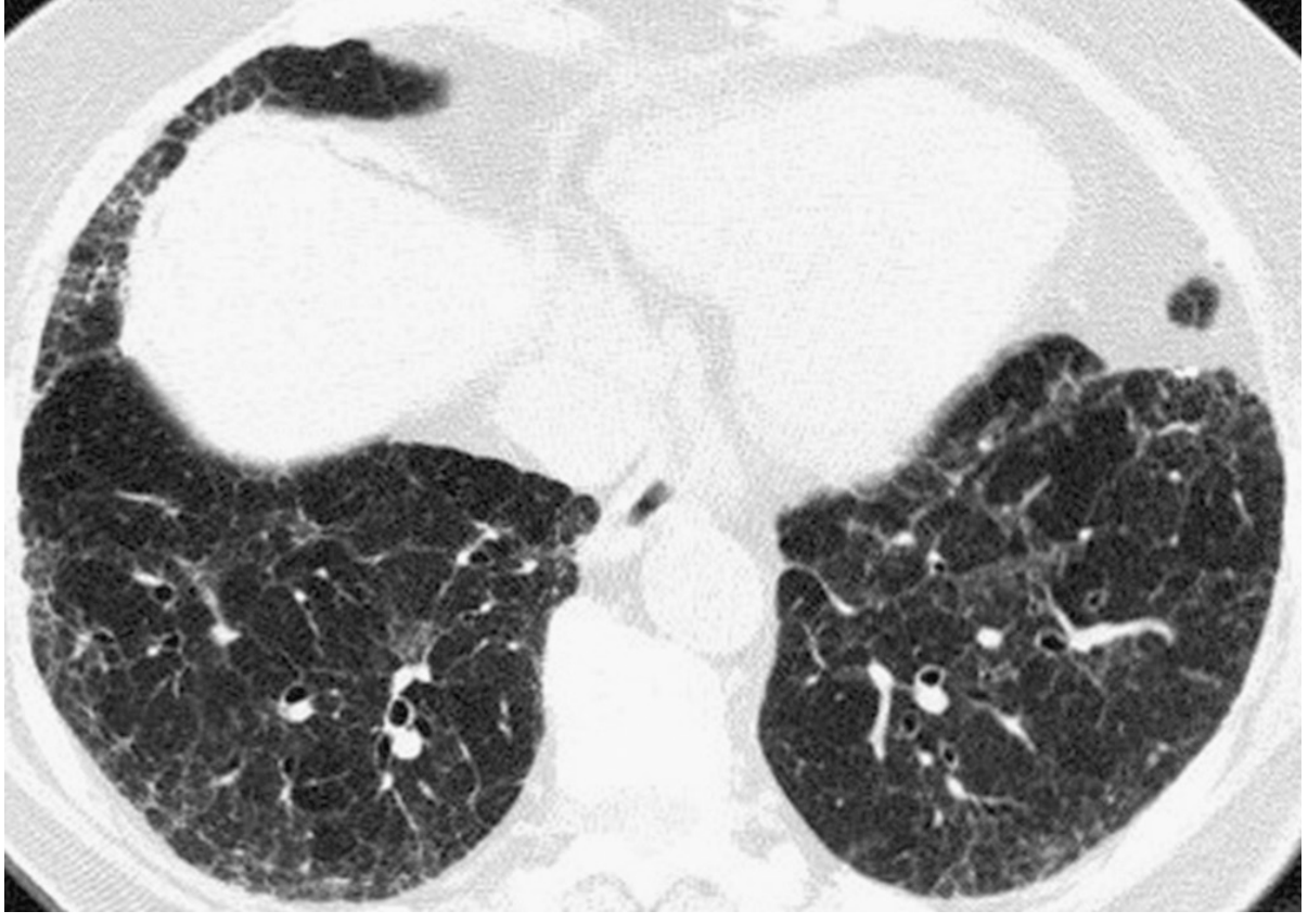
- Bilateral subpleural reticulation (increased fine lines in peripheral lung)
- Architectural distortion
- Traction bronchiectasis - dilated, irregular bronchi within fibrotic tissue
- No discrete honeycombing at this stage
CASE 2 - IPF: Coronal View Demonstrating Basal Predominance
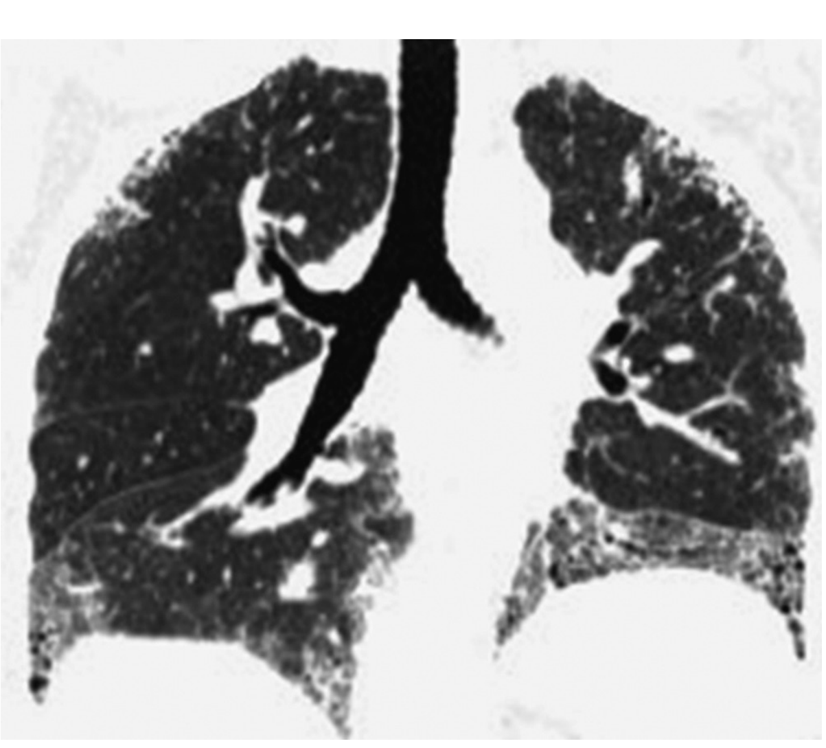
- Coronal view dramatically demonstrates basal predominant distribution
- Subpleural reticulation most dense at lung bases
- The upper lobes show much less disease - this basal gradient is pathognomonic of UIP
- Upper lung zones are relatively spared
CASE 3 - IPF: Classic Honeycombing on Prone HRCT
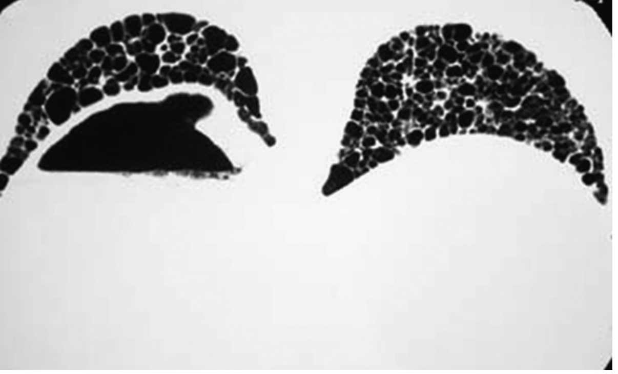
- Extensive subpleural cystic change = honeycombing
- Cyst walls are visible and mildly thickened (1-2 mm)
- Cysts share walls with one another - the "clustered" arrangement is key
- Prone position confirms these are not dependent atelectasis
| Feature | Honeycombing | LAM cysts | LCH cysts |
|---|---|---|---|
| Wall | Visible, ~1-2 mm, shared | Very thin, uniform | Thick, bizarre shaped |
| Location | Subpleural, basal | Diffuse, uniform | Upper lobes |
| Distribution | Layered/stacked | Random | Spares bases |
| Patient | Older adult, M>F | Young woman | Smoker |
CASE 4 - Combined Pulmonary Fibrosis and Emphysema (CPFE)
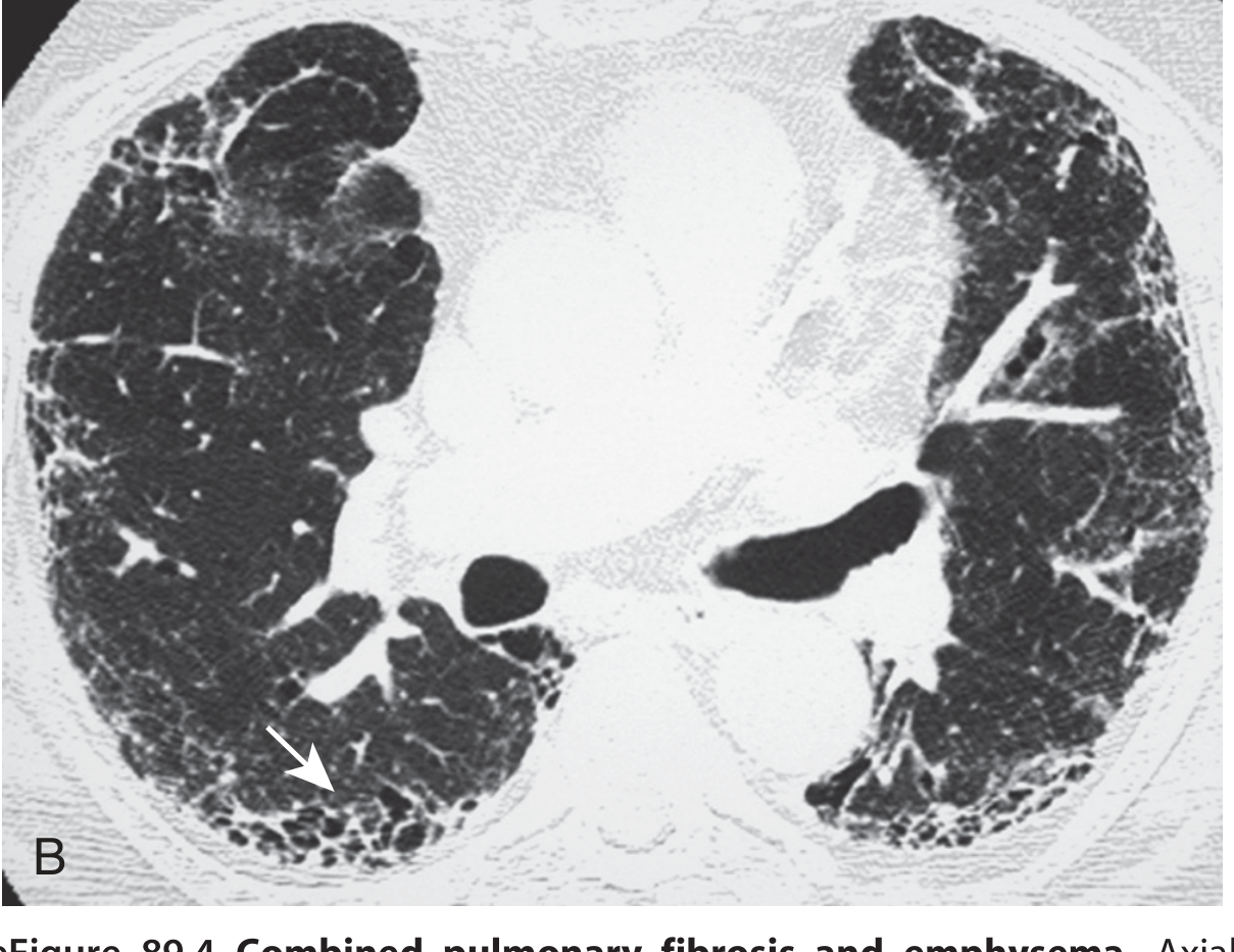
- Upper lobes: centrilobular (single arrowhead) and paraseptal (double arrowhead) emphysema
- Lower lobes (this image): basal reticulation + architectural distortion + honeycombing (arrow)
- The two processes occupy different zones
CASE 5 - Sarcoidosis: Perilymphatic Nodule Distribution
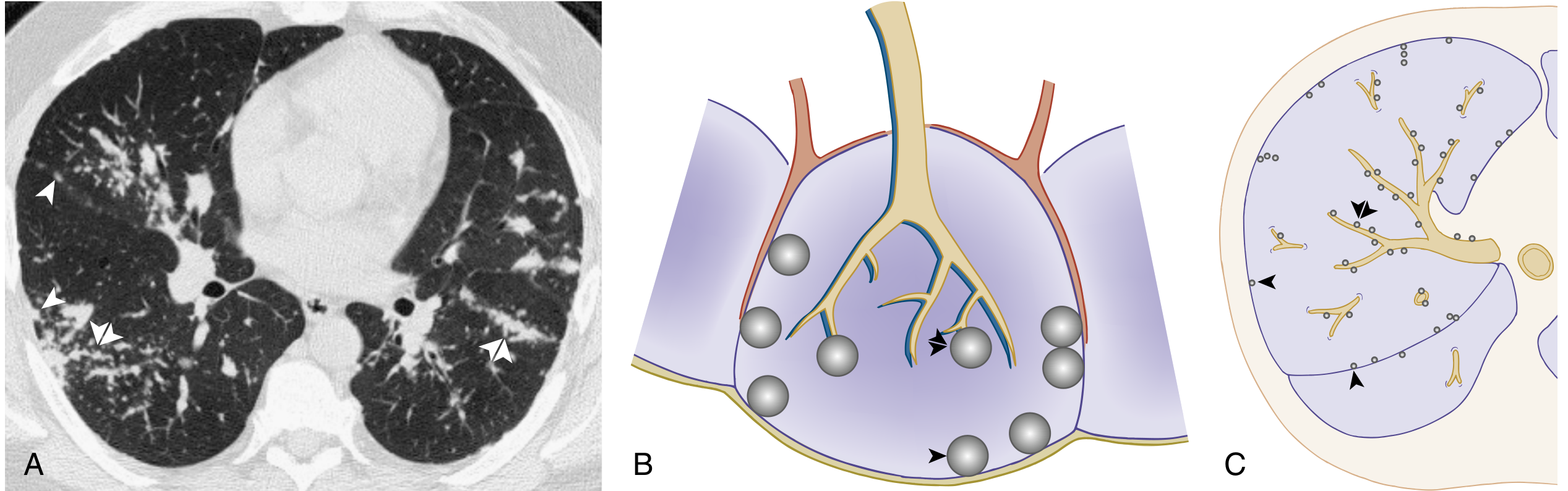
- Numerous subpleural nodules along the costal and fissural pleural surfaces (single arrowheads)
- Nodules along bronchovascular bundles (double arrowheads)
- Upper zone predominance (not shown but typical)
- Perilymphatic micronodules (1-5 mm), bilateral, upper zone
- Bilateral hilar + right paratracheal lymphadenopathy
- Perihilar distribution ("galaxy sign" in advanced disease)
- Upper lobe fibrosis in stage IV (end-stage)
CASE 6 - Miliary TB: Random Nodule Distribution
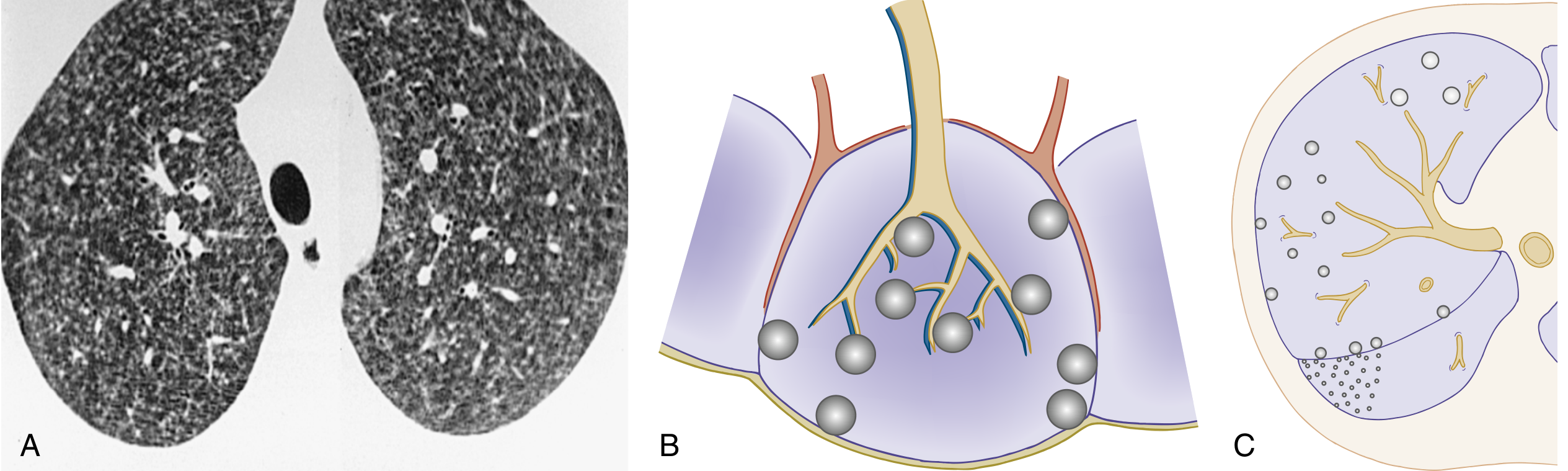
- Numerous small circumscribed nodules equally distributed throughout both lungs bilaterally
- Nodules seen in contact with fissural pleural surfaces as well as sparing fissural surfaces (random)
- No zonal preference - upper, mid and lower zones equally affected
- Miliary TB (fever + endemic area + immunocompromise)
- Miliary fungal infection (histoplasmosis, coccidioidomycosis)
- Hematogenous metastases (known primary malignancy)
- Miliary sarcoidosis (rare)
CASE 7 - Diffuse Pulmonary Haemorrhage: Centrilobular Nodules
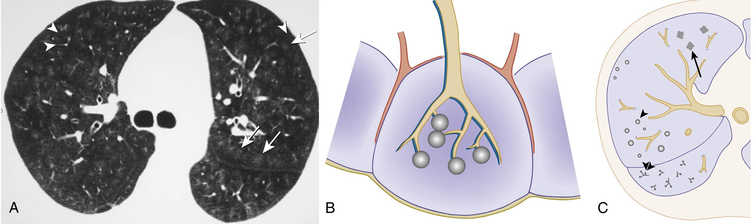
- Numerous bilateral poorly defined GGO nodules (arrows)
- Nodules approach but do not touch the pleura - note the "gap" between nodules and the major fissure (left side)
- Nodules spared from the interlobular septa and small pulmonary veins (arrowheads)
- This "sparing" around the pleural surfaces is the hallmark of centrilobular distribution
- Hypersensitivity pneumonitis (subacute phase, bird/mold exposure)
- Endobronchial spread of infection (TB, NTM, bacterial: "tree-in-bud")
- Respiratory bronchiolitis-ILD (smoker, GGO centrilobular nodules)
- Pulmonary oedema
Key Teaching Points from These Cases
| Case | Key Finding | Lesson |
|---|---|---|
| 1 - Early IPF | Subpleural reticulation + traction bronchiect., no honeycombing | Probable UIP - no biopsy needed |
| 2 - IPF coronal | Basal gradient on coronal view | Always view coronal for zonal distribution |
| 3 - IPF Honeycombing | Clustered subpleural cysts, shared walls | Typical UIP - honeycombing cements diagnosis |
| 4 - CPFE | Upper emphysema + lower honeycombing | Spirometry can be normal; DLCO is key |
| 5 - Sarcoidosis | Perilymphatic nodules + hilar nodes | Pleural + bronchovascular = perilymphatic |
| 6 - Miliary TB | Equal nodules all zones, random | Random = hematogenous spread |
| 7 - DAH | Centrilobular GGO nodules, pleura spared | Sparing of pleura = centrilobular |
- Radiopaedia Cases - HRCT Chest - free case library with scroll-through CT
- Radiology Assistant - Common HRCT Diagnoses - excellent annotated teaching cases for HP, sarcoid, UIP, LAM
- AJR - Hypersensitivity Pneumonitis Spectrum - classic teaching article with clinical-radiological-pathological correlation
Please give images disease wise
HRCT Chest - Images by Disease
1. EMPHYSEMA
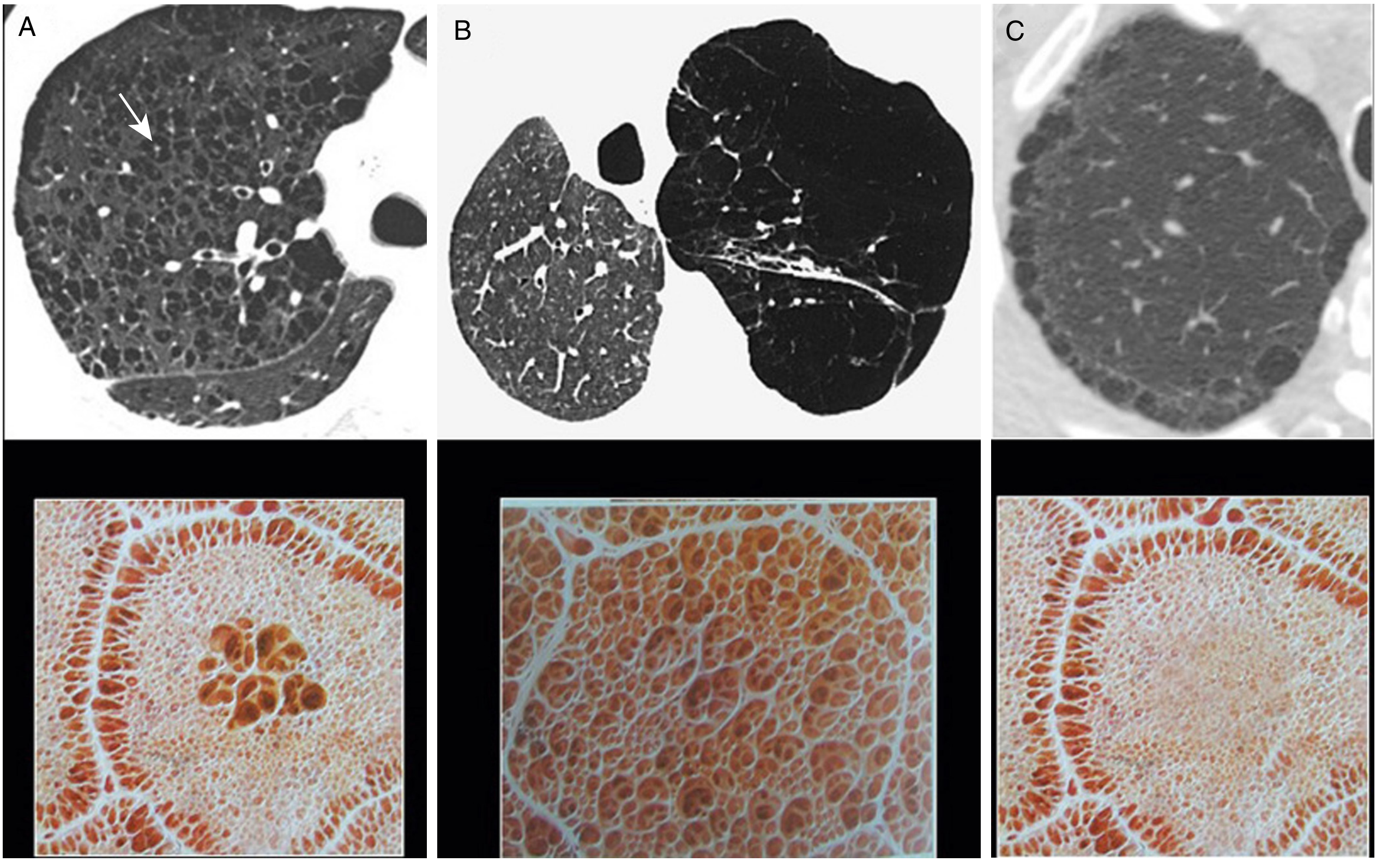
| Type | HRCT Feature | Distribution | Cause |
|---|---|---|---|
| Centrilobular (A) | "Punched-out" lucencies WITHOUT walls; central artery dot | Upper lobe, centrilobular | Smoking |
| Panlobular (B) | Diffuse low attenuation, simplified architecture, fewer vessels | Lower lobe predominant | Alpha-1 antitrypsin deficiency |
| Paraseptal (C) | Single row of subpleural thin-walled cysts | Upper lobe, subpleural | Spontaneous pneumothorax risk |
2. CYSTIC LUNG DISEASES (Multiple in One Panel)
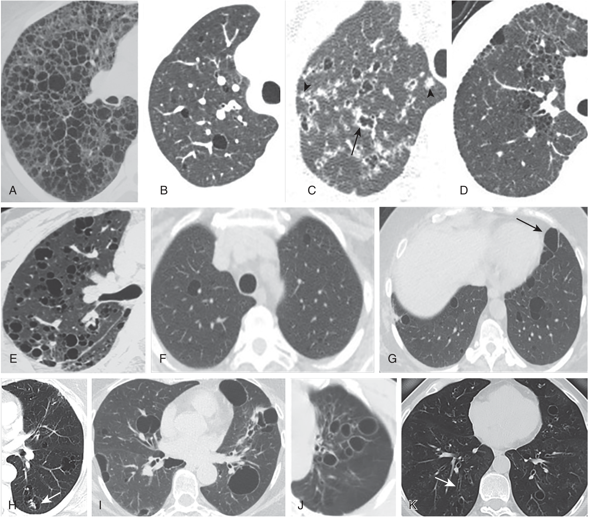
| Disease | Cyst Shape | Distribution | Clue |
|---|---|---|---|
| LAM | Round, uniform, thin walls | Diffuse throughout both lungs | Young woman, pneumothorax |
| LCH | Bizarre shapes, thick walls | Upper lobe, spares bases | Smoker, nodules early stage |
| LIP/Sjogren | Round, uniform | Midlung / subpleural | Autoimmune, dry eyes/mouth |
| Birt-Hogg-Dube | Oblong, septated | Lower lobe, subpleural, paramediastinal | Skin fibrofolliculomas, renal tumours |
| Amyloidosis | Variable | Any | Calcified nodule within cyst (pathognomonic) |
3. IDIOPATHIC PULMONARY FIBROSIS / UIP
3a - Early IPF (Probable UIP): Axial View
3b - IPF: Coronal View Showing Basal Predominance
3c - IPF: Honeycombing (Typical UIP)
3d - Combined Pulmonary Fibrosis + Emphysema (CPFE)
4. HYPERSENSITIVITY PNEUMONITIS (HP)
4a - Fibrotic HP: GGO + Reticulation + Traction Bronchiectasis
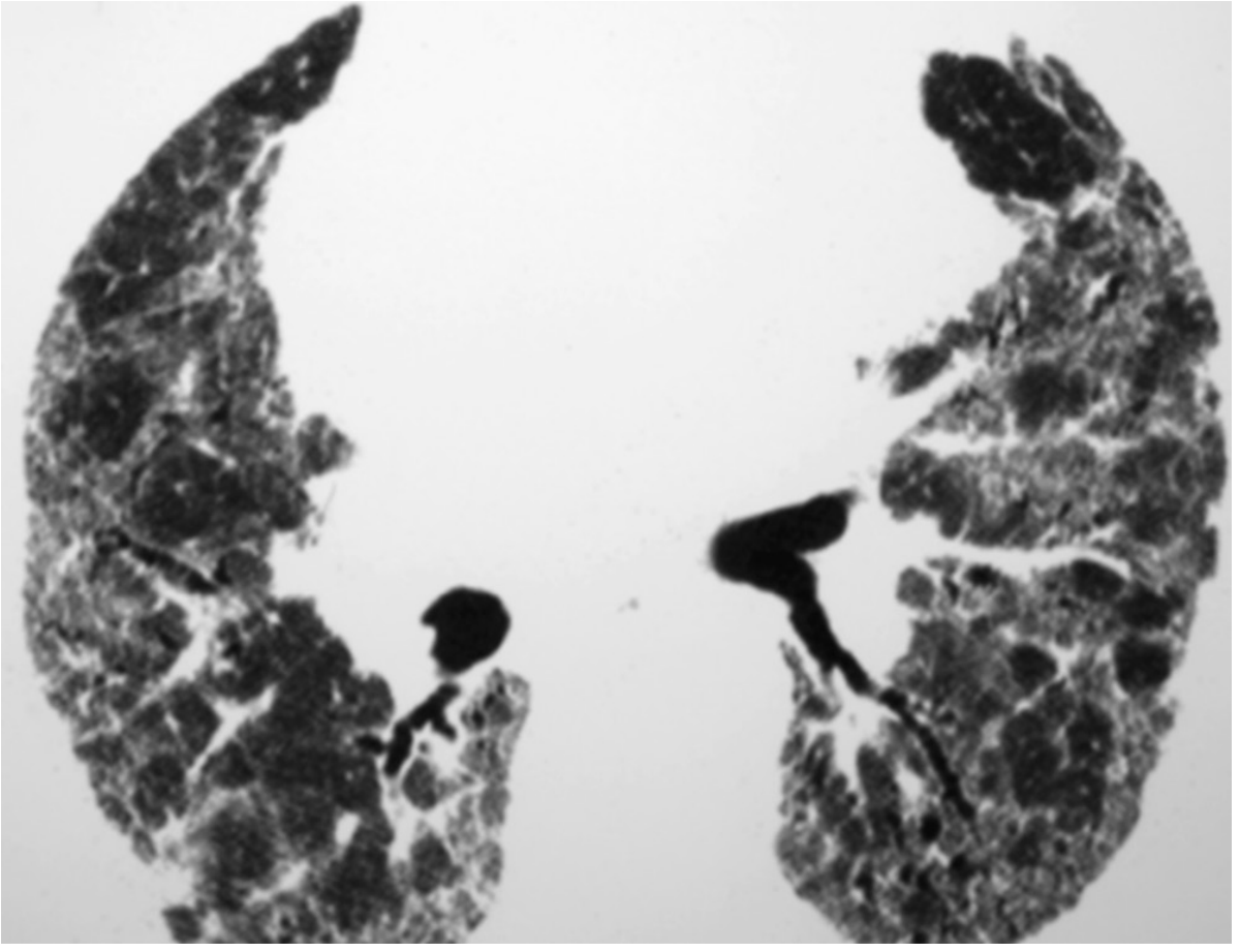
4b - HP: The "Headcheese" Sign
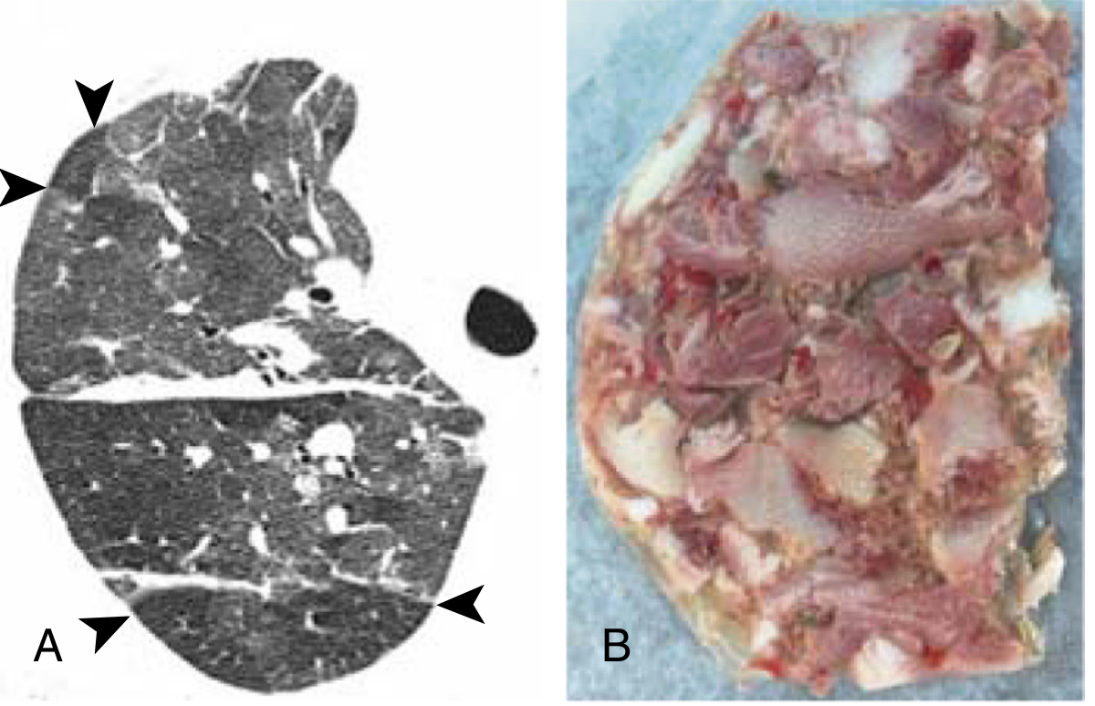
- White/grey = GGO (infiltration/inflammation)
- Normal grey = unaffected lung
- Dark lobules (arrowheads) = air trapping / mosaic perfusion
| Feature | HP | IPF/UIP |
|---|---|---|
| Zone | Mid/upper, or diffuse | Basal + subpleural |
| GGO | Prominent, diffuse | Minimal |
| Air trapping | YES (on expiratory scan) | Absent |
| Centrilobular nodules | YES (subacute phase) | No |
| Headcheese sign | YES (highly specific) | No |
| Exposure history | Bird/mould/farmer | None |
5. SARCOIDOSIS (Scadding Stages 1-4)
5a - Stage 1: Bilateral Hilar Lymphadenopathy Only
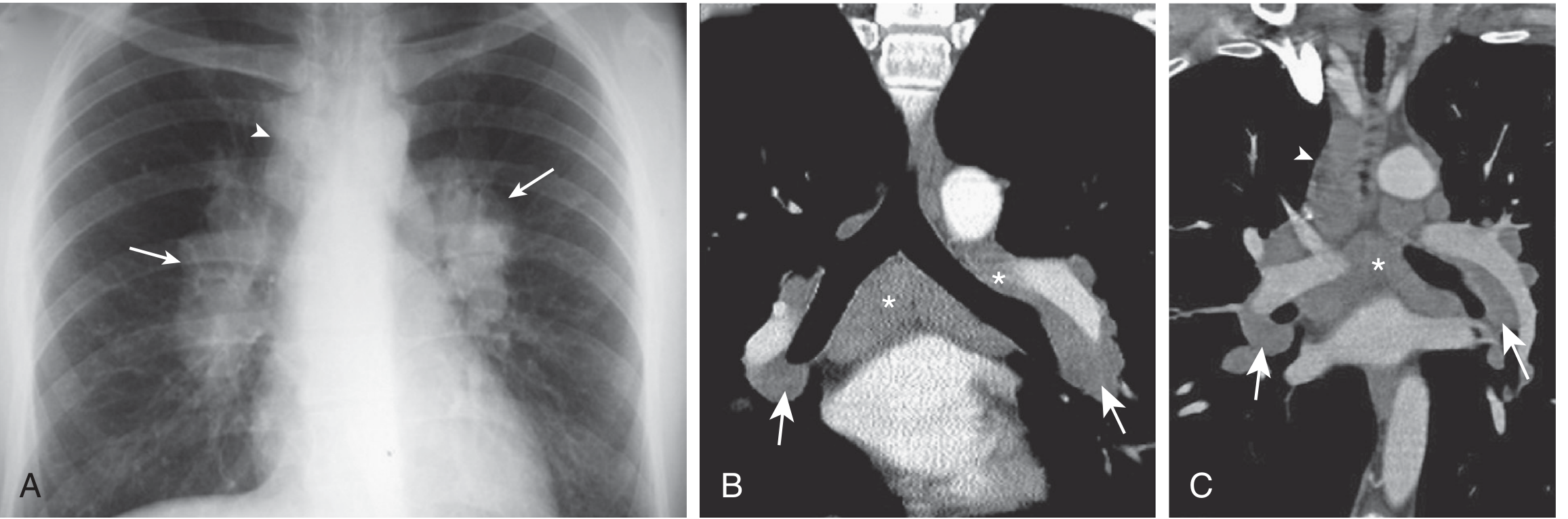
5b - Stage 2: BHL + Parenchymal Nodules
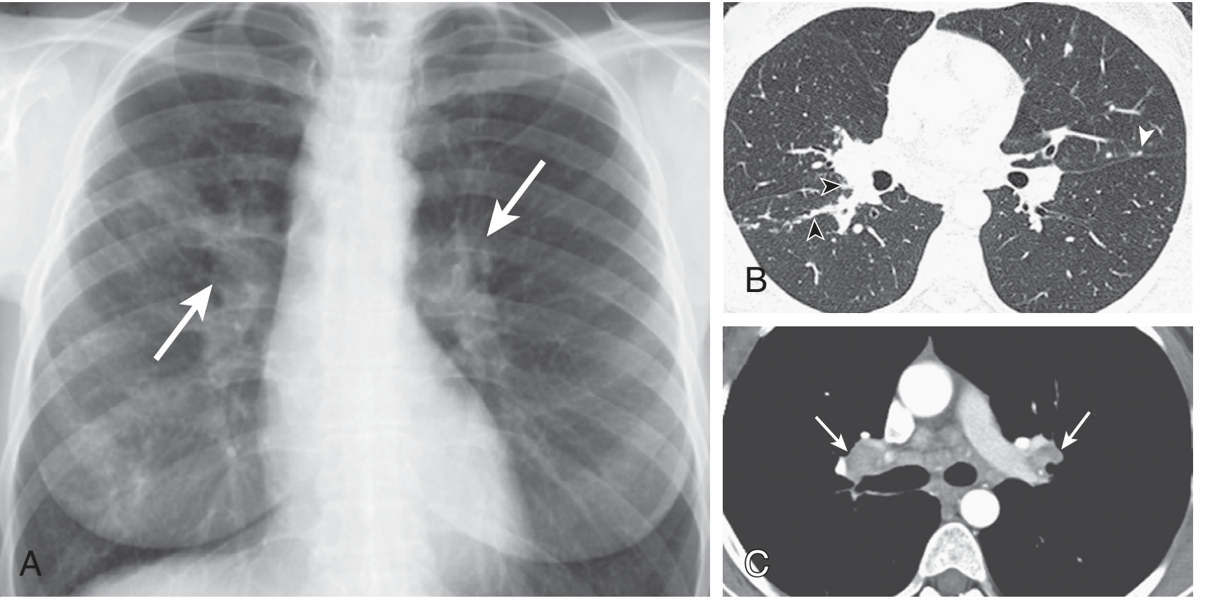
- Nodules along fissural surfaces (perilymphatic, white arrowhead)
- "Beaded" appearance of bronchovascular bundles (black arrowheads) - pathognomonic of sarcoidosis
5c - Stage 3: Parenchymal Disease Only (No BHL)
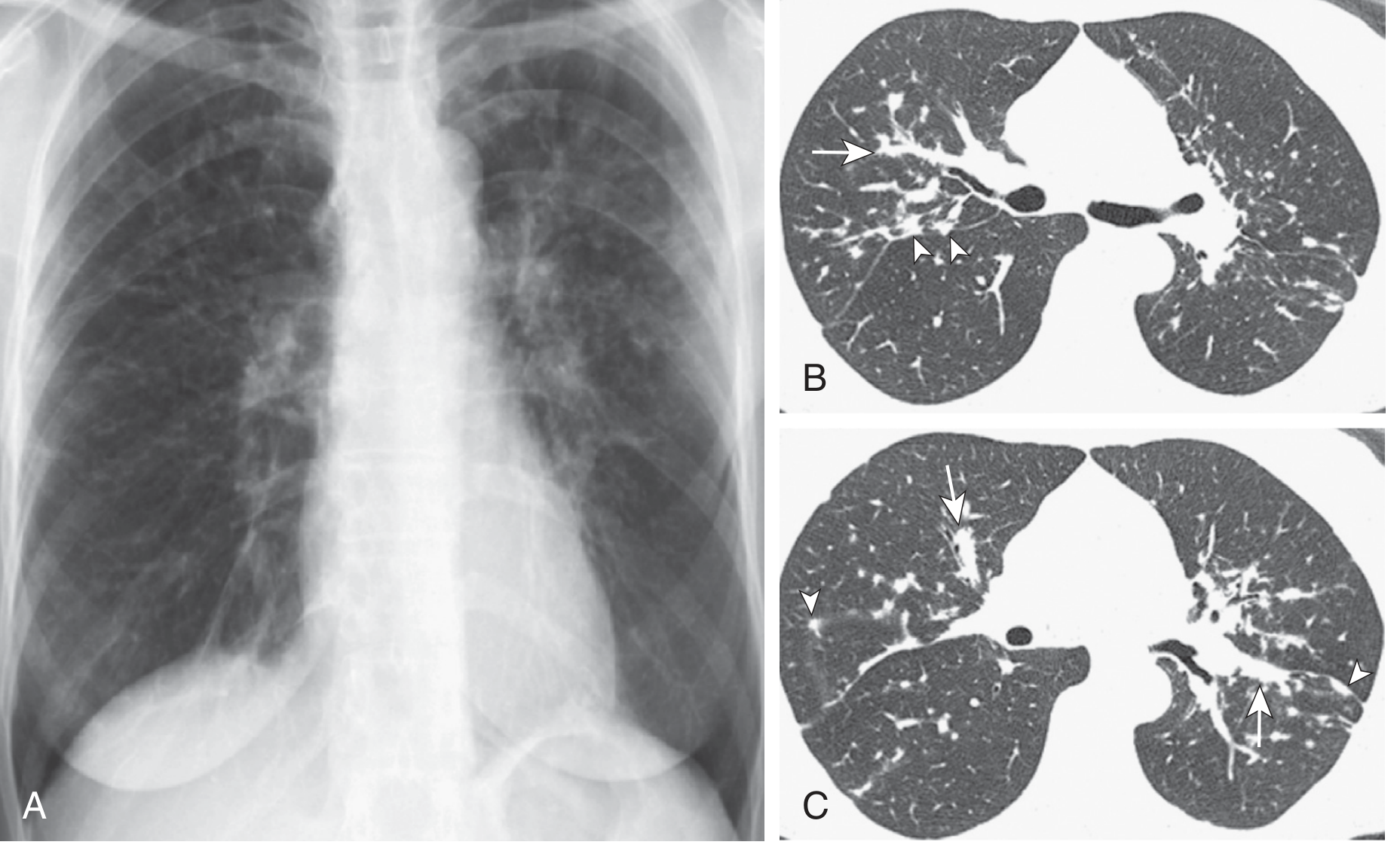
5d - Stage 4: Fibrotic Sarcoidosis
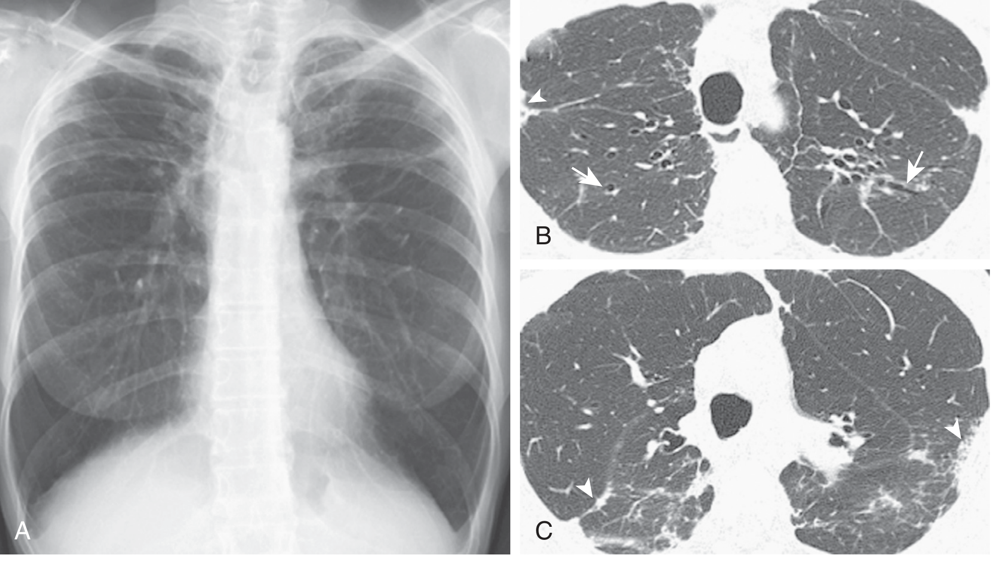
6. NODULE DISTRIBUTION: PERILYMPHATIC vs RANDOM vs CENTRILOBULAR
6a - Perilymphatic (Sarcoidosis)
6b - Random (Miliary TB)
6c - Centrilobular (Pulmonary Haemorrhage / HP)
Quick Summary Table
| Disease | Dominant Pattern | Zone | Key Sign | Avoid Confusing With |
|---|---|---|---|---|
| IPF (UIP) | Reticulation + Honeycombing | Basal + Subpleural | Traction bronchiectasis, no GGO | NSIP (has GGO + subpleural sparing) |
| Emphysema | Low attenuation (no walls) | Upper (centrilobular) | Central dot sign | LAM (has visible walls) |
| HP | GGO + Centrilobular nodules + Air trapping | Mid/upper or diffuse | Headcheese sign | IPF (HP has air trapping + GGO) |
| Sarcoidosis | Perilymphatic nodules | Upper, perihilar | Beaded vessels, 1-2-3 sign | Lymphangitic carcinomatosis |
| Miliary TB | Random tiny nodules | Uniform, all zones | No zonal preference | Hematogenous mets (need clinical context) |
| LAM | Multiple round thin-walled cysts | Diffuse | Uniform cysts in young woman | LCH (bizarre shapes), emphysema (no walls) |
| LCH | Bizarre-shaped cysts + nodules | Upper, spares bases | Smoker, upper predominance | LAM (uniform), emphysema |
Heart failure according tin tintinalli textbook
Heart Failure — Tintinalli's Emergency Medicine (Chapter 53)
INTRODUCTION & EPIDEMIOLOGY
- >80% of ED visits for AHF result in hospital admission
- ~50% of patients die within 5 years of diagnosis
- Hospitalization is an inflection point - hospitalised patients have higher mortality than matched non-hospitalised cohort
- Prevalence is increasing due to aging population and improved AMI survival
PATHOPHYSIOLOGY
Neurohormonal Cascade
- Renin-angiotensin-aldosterone system (RAAS) and sympathetic nervous system activate
- Norepinephrine, vasopressin, endothelin (potent vasoconstrictor), TNF-α are released
- Net effect: Na+ and water retention + increased SVR
- This maintains BP and perfusion short-term but increases myocardial workload, wall tension, and O2 demand
- Leads to cardiac remodelling and progressive dysfunction
Natriuretic Peptides (Counterregulatory Response)
| Peptide | Source | Half-life |
|---|---|---|
| ANP (Atrial Natriuretic Peptide) | Atria | Short |
| BNP (B-type Natriuretic Peptide) | Cardiac ventricle | ~20 min |
| NT-proBNP (inactive cleavage product) | Ventricle | ~2 hours |
| CNP | Endothelium | - |
Acute Pulmonary Oedema Mechanism
- CO drops → SVR rises (compensatory vasoconstriction)
- ↑ SVR → further drops CO
- Even small BP elevations can trigger this spiral in susceptible patients
- Can be abrupt, severely symptomatic, and rapidly fatal
CLASSIFICATION (6 Phenotypes - Table 53-1)
| Classification | Characteristics |
|---|---|
| Hypertensive AHF | AHF signs + BP >140 mmHg + preserved LV function; CXR: pulmonary oedema; onset <48 h; fluid redistribution more than overload |
| Pulmonary Oedema | Respiratory distress + rales + reduced SpO2 + CXR findings |
| Cardiogenic Shock | Tissue hypoperfusion; SBP typically <90 mmHg (see Ch. 50) |
| Acute-on-Chronic HF | Mild-moderate AHF; SBP 90-140 mmHg; increased peripheral oedema; onset over days |
| High-Output Failure | High CO, tachycardia, warm extremities, pulmonary congestion; caused by anaemia, thyrotoxicosis |
| Right Heart Failure | Low-output syndrome; JVD + hepatomegaly ± hypotension; from pulmonary disease, TR, OSA; no pulmonary congestion |
SYSTOLIC vs. DIASTOLIC HEART FAILURE
| HFrEF (Systolic) | HFpEF (Diastolic) | |
|---|---|---|
| EF | <50% | Preserved (≥50%) |
| Mechanism | Ventricle fails to eject blood | Ventricle fails to receive blood |
| Problem | Afterload sensitivity, ↑ intracardiac volume | ↓ LV compliance, preload sensitivity |
| Common causes | Ischaemic CM, dilated CM | Hypertension, LVH, ageing |
| Key feature | ↑ LV volumes, dilated | Normal or ↑ wall thickness, stiff |
DIAGNOSIS
"There is no single diagnostic test for heart failure; it is a clinical diagnosis based on all clinical data, especially history and physical examination."
History
| Finding | Sensitivity | Specificity | LR+ |
|---|---|---|---|
| History of HF | 56% | 80% | 2.7 |
| Dyspnoea on exertion | 84% (highest sensitivity) | - | - |
| Paroxysmal nocturnal dyspnoea | Lower | 76-84% (most specific) | - |
| Orthopnoea | Lower | 76-84% | - |
| Oedema | Lower | 76-84% | - |
Physical Examination
| Finding | LR+ | LR- | Note |
|---|---|---|---|
| S3 gallop | 4.0 (highest LR+) | 0.91 | Not useful if absent; inter-rater reliability poor |
| Overall clinical gestalt | Sensitivity 61%, specificity 86% | - | Best when >80% confident |
Common Precipitants of AHF (Table 53-2)
- Excess salt or fluid intake
- Medication non-adherence
- Acute coronary syndrome
- New-onset or poorly controlled arrhythmias (especially AF)
- Worsening valvular disease
- Hypertensive crisis
- Renal failure (especially missed dialysis)
- Substance abuse (cocaine, methamphetamines, ethanol)
- Poorly controlled hypertension
- Recent addition of negative inotropes (CCB, beta-blockers)
- NSAID use
- Infections (sepsis, pneumonia)
- Pulmonary embolism
- Anaemia/thyrotoxicosis (high-output)
Diagnostic Testing
- Most useful objective test in undifferentiated dyspnoea
- BNP <100 pg/mL = very unlikely HF; BNP >400 pg/mL = highly likely HF
- NT-proBNP: age-adjusted cutoffs (>450 pg/mL in <50 yrs; >900 in 50-75 yrs; >1800 in >75 yrs)
- Elevated troponin in AHF = high-risk marker (myocardial injury component)
- Cardiomegaly (CTR >0.5)
- Pulmonary vascular redistribution (upper lobe diversion)
- Interstitial oedema (Kerley B lines)
- Alveolar oedema ("butterfly/bat-wing" pattern)
- Pleural effusions (right > left)
- Not diagnostic but identifies precipitants: AF, ischaemia, LVH, bundle branch blocks
- IVC: collapsibility assesses volume status
- B-lines on lung US: ≥3 per zone = interstitial oedema (sens ~90%)
- LV function: gross assessment of systolic function
- Pericardial effusion/tamponade
TREATMENT
Initial Measures (All AHF)
- Supplemental oxygen to keep SpO2 ≥95%
- Cardiac monitoring + IV access
- Frequent vital signs
- Urinary catheter only for severely ill or incontinent (avoid routine use)
1. Noninvasive Ventilation (NIV)
- CPAP or BiPAP - decreases work of breathing, avoids intubation
- Indicated for severe dyspnoea, pulmonary oedema with hypoxia
- Reduces need for intubation and improves short-term survival (Tables 53-3 & 53-4)
2. HYPERTENSIVE AHF: Vasodilators First
"Initiate nitrates BEFORE diuretics in hypertensive AHF"
Nitroglycerin (NTG)
- Short-acting, rapid-onset venous and arterial dilator
- Decreases preload; at high doses, reduces afterload
- May have coronary vasodilatory benefit (↓ myocardial ischaemia)
| Route | Dose | Notes |
|---|---|---|
| Sublingual | 0.4 mg (400 mcg) spray/tablet q 1-5 min | Easily administered, rapidly bioavailable |
| IV | Start 0.5-0.7 mcg/kg/min; titrate up to 200 mcg/min | Based on BP + symptoms |
| Transdermal | 0.5-2 inches to chest wall | Slow onset - only after initial improvement |
Nitroprusside
- Start 0.3 mcg/kg/min; max 10 mcg/kg/min
- Complications: Hypotension, cyanide/thiocyanate toxicity, coronary steal
3. DIURETICS (Table 53-6)
| Drug | IV Dose | Onset | Duration |
|---|---|---|---|
| Furosemide | No prior use: 20-40 mg IVP; Prior use: total daily oral dose × 1-2.5, given as IV boluses q12h | 15-20 min | 4-6 h |
| Bumetanide | 1-3 mg IV (40:1 furosemide equivalent) | 10 min | Peak at 60 min |
| Torsemide | 10-20 mg IV (2:1 furosemide equivalent) | 10 min | Peak 1-2 h |
4. NORMOTENSIVE / ACUTE-ON-CHRONIC HF
- Oxygen + monitoring
- BNP, troponin, renal function, electrolytes
- Loop diuretic (furosemide)
- Consider NIV if dyspnoeic
- Assess for ischaemia/arrhythmia as precipitant
5. DRUGS TO AVOID / USE CAUTIOUSLY
| Drug | Reason to Avoid in Acute HF |
|---|---|
| Morphine | ↑ need for mechanical ventilation, prolonged hospitalisation, ICU admission, mortality. If used, small titrated doses (2-4 mg IV) only |
| NSAIDs | Na+/water retention, blunt diuretic effect, ↑ morbidity/mortality |
| CCBs (oral) | Myocardial depressant activity; no benefit or worse outcomes |
| Beta-blockers | Not initiated in acute setting (except rate control); do not start de novo |
| IV ACE-I/ARBs | Insufficient ED data; continue oral if already on them |
HIGH-RISK FEATURES (Admission Criteria - Table 53-7/53-8)
- Hypotension (SBP <90 mmHg) - cardiogenic shock
- Persistent hypoxia despite NIV
- Altered mental status
- New ischaemia: elevated troponin, ischaemic ECG changes
- Tachycardia (new AF or refractory arrhythmia)
- Severe renal dysfunction (elevated creatinine, low Na+)
- Need for invasive monitoring or procedures → ICU
- Elevated troponin or natriuretic peptides
- Low serum sodium
- Renal dysfunction (elevated creatinine)
- Low blood pressure
DISPOSITION ALGORITHM
AHF patient responds to ED therapy?
│
YES ─────────────────────────────── NO
│ │
High-risk features present? Admit to hospital
│
NO ──────────────── YES
│ │
Observation unit Hospital admission
(12-24 h, monitor (consider ICU if needs
vitals, UO, weight, invasive monitoring)
further testing)
│
Discharge if improved + no high-risk features
+ outpatient follow-up within 5 days
- ~75% of AHF patients respond to therapy, have no high-risk features, and can be discharged
- Outpatient follow-up within 5 days decreases readmissions
- Observation unit strategy reduces costs for selected lower-risk patients
SPECIAL SITUATIONS
Hypertensive Acute Pulmonary Oedema
- Treat aggressively with nitrates + NIV first
- Failing heart very sensitive to afterload - even SBP 150 mmHg can trigger pulmonary oedema
- Prompt vasodilation can avoid intubation
High-Output Heart Failure
- Normal/elevated EF; tachycardia; warm extremities
- Causes: anaemia, thyrotoxicosis, AV fistula, Paget's, sepsis
- Treat the underlying cause - volume removal may worsen if low-output coexists
Right Heart Failure
- JVD + hepatomegaly + peripheral oedema; no pulmonary oedema
- Causes: pulmonary hypertension, COPD, PE, TR, OSA
- Do NOT aggressively diurese - RV is volume-dependent
- Treat precipitant; cautious fluids if needed
KEY PEARLS FROM TINTINALLI'S
- No single test diagnoses AHF - it is a clinical diagnosis
- Dyspnoea on exertion = most sensitive symptom (84%); S3 gallop = most specific sign (LR+ 4.0)
- Nitrates before diuretics in hypertensive AHF
- Avoid morphine - associated with worse outcomes
- NIV (CPAP/BiPAP) reduces intubation and improves survival in respiratory distress
- BNP <100 makes HF unlikely; >400 makes it likely
- High-risk markers absent in 50% of patients - use clinical judgment for disposition
- 5-day follow-up after discharge reduces readmissions
- In normotensive AHF, look for and treat the precipitant (ACS, AF, infection, missed dialysis, non-compliance)
- Calcium channel blockers are contraindicated in systolic HF
- Tintinalli's Emergency Medicine: A Comprehensive Study Guide, Chapter 53 - Acute Heart Failure