
One lung ventilation for 10 marks anesthesia residency
Reading File
Finding Sources
Finding Sources
Reading File
Reading File
Reading File
Reading File
Reading File
Reading File
Reading File
Reading File
Reading File
Loading Image
Searching Images
double lumen tube endobronchial placement diagram one lung ventilation
I now have comprehensive material from Miller's, Morgan & Mikhail's, and Barash's to write a thorough 10-mark answer. Let me compile it.
One-Lung Ventilation (OLV)
Sources: Miller's Anesthesia 10e, Morgan & Mikhail's Clinical Anesthesiology 7e, Barash's Clinical Anesthesia 9e
Definition
One-lung ventilation (OLV) is a technique in which only one lung is ventilated while the other is intentionally allowed to collapse, primarily to facilitate thoracic surgery. The non-ventilated lung is deliberately deflated but continues to be perfused, creating an obligatory right-to-left intrapulmonary shunt.
Indications
Divided into absolute and relative:
Absolute (Lung Isolation)
- Protection from contamination - massive hemoptysis, lung abscess, empyema (to prevent spillage to the healthy lung)
- Control of distribution of ventilation - bronchopleural fistula, bronchopleural cutaneous fistula, unilateral cyst/bullae, major bronchial disruption
- Unilateral bronchopulmonary lavage (e.g., pulmonary alveolar proteinosis)
- Video-assisted thoracoscopic surgery (VATS)
Relative (Lung Separation for Surgical Exposure)
| Priority | Examples |
|---|---|
| High priority | Thoracic aortic aneurysm, pneumonectomy, lung volume reduction, upper lobectomy, minimally invasive cardiac surgery |
| Low priority | Esophageal surgery, middle/lower lobectomies, mediastinal mass resection, thymectomy, bilateral sympathectomies |
Key distinction: Lung isolation (protects against cross-contamination) requires DLT; lung separation (surgical exposure) can use either DLT or bronchial blockers.
- Barash's Clinical Anesthesia 9e, p. 3173
Physiology of OLV
Two-Lung vs One-Lung Ventilation
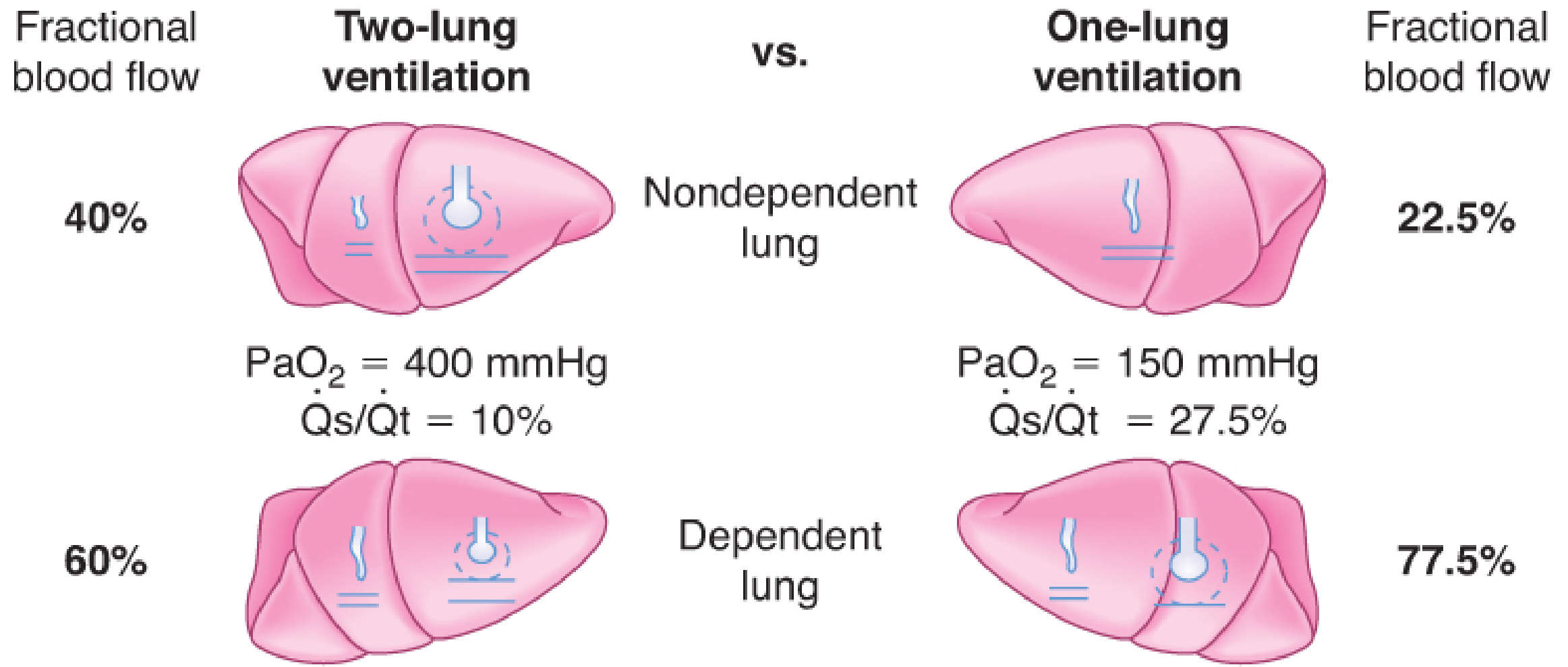
During two-lung ventilation:
- Nondependent lung: 40% of blood flow; dependent lung: 60%
- PaO2 ~400 mmHg; Qs/Qt ~10%
During OLV (lateral decubitus):
- Nondependent (collapsed) lung: ~22.5%; dependent lung: ~77.5%
- PaO2 falls to ~150 mmHg; Qs/Qt rises to ~27.5%
The increase in Qs/Qt (intrapulmonary shunt) from ~10% to ~27.5% is the fundamental cause of hypoxemia during OLV.
Two Main Causes of Impaired Oxygenation During OLV
- Persisting blood flow through the non-ventilated (nondependent) lung → obligatory shunt
- Development of atelectasis in the dependent (ventilated) lung → additional local shunt and low V/Q areas
- Miller's Anesthesia 10e, p. 1321
Role of Hypoxic Pulmonary Vasoconstriction (HPV)
HPV is a compensatory mechanism that reduces blood flow to the non-ventilated lung, limiting the degree of shunt. Without HPV, OLV would result in a shunt fraction of ~35-40%; with HPV it is typically 20-30%. HPV reduces blood flow to the nondependent collapsed lung by ~50%.
Factors that INHIBIT HPV (worsen oxygenation):
- Pulmonary hypertension
- Hypocapnia, alkalosis
- Increased cardiac output; increased mixed venous PO2
- Hypothermia
- Vasodilators: nitroglycerin, nitroprusside, nitric oxide
- Phosphodiesterase inhibitors (milrinone, enoximone)
- Beta-adrenergic agonists
- Calcium channel blockers
- Inhalation anesthetics (volatile agents at >1 MAC)
Factors that decrease blood flow to the VENTILATED (dependent) lung (also worsen hypoxemia):
- High mean airway pressures (excessive PEEP, hyperventilation, high PIP)
- Low FiO2 in the ventilated lung (triggers HPV in ventilated lung)
- Vasoconstrictors (greater effect on normoxic than hypoxic vessels)
- Intrinsic PEEP from inadequate expiratory time
- Morgan & Mikhail's Clinical Anesthesiology 7e, p. 1024
Techniques for Achieving OLV
Four techniques are available:
1. Double-Lumen Endobronchial Tube (DLT) - Most Commonly Used
- Two bonded endotracheal tubes: one lumen enters a mainstem bronchus, one ends in the distal trachea
- Two cuffs: a proximal tracheal cuff and a distal bronchial cuff
- Left-sided DLT preferred in most situations (longer left mainstem bronchus; safer margin from carina)
- Right-sided DLT used when: left pneumonectomy, left-sided tracheobronchial disruption, left mainstem stenosis, left-sided tumor at the carina
- Sizes: 35, 37, 39, 41 Fr (generally 39-41 Fr for men; 35-37 Fr for women)
- Fiberoptic bronchoscopy is the gold standard for confirming position
- Advantages: reliable, fast placement, allows suctioning, CPAP/PEEP to either lung independently
- Disadvantage: larger tube, more airway trauma, higher rate of hoarseness (44% vs 17% with blockers)
2. Bronchial Blockers
- Single-lumen ETT + balloon-tipped blocker (e.g., Arndt, Cohen, EZ-blocker)
- Useful in: difficult airway, tracheostomy patients, existing ETT
- Disadvantage: unreliable seal (not suitable for true lung isolation/protection), difficult suctioning, slower collapse, higher dislodgement risk
3. Mainstem Intubation with Single-Lumen ETT
- Conventional ETT advanced into a mainstem bronchus
- Simplest but least versatile; limited to emergencies
4. Tubeless Techniques
- For select VATS procedures; no intubation; relies on spontaneous ventilation
- Barash's Clinical Anesthesia 9e, pp. 3175-3186
Management of OLV: Lung-Protective Ventilation Strategy
The focus has shifted from merely preventing hypoxemia to preventing acute lung injury (ALI) - which occurs in 2.5% of all resections and up to 7.9% post-pneumonectomy, with ~40% mortality/major morbidity.
Recommended ventilation parameters during OLV:
| Parameter | Target |
|---|---|
| Tidal volume | 4-5 mL/kg predicted body weight (NOT the same as two-lung TV) |
| FiO2 | Start at 1.0; can reduce to 50-80% if saturations are adequate |
| Plateau pressure | <25 cmH2O |
| Peak airway pressure | <35 cmH2O |
| PEEP | Titrated to optimal compliance in ventilated lung |
| Ventilatory mode | Pressure-controlled ventilation preferred (limits peak pressures, more homogenous gas distribution) |
Pre-OLV preparation:
- Before commencing OLV, ventilate with 100% O2 to de-nitrogenate the operative lung (nitrogen's low blood-gas solubility delays collapse)
- Perform a recruitment maneuver on the dependent lung immediately after starting OLV (sustain 20 cmH2O for 15-20 seconds) to clear atelectasis that developed during two-lung anesthesia
- Morgan & Mikhail's Clinical Anesthesiology 7e, pp. 1045-1046
Management of Hypoxemia During OLV
Target: SpO2 ≥90% (PaO2 >60 mmHg). In patients with CAD, cerebrovascular disease, or anemia, maintain a higher threshold.
Stepwise management (escalating):
| Step | Action |
|---|---|
| 1 | Check DLT/blocker position with FOB - surgical manipulation may dislodge it; suction both lumens |
| 2 | Increase FiO2 to 1.0 |
| 3 | Optimize cardiac output - ensure no IVC compression by surgeon; stop vasodilators; reduce volatile agent to ≤1 MAC |
| 4 | Recruitment maneuver of the ventilated lung (20 cmH2O for 15-20 sec; causes transient worsening before improvement) |
| 5 | Apply PEEP to the ventilated lung (avoid in emphysematous patients - risk of intrinsic PEEP) |
| 6 | Apply CPAP 1-2 cmH2O to the non-ventilated lung (first do a recruitment maneuver on it) |
| 7 | Apneic oxygen insufflation to non-ventilated lung |
| 8 | Partial ventilation of non-ventilated lung (intermittent PPV, small tidal volumes, selective lobar collapse) |
| 9 | Pharmacologic: almitrine (potentiates HPV; improves PaO2 without altering PAP or CO) + inhaled NO to ventilated lung |
| 10 | Ask surgeon to clamp ipsilateral pulmonary artery |
| 11 | Resume two-lung ventilation (if severe/precipitous desaturation) |
| Last resort | Venovenous ECMO |
- Miller's Anesthesia 10e, pp. 7212-7213; Morgan & Mikhail's 7e, pp. 1046-1047
Lung Injury Mechanisms During OLV
Both lungs are at risk:
- Dependent (ventilated) lung: hyperperfusion (77.5% of cardiac output) + ventilator-induced lung injury (VILI) from large tidal volumes and high pressures
- Non-dependent (collapsed) lung: ischemia-reperfusion injury + direct surgical trauma
This is why lung-protective ventilation is so critical - tidal volumes >8 mL/kg used historically (same as two-lung TV) are now strongly discouraged.
Pharmacological Adjuncts
- Almitrine (IV pulmonary vasoconstrictor): potentiates HPV in non-ventilated lung; at low doses does not increase PA pressure or reduce CO; best used in combination with inhaled NO
- Inhaled NO (pulmonary vasodilator): when given to ventilated lung, increases blood flow to already-ventilated areas; minimal effect alone but synergistic with almitrine
- Volatile anesthetics: inhibit HPV at >1 MAC; use ≤1 MAC or switch to TIVA (propofol) if hypoxemia is refractory
- Miller's Anesthesia 10e, p. 1322
Monitoring During OLV
- SpO2 (continuous)
- Arterial line for serial ABGs - especially for permissive hypercapnia monitoring
- End-tidal CO2: useful as a trend monitor but arterial-to-end-tidal CO2 gradient is unpredictable and increases during OLV due to dead space ventilation
- Peak/plateau airway pressures: continuous monitoring to limit VILI
- Periodic fiberoptic bronchoscopy to confirm DLT position, especially after patient repositioning
Postoperative Considerations
- Operative lung is reinflated gradually to PIP <30 cmH2O to avoid disruption of staple lines
- During reinflation, clamping the dependent lung lumen prevents overdistention
- V/Q matching may take time to normalize; expect impaired oxygenation early postoperatively
- Pain control (thoracic epidural, paravertebral block) is essential to prevent splinting and atelectasis
Summary table for exam:
| Feature | Key Point |
|---|---|
| Shunt fraction during OLV | ~27.5% (up from 10%) |
| Key compensatory mechanism | HPV (reduces non-ventilated lung flow by 50%) |
| Best technique for lung isolation | Left-sided DLT confirmed with FOB |
| Tidal volume during OLV | 4-5 mL/kg IBW |
| First step for hypoxemia | Check DLT position with FOB |
| Best pharmacologic combo | Almitrine + inhaled NO |
| ALI incidence post-pneumonectomy | 7.9%; mortality 40% |
| Acceptable SpO2 lower limit | ≥90% (PaO2 >60 mmHg) |
This is a shared conversation. Sign in to Orris to start your own chat.