
Cerebellum in physiology
Reading File
Now let me search the medical library for cerebellum physiology content in parallel.
Finding Sources
Finding Sources
The best sources are Costanzo Physiology, Ganong's, and Neuroscience: Exploring the Brain. Let me read from the top physiology sources in parallel.
Reading File
Reading File
Reading File
Good, I have initial content. Now let me read the dedicated cerebellum sections in Costanzo and Ganong more deeply, plus check for images.
Reading File
Reading File
Excellent content. I can see a diagram image URL in Costanzo. Let me fetch that image and continue reading for the output pathways and clinical features sections.
Loading Image
Reading File
I now have comprehensive content from Costanzo Physiology and Ganong's. I have all the major topics covered. Let me now compile the full, well-structured answer.
Cerebellum - Physiology
Overview and Location
The cerebellum ("little brain") sits in the posterior fossa, dorsal to the pons and medulla, just below the occipital lobe. Despite comprising only ~10% of brain weight, its surface area is about 75% that of the cerebral cortex due to dense foliation. It is connected to the brainstem by three cerebellar peduncles:
| Peduncle | Direction | Main Contents |
|---|---|---|
| Superior cerebellar peduncle | Efferent (mostly) | Deep cerebellar nuclei → brainstem, red nucleus, thalamus |
| Middle cerebellar peduncle | Afferent only | Contralateral pontine nuclei → cerebellum |
| Inferior cerebellar peduncle | Mixed | Spinocerebellar/brainstem afferents + efferents to vestibular nuclei |
Its core functions are:
- Coordination of movement (rate, range, force, direction = synergy)
- Planning and initiation of movement
- Maintenance of posture and balance
- Coordination of head and eye movements
- Certain forms of motor learning
In the motor control hierarchy (Neuroscience: Exploring the Brain), the cerebellum operates at the tactics level - translating strategic goals (from the cortex/basal ganglia) into precise sequences of muscle contractions, arranged in space and time, for smooth and accurate execution.
Anatomical Divisions and Functional Regions
The cerebellum has three functional divisions, each dominated by a different input:
| Division | Dominant Input | Primary Function |
|---|---|---|
| Vestibulocerebellum (flocculonodular lobe) | Vestibular organs | Balance and eye movements |
| Spinocerebellum (vermis + intermediate hemisphere) | Spinal cord (proprioception) | Synergy of ongoing movement |
| Pontocerebellum (lateral cerebellar hemispheres) | Cerebral cortex via pontine nuclei | Planning and initiation of movement |
Anatomically, the cerebellum is divided by two transverse fissures:
- Posterolateral fissure separates the flocculonodular lobe (vestibulocerebellum)
- Primary fissure divides the remainder into anterior and posterior lobes
There are four deep cerebellar nuclei (from lateral to medial): dentate, emboliform, globose (emboliform + globose = interpositus nucleus), and fastigial.
Cerebellar Cortex - Layers and Cell Types
The cerebellar cortex has three layers, organized around its output cell - the Purkinje cell:
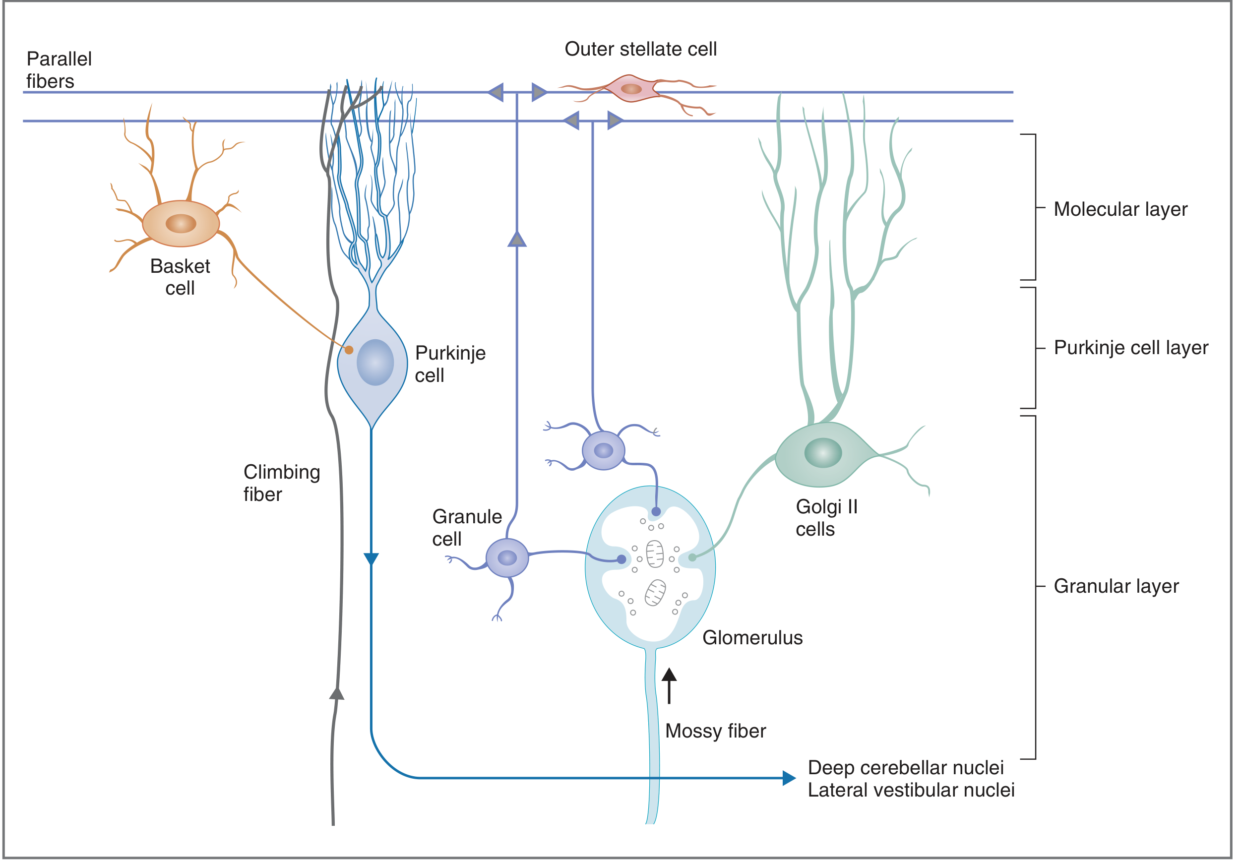
1. Granular Layer (innermost)
- Contains granule cells, Golgi II cells, and glomeruli
- Glomeruli = synaptic complexes where mossy fiber axons contact granule cell and Golgi II cell dendrites
- Granule cells are the only excitatory interneurons in the cerebellar cortex
2. Purkinje Cell Layer (middle)
- A single layer of large Purkinje cells - among the largest neurons in the CNS
- These cells have extensively branched, flat dendritic arbors in the molecular layer
- Their output is always inhibitory (GABA)
- The only output pathway from the cerebellar cortex
3. Molecular Layer (outermost)
- Contains basket cells, outer stellate cells, dendrites of Purkinje and Golgi II cells
- Contains parallel fibers (axons of granule cells that bifurcate into a "T" shape and run long distances)
- Parallel fibers run at right angles to the flat Purkinje dendritic trees - one Purkinje cell may receive input from up to 250,000 parallel fibers
Input to the Cerebellar Cortex
Two major afferent fiber systems:
1. Climbing Fibers
- Originate from the inferior olive of the medulla
- Project directly onto Purkinje cell dendrites
- Each Purkinje cell receives input from only one climbing fiber (but each fiber makes many synaptic contacts)
- Extremely powerful: one climbing fiber action potential elicits complex spikes (multiple excitatory bursts) in the Purkinje cell
- Role: "conditioning" Purkinje cells, modulating responses to mossy fiber input, and motor learning
2. Mossy Fibers
- The majority of cerebellar input
- Includes vestibulocerebellar, spinocerebellar, and pontocerebellar afferents
- Project onto granule cells in the glomeruli
- Granule cell axons → parallel fibers → synapse on Purkinje cell dendrites → produce simple spikes (single action potentials)
- Each mossy fiber creates a "beam" of excitation across many Purkinje cells along a row
Both fiber systems also send collateral branches directly to the deep cerebellar nuclei, bypassing the cortex.
Cerebellar Interneurons (Modulators of Purkinje Output)
All interneurons except granule cells are inhibitory:
| Cell | Effect | Target |
|---|---|---|
| Granule cells | Excitatory | Basket cells, stellate cells, Golgi II cells, Purkinje cells (via parallel fibers) |
| Basket cells | Inhibitory | Purkinje cells |
| Stellate cells | Inhibitory | Purkinje cells |
| Golgi II cells | Inhibitory | Granule cells (feedback inhibition, reduces excitation of Purkinje cells) |
Output of the Cerebellum
- The only output of the cerebellar cortex is via Purkinje cell axons (inhibitory, GABA)
- Purkinje cells project topographically to the deep cerebellar nuclei and lateral vestibular nuclei
- Deep cerebellar nuclei (especially dentate nucleus) are the main output relay - they receive tonic excitatory input from collaterals of mossy and climbing fibers, which is modulated by Purkinje cell inhibition
- Final output from deep nuclei travels via the superior cerebellar peduncle to:
- Ventrolateral thalamus → primary motor cortex (for voluntary movement coordination)
- Red nucleus → rubrospinal tract
- Brainstem reticular formation → reticulospinal tract
This output regulates the rate, range, force, and direction of movement (synergy).
Cerebellar Peduncle Summary
| Peduncle | Afferent Inputs | Efferent Outputs |
|---|---|---|
| Superior | Spinocerebellar fibers (anterior) | Deep cerebellar nuclei → red nucleus, thalamus |
| Middle | Pontocerebellar (corticopontocerebellar pathway) | None |
| Inferior | Spinocerebellar (posterior), vestibular, cuneocerebellar, olivocerebellar (climbing fibers) | Fastigial nucleus → vestibular nuclei |
Cerebellar Motor Learning
The climbing fiber system is thought to be critical for cerebellar motor learning (e.g., adaptation of reflexes and acquired motor skills). When a movement produces an error signal, climbing fibers signal the error to Purkinje cells and induce long-term depression (LTD) at parallel fiber-Purkinje cell synapses, modifying future movement output. This is the cellular basis of learned motor adaptation.
Disorders of the Cerebellum - Clinical Features
Cerebellar lesions produce ipsilateral signs (unlike upper motor neuron lesions) due to double crossing of pathways. The core clinical syndrome is cerebellar ataxia - impaired coordination due to errors in rate, range, force, and direction of movement.
| Sign | Description |
|---|---|
| Ataxia | Incoordinated, unsteady gait or limb movements |
| Dysmetria | Limb overshoots (past-pointing) or undershoots its target |
| Dysdiadochokinesia | Inability to perform rapid alternating movements (e.g., pronation/supination) |
| Intention tremor | Tremor that increases as the limb approaches the target (perpendicular to movement direction; absent at rest - differs from Parkinson's resting tremor) |
| Rebound phenomenon | Inability to stop a movement; if forearm flexion against resistance is suddenly released, the limb flies up (failure to activate antagonist muscles in time) |
| Nystagmus | Due to vestibulocerebellum involvement |
| Dysarthria | Scanning (staccato) speech from incoordination of speech muscles |
| Hypotonia | Decreased muscle tone (reduced activation of motor neurons) |
Sources:
- Costanzo Physiology, 7th Ed., pp. 113-116
- Ganong's Review of Medical Physiology, 26th Ed., pp. 253-256
- Neuroscience: Exploring the Brain, 5th Ed., Ch. 14
This is a shared conversation. Sign in to Orris to start your own chat.