
Explain me about FNAC technique in histo pathology
FNAC fine needle aspiration cytology technique
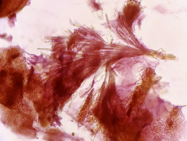
Fine-needle aspiration cytology (FNAC) of a soft tissue lesion around the ankle/toes was performed using a 23-gauge needle. The aspirate shows amorphous granular material with cohesive aggregates of needle-shaped crystals possessing pointed tips, consistent with monosodium urate crystals in a gouty tophus. The background contains inflammatory cells and multinucleated giant cells, reflecting a granulomatous reaction to crystal deposition. Stained cytology commonly employs Papanicolaou or Romanowsky stains to highlight needle crystals, which appear slender with sharp ends and may form rosettes or fragile bundles. These features distinguish tophi from other crystal deposits and inflammatory masses. Clinically, this cytologic profile supports a diagnosis of chronic tophaceous gout involving soft tissues around the ankle and digits, particularly in patients with hyperuricemia or prior gout attacks. Differential considerations include calcium pyrophosphate dihydrate (CPPD) crystals (rhomboid shapes), cholesterol crystals, or infectious/inflammatory nodules; however, the characteristic needle-shaped, negatively birefringent urate crystals are highly specific. Diagnostic significance lies in definitive cytologic demonstration of monosodium urate within inflammatory stroma, guiding urate-lowering therapy and anti-inflammatory management. Correlate with serum uric acid, renal function, and imaging studies for comprehensive disease assessment and therapeutic planning; case contributed by Dr. Dhiraj Nikumbh. This case highlights diagnostic utility for aspiration cytology and treatment.
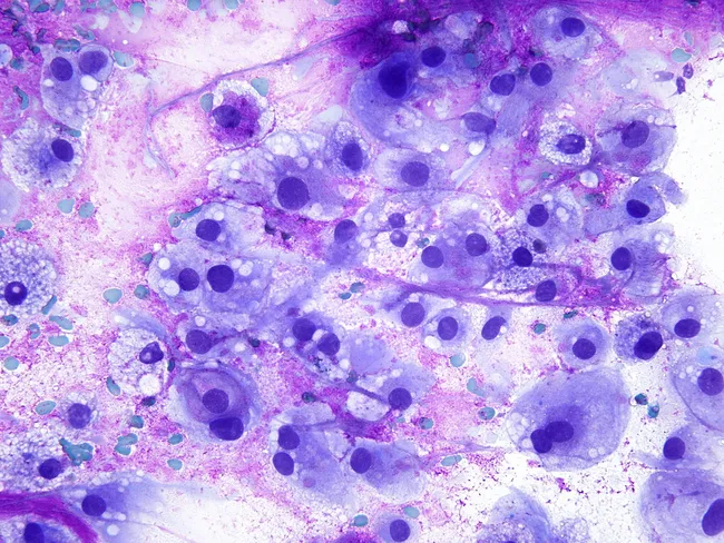
Fine-needle aspiration cytology (FNAC) of a paraspinal soft tissue mass demonstrates cells with voluminous, bubbly cytoplasm and large, bland hyperchromatic nuclei set in a myxoid, occasionally granular background. The cytologic smear is highly cellular, with scattered physaliferous cells featuring vacuolated, sometimes foamy cytoplasm and small-to-moderate nuclei. Many cells appear columnar-oval with a discohesive affinity, and occasional binucleation is seen. The distinctive bubbly cytoplasmic vacuoles and the presence of cartilaginous or chondroid–like matrix support a chordoma diagnosis, though differential includes clear cell renal cell carcinoma and other chondroid neoplasms. The staining used is Giemsa, which highlights the cytoplasmic vacuolization and the delicate, lace-like myxoid stroma around the tumor cells. Overall, the image demonstrates a neoplasm with notochordal differentiation, consistent with chordoma arising in the paraspinal region. Diagnostic significance lies in recognizing physaliferous cells and their abundant cytoplasm, which, in the appropriate clinical and radiologic context, directs confirmation by immunohistochemistry (brachyury positivity) and correlation with imaging to define extent. Differential diagnoses include metastatic RCC, chondrosarcoma, and chordoid variants; immunostaining and radiologic correlation are essential. This cytology image has educational value for surgical pathology, cytology practice, and differential diagnosis training in spine-associated tumors.
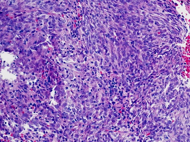
Fine-needle aspiration cytology (FNAC) of an abdominal mass was prepared as a conventional smear and stained with Hematoxylin and Eosin (H&E). The smear is moderately cellular, composed of interlacing sheets and fascicles of spindle-shaped and epithelioid cells with elongated to oval nuclei, finely textured chromatin, and occasional small nucleoli. The cytoplasm is variable, pale to eosinophilic, with occasional binucleation; cells are embedded in a prominent inflammatory background containing lymphocytes and plasma cells, with rare macrophages. Mitoses are infrequent; necrosis is not evident. The overall morphology shows a dendritic cell-like constellation with broad cytoplasmic processes and cohesive clusters that can mimic other spindle cell neoplasms. Immunohistochemistry (performed on ancillary material) reveals strong and diffuse CD21 positivity, supporting a follicular dendritic cell lineage. The cytologic features, together with CD21 expression, favor follicular dendritic cell sarcoma (FDC sarcoma) over other spindle cell tumors such as fibrosarcoma, inflammatory myofibroblastic tumor, sarcomatoid carcinoma, or melanoma variants. Clinically, this diagnosis is important because it informs surgical planning and adjuvant therapy considerations and requires correlative histopathology for definitive confirmation. The image documents cytomorphology with dendritic cell-rich background and immunophenotype concordance; additional markers (CD23, CD35) can increase diagnostic confidence in FDC sarcoma.
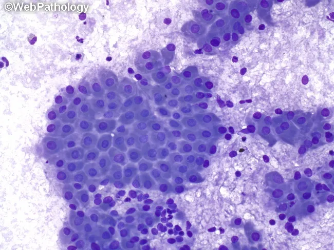
Fine-needle aspiration cytology (FNAC) of a parotid gland lesion demonstrates cohesive sheets and clusters of oncocytic epithelial cells embedded in a lymphoid-rich background. Diff-Quik stained smears reveal cells with abundant granular eosinophilic cytoplasm, round to oval nuclei with smooth nuclear chromatin, and scant cytoplasm at the periphery; occasional mitoses are absent or rare. The inflammatory background consists of mature lymphocytes, plasma cells, and occasional mucus-containing fluid, consistent with Warthin’s tumor (papillary cystadenoma lymphomatosum). The architectural pattern shows double-layered oncocytic epithelium with rosette-like clusters and microcystic spaces; however, cytology often lacks intact glandular ducts, making full histology difficult. Clinically, these findings strongly support a benign parotid tumor; the differential includes pleomorphic adenoma and mucoepidermoid carcinoma, particularly if mucous-producing cells or cytologic atypia are present. The diagnostic significance is high when cytology presents characteristic oncocytic cells and a lymphoid stroma; correlation with imaging and clinical features is essential. Management implications include surgical planning for superficial parotidectomy with facial nerve preservation. In cases with cystic change or hemorrhagic background, FNAC may yield background mucus or thick fluid; the presence of numerous oncocytic cells with dense lymphoid stroma is highly suggestive of Warthin’s tumor. Typical demographic associations include older male patients and parotid localization.
Fine Needle Aspiration Cytology (FNAC) - Complete Overview
What is FNAC?
Indications
| Site | Common Applications |
|---|---|
| Neck | Lymphadenopathy, thyroid nodules, salivary gland swellings |
| Breast | Palpable masses, cysts, axillary nodes |
| Thyroid | Nodules (solid vs. cystic, benign vs. malignant) |
| Lymph nodes | Distinguishing reactive hyperplasia, lymphoma, metastasis |
| Soft tissue | Lipomas, sarcomas, inflammatory swellings |
| Abdominal/pelvic masses | With CT/ultrasound guidance |
| Bone | With image guidance for deep lesions |
Equipment Required
- Needle: 22- or 23-gauge (fine) needle; for cystic lesions an 18-20 gauge may be used
- Syringe: 10 mL to 20 mL (sometimes up to 30 mL for cystic lesions), fitted tightly to the needle
- Glass slides: for smear preparation
- Fixative: absolute alcohol (for Papanicolaou stain) or air-dried (for Giemsa/Diff-Quik stain)
- Local anaesthetic (optional, 1-2 mL infiltrated subcutaneously)
- Sterile swabs, antiseptic solution
Step-by-Step Technique
1. Patient Preparation
- Informed consent obtained
- Patient positioned comfortably (recumbent or sitting depending on site)
- Skin over puncture site cleaned with antiseptic
- Local anaesthesia infiltrated into the skin (optional but useful for deeper or sensitive lesions)
2. Mass Stabilization
- The mass is stabilized between the thumb and index finger of the non-dominant hand (the "three-finger technique")
- This prevents the lesion from rolling away during needle insertion
3. Needle Insertion and Aspiration
- The needle (attached to the syringe) is advanced into the mass
- Full suction (maximum negative pressure) is applied by pulling back the syringe plunger
- While maintaining suction, the needle is passed through the mass in different planes - typically 3 to 5 passes in different directions to obtain an adequate and representative sample
- This fanning motion dislodges cells into the needle lumen
4. Releasing Suction
- Before withdrawing the needle, suction is released (plunger allowed to return to neutral position)
- This step is critical - failing to release suction before withdrawal causes the aspirate to be sucked into the syringe barrel and lost
- The needle is then withdrawn from the mass
5. Smear Preparation
- The cellular material (often in the needle hub alone) is expressed onto a glass slide
- A second slide is used to spread the material in a thin, even smear (similar to a blood film technique)
- Multiple smears are made from each pass
6. Fixation and Staining
| Method | Fixation | Stain Used | Best For |
|---|---|---|---|
| Wet-fixed smear | Immediately placed in absolute alcohol | Papanicolaou (PAP) stain | Nuclear detail, epithelial lesions |
| Air-dried smear | Allowed to air dry at room temperature | Giemsa / Diff-Quik (Romanowsky) | Lymphoid lesions, myxoid stroma, crystals |
Image-Guided FNAC
- Deep-seated (e.g., retroperitoneal, mediastinal)
- Small or non-palpable
- Adjacent to vital structures
Cytological Interpretation: What is Seen
- Nuclear features: size, shape, chromatin pattern, nucleoli (key for malignancy assessment)
- Cytoplasmic features: amount, vacuolation, granularity
- Cell arrangements: cohesive sheets vs. discohesive single cells
- Background: necrosis, inflammatory cells, mucus, colloid, crystals
- Special findings: e.g., colloid in thyroid; lymphocytes in lymphoma; physaliferous cells in chordoma; urate crystals in gout
FNAC vs. Core Needle Biopsy
| Feature | FNAC | Core Needle Biopsy |
|---|---|---|
| Needle size | Fine (22-25 G) | Larger (14-18 G) |
| Sample | Cells only (cytology) | Tissue core (histology) |
| Architecture preserved? | No | Yes |
| Immunohistochemistry? | Limited | Full IHC panel possible |
| Pain/risk | Minimal | Slightly more |
| Best for | Cysts, lymph nodes, thyroid | Lymphoma subtyping, soft tissue, HPV/EBV testing |
Advantages of FNAC
- Minimally invasive - no incision required
- Performed as an outpatient procedure
- Rapid results (same day in rapid on-site evaluation - ROSE)
- Inexpensive
- Can be repeated if non-diagnostic
- Guides further management before surgery
- The "triple test" (clinical exam + imaging + FNAC) has a false-negative rate approaching that of surgical biopsy for breast masses - Textbook of Family Medicine, 9th Edition
Limitations of FNAC
- Provides cytology only - cannot assess tissue architecture (e.g., cannot distinguish ductal carcinoma in situ from invasive breast carcinoma)
- Non-diagnostic rate of 3-10% even in experienced hands
- Cannot reliably sub-classify lymphoma (core biopsy preferred)
- Operator-dependent
- Cannot evaluate microcalcifications (core biopsy needed)
- Risk of sampling error in heterogeneous tumours
Contraindications
- Overlying skin infection at puncture site
- Underlying pulsatile vascular mass (risk of haemorrhage)
- Bleeding disorders / anticoagulation
- Uncooperative patient
Clinical Examples
FNAC Cytology Images
Summary
- S Das: A Manual on Clinical Surgery, 13th Edition
- Bailey and Love's Short Practice of Surgery, 28th Edition
- Textbook of Family Medicine, 9th Edition (Pfenninger/Fowler)
- Scott-Brown's Otorhinolaryngology Head & Neck Surgery