

What s this
What the diagram depicts
The two real procedures being parodied
-
Cystoscopy - a real procedure where a thin camera scope is passed through the urethra to examine the bladder and urethra (used to diagnose bladder cancer, stones, strictures, etc.)
-
Digital Rectal Exam (DRE) - a real clinical exam where a physician inserts a gloved finger into the rectum to feel the prostate for enlargement, nodules, or abnormalities (used to screen for prostate cancer or BPH)
Why it's funny
Hyperandronism in female
hyperandrogenism female hirsutism virilization causes
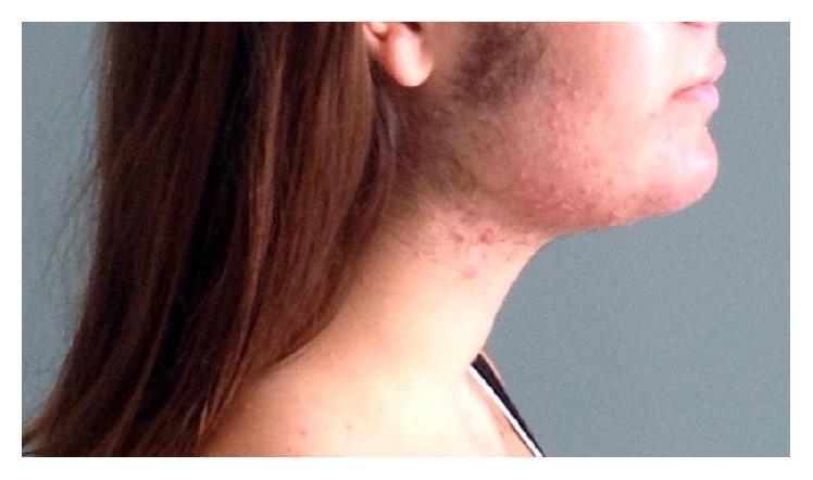
A clinical photograph of a female patient's face and neck in a lateral profile view, demonstrating clinical signs of hyperandrogenism and virilization. The image reveals prominent hirsutism, characterized by coarse, dark hair growth in an androgen-dependent distribution, specifically involving the preauricular (sideburn) area, cheeks, and along the mandibular jawline. Accompanying these findings is an inflammatory acneiform eruption, presenting as erythematous papules and pustules clustered around the jaw and upper neck. This combination of acne and hirsutism is highly suggestive of an underlying hyperandrogenic state. The educational focus of this image is the recognition of physical manifestations of excessive androgen levels in female patients, which may be associated with conditions such as polycystic ovary syndrome (PCOS) or, in this clinical context, pregnancy-related luteoma.
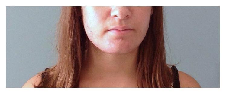
This clinical photograph displays the lower face and neck of a female patient six months postpartum, illustrating symptoms of virilization. The primary visual finding is hirsutism, characterized by prominent, dark terminal hair growth along the cheeks, jawline, and chin. The skin in the perioral and mandibular regions shows subtle textural irregularities and evidence of acneic lesions, which are common dermatological manifestations of hyperandrogenism. This image serves as an educational example of the clinical presentation of androgen-secreting conditions during or after pregnancy, such as a pregnancy luteoma. The photograph documents the residual physical effects of severe hyperandrogenemia (elevated testosterone and androstenedione) as it gradually resolves following medical or surgical intervention.
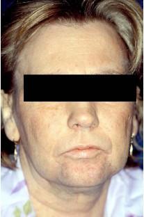
This clinical photograph displays androgenized facial features in a 41-year-old female patient, illustrating the physical manifestations of hyperandrogenism and virilization. Key visible findings include hirsutism, characterized by coarse, dark terminal hair growth on the upper lip, chin, and jawline in a male-pattern distribution. The facial skin exhibits signs of thickening and a roughened texture. The jawline appears relatively prominent and squared, contributing to the overall masculinization of the facial phenotype. Such features in a female clinical context are often associated with systemic endocrine dysfunction, such as paraneoplastic syndromes secondary to hormone-secreting tumors (e.g., Krukenberg tumors or ovarian/adrenal malignancies). This image serves as an educational example of virilization symptoms used in the differential diagnosis of androgen-secreting pathologies in endocrinology and oncology.
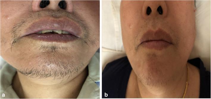
This clinical comparison photograph illustrates the resolution of hirsutism in a 69-year-old female patient following the surgical removal of a Leydig Cell Tumor (LCT). Image (a) represents the pre-operative state, showing prominent male-pattern terminal hair growth on the upper lip (mustache area) and chin. The hair is dark, coarse, and relatively dense, characteristic of hyperandrogenism. Image (b) shows the post-operative result, demonstrating a significant reduction in terminal hair density and thickness in the same facial regions. The remaining hair is sparse and fine, indicating clinical improvement of virilization symptoms. The images serve as an educational example of the cutaneous manifestations of androgen-secreting ovarian tumors and the subsequent reversal of secondary sexual characteristics after hormonal normalization. Key educational concepts include the clinical signs of hyperandrogenemia in postmenopausal women and the diagnostic importance of recognizing sudden-onset hirsutism as a marker for underlying endocrine-active neoplasms like sex cord-stromal tumors.
hyperandrogenism women diagnosis management
Hyperandrogenism in Females
Androgens in Women - Normal Physiology
- Testosterone (principal circulating androgen)
- Androstenedione
- DHEA and DHEAS (adrenal markers)
Clinical Manifestations
1. Hirsutism
2. Acne
- Jawline/lower face distribution
- Severe or sudden onset acne in women should raise suspicion for HA
3. Androgenic Alopecia
- Scalp hair thinning or male-pattern baldness (frontal-parietal)
4. Virilization (severe HA)
- Deepening of voice
- Breast atrophy
- Increased muscle bulk
- Clitoromegaly
- Increased libido
- Acanthosis nigricans (sign of insulin resistance)
Clinical Photos of Hyperandrogenism
Causes (Etiology)
OVARIAN causes (most common)
| Cause | Notes |
|---|---|
| PCOS (most common overall) | Accounts for ~70-80% of all HA cases |
| Functional ovarian hyperandrogenism | Exaggerated ovarian androgen response to LH |
| Hyperthecosis / Stromal hyperplasia | Benign theca/stroma proliferation; can cause virilization |
| Androgen-secreting ovarian neoplasms | Sertoli-Leydig tumors, granulosa cell tumors, lipid cell tumors |
| Syndromes of extreme insulin resistance | Insulin stimulates ovarian androgen production |
ADRENAL causes
| Cause | Notes |
|---|---|
| Congenital Adrenal Hyperplasia (CAH) | 21-hydroxylase deficiency in >90%; classic and non-classic forms |
| Cushing syndrome | Glucocorticoid excess + adrenal androgen excess |
| Functional adrenal hyperandrogenism | Excess adrenal androgen without clear enzyme defect |
| Adrenal neoplasms (adenoma/carcinoma) | DHEAS >18.5 μmol/L (>7000 μg/L) suggests this |
| Premature adrenarche | Early adrenal activation in childhood |
OTHER causes
| Cause | Notes |
|---|---|
| Hyperprolactinemia | Prolactin can stimulate adrenal androgens |
| Acromegaly | GH excess |
| Obesity | Increased peripheral conversion of androgens; reduces SHBG |
| Drugs | Androgens, anabolic steroids, valproic acid, cyclosporine, phenytoin, minoxidil, danazol |
| Idiopathic hirsutism | Normal androgens, increased 5α-reductase activity |
| Pregnancy-related | Luteoma, hyperreactio luteinalis |
- Harrison's Internal Medicine 22E, p. 946-980; Berek & Novak's Gynecology, p. 1909
Diagnostic Evaluation
- Rapid onset and rapid progression of hirsutism
- Signs of virilization (clitoromegaly, voice deepening)
- Total testosterone >12 nmol/L (>3.5 ng/mL) = likely androgen-secreting tumor
- Total testosterone >7 nmol/L (>2 ng/mL) = suggestive of tumor (also seen in hyperthecosis)
- DHEAS >18.5 μmol/L (>7000 μg/L) = suggests adrenal tumor
- Total testosterone (by LC/MS assay)
- Free testosterone (calculated from total T + SHBG, or by LC/TMS)
- DHEAS (adrenal androgen marker)
- SHBG
- FSH/LH (LH:FSH ratio >2 in PCOS)
- Prolactin, TSH
- 17-OHP (for CAH - elevated in 21-hydroxylase deficiency)
- 24-hour urine cortisol or dexamethasone suppression test (if Cushing suspected)
- Pelvic ultrasound (ovarian morphology, PCO appearance)
- CT/MRI abdomen-pelvis (if adrenal or ovarian tumor suspected)
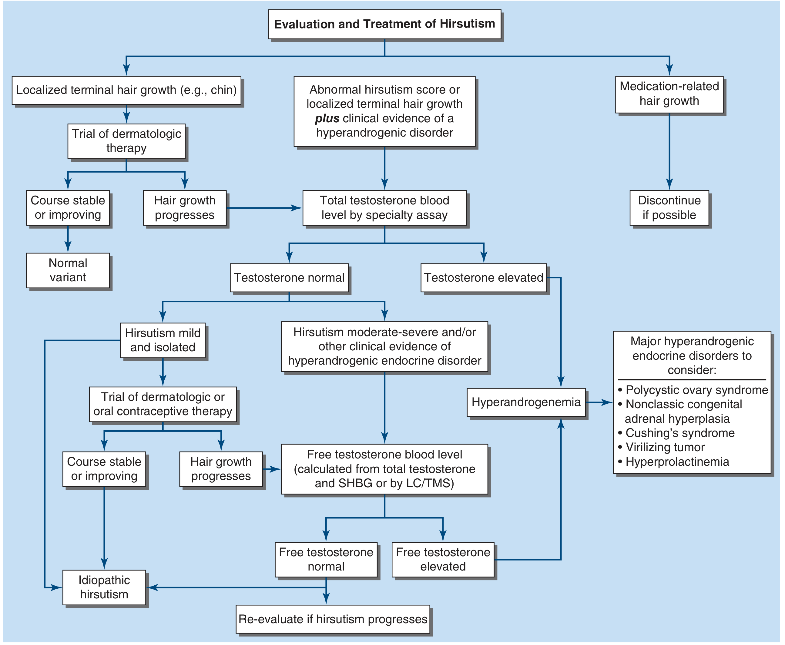
Diagnostic Algorithm
PCOS - The Most Common Cause
- Clinical or biochemical hyperandrogenism
- Oligo/anovulation (menstrual irregularity, <8 cycles/year)
- Polycystic ovarian morphology on ultrasound (>12 follicles per ovary OR ovarian volume >10 mL)
Treatment
Non-pharmacological
- Weight loss (5-7% body weight over 6 months) reduces free testosterone and can restore ovulation in >75% of women with PCOS; reduces insulin resistance. - Berek & Novak's Gynecology, p. 1935
Pharmacological
| Category | Agent | Mechanism |
|---|---|---|
| Hormonal suppression | Combined oral contraceptives (COCs) | Suppress LH → reduce ovarian androgens; increase SHBG; inhibit 5α-reductase in skin |
| Medroxyprogesterone | Progestational suppression | |
| GnRH analogues | Suppress LH/FSH → reduce ovarian androgens | |
| Glucocorticoids | Suppress ACTH → reduce adrenal androgens (in CAH) | |
| Antiandrogens | Spironolactone | Androgen receptor blocker + weak inhibitor of steroidogenesis (most commonly used) |
| Cyproterone acetate | Androgen receptor blocker (not available in USA) | |
| Flutamide | Androgen receptor blocker | |
| 5α-Reductase inhibitors | Finasteride | Blocks testosterone → DHT conversion at PSU |
| Insulin sensitizers | Metformin | Reduces insulin → reduces ovarian androgen stimulation; may restore ovulation |
| GLP-1 agonists | Semaglutide, liraglutide | Recent evidence supports weight loss + hormonal improvement in PCOS with obesity (PMID 39178623) |
| Steroidogenic enzyme inhibitors | Ketoconazole | Inhibits androgen synthesis (rarely used due to hepatotoxicity) |
Mechanical hair removal
- Temporary: Shaving, waxing, threading, depilatory creams
- Permanent: Laser hair removal, electrolysis
Note for patients desiring pregnancy: Antiandrogens are teratogenic (contraindicated); focus shifts to ovulation induction (clomiphene, letrozole, gonadotropins). COCs cannot be used in this group.
Summary Table
| Feature | Hirsutism only | + Oligomenorrhea | + Virilization | Rapid onset |
|---|---|---|---|---|
| Most likely | Idiopathic / PCOS | PCOS | Tumor / Hyperthecosis | Androgen-secreting tumor |
| Key test | Free testosterone | LH:FSH, pelvic USS | Total T, CT/MRI | Total T >12 nmol/L → imaging |
Recent evidence note: A 2025 meta-analysis (PMID 40806019) found intermittent fasting significantly improves anthropometric measures and hormonal profiles in women with PCOS. GLP-1 receptor agonists also show promising results for weight and androgen reduction in obese PCOS patients (PMID 39178623).