
Animia kya hai kasa hoti hai hindi lacture
anemia types causes red blood cell diagram
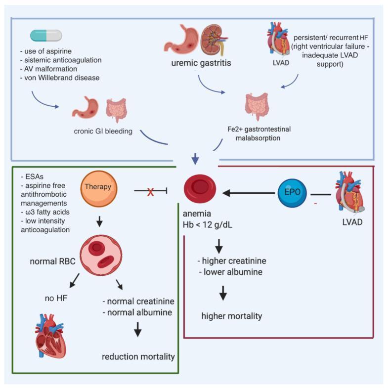
This pathophysiology diagram illustrates the multifactorial causes and consequences of anemia in patients with heart failure (HF) and a left ventricular assist device (LVAD). The top section identifies contributors to anemia, including chronic gastrointestinal (GI) bleeding secondary to aspirin use, systemic anticoagulation, arteriovenous (AV) malformations, and von Willebrand disease. Other contributing factors shown are uremic gastritis, iron (Fe2+) malabsorption, and persistent right ventricular failure. Central to the diagram is the manifestation of anemia (defined as Hemoglobin < 12 g/dL), which correlates with elevated creatinine, decreased albumin, and increased mortality. The diagram also highlights therapeutic interventions such as erythropoiesis-stimulating agents (ESAs), aspirin-free antithrombotic management, omega-3 fatty acids, and low-intensity anticoagulation. Successful therapy is depicted as leading to normal red blood cell (RBC) counts, resolution of heart failure symptoms, and reduced mortality. The role of erythropoietin (EPO) is also noted in the context of LVAD-related anemia management.
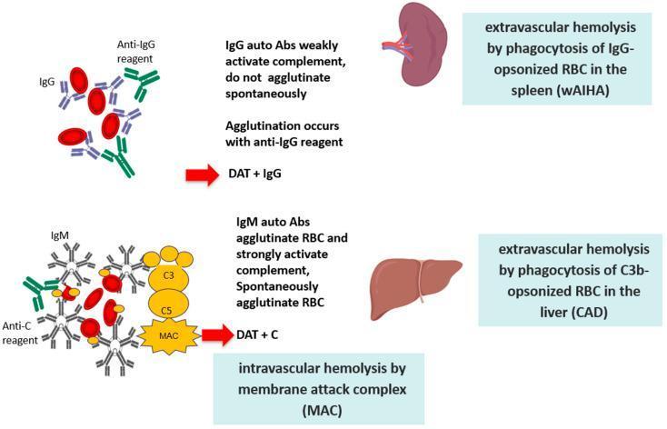
This pathophysiology diagram illustrates the two primary mechanisms of red blood cell (RBC) destruction in Autoimmune Hemolytic Anemia (AIHA). The top section depicts Warm AIHA (wAIHA), characterized by IgG autoantibodies. These monomeric antibodies weakly activate the complement system and do not cause spontaneous RBC agglutination; instead, agglutination occurs only with an anti-IgG reagent, resulting in a positive Direct Antiglobulin Test (DAT + IgG). This pathway leads to extravascular hemolysis via phagocytosis of IgG-opsonized RBCs primarily in the spleen. The bottom section depicts Cold Agglutinin Disease (CAD), mediated by pentameric IgM autoantibodies. These antibodies cause spontaneous RBC agglutination and strongly activate the complement cascade. The diagram shows the binding of C3, C5, and the formation of the Membrane Attack Complex (MAC), leading to intravascular hemolysis. Additionally, it highlights extravascular hemolysis occurring via phagocytosis of C3b-opsonized RBCs in the liver, with a positive Direct Antiglobulin Test for complement (DAT + C). Key anatomical landmarks include the spleen for IgG-mediated clearance and the liver for C3b-mediated clearance.
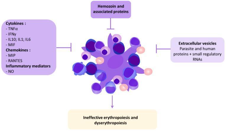
A pathophysiology diagram illustrating the human and parasite factors that contribute to malarial anemia. At the center is a complex cellular cluster, depicting a bone marrow erythroblastic island consisting of a central macrophage (dark purple, star-burst shape) surrounded by developing erythroblasts and red blood cells in various shades of purple and pink. Four key external factors are shown impacting this central unit via inhibitory or causal arrows: 1) Pro-inflammatory mediators (left) including cytokines (TNFα, IFNγ, IL1, IL6, IL10, MIF), chemokines (MIP, RANTES), and nitric oxide (NO); 2) Hemozoin and associated parasite proteins (top); 3) Extracellular vesicles containing parasite/human proteins and regulatory RNAs/miRNAs (right); and 4) The resulting clinical outcome (bottom) categorized as ineffective erythropoiesis and dyserythropoiesis. The diagram uses a logical flow to explain how Plasmodium-induced systemic inflammation and parasite-derived products disrupt normal red blood cell production within the hematological system.
iron deficiency anemia blood smear microcytic hypochromic
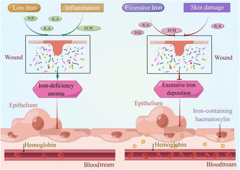
A comparative medical illustration detailing the effects of iron dysregulation on wound healing and systemic states. The left panel depicts a 'Low Iron' and 'Inflammation' scenario where a cutaneous wound is associated with 'Iron-deficiency anemia'. Key signaling molecules shown include TGF, IL-6, IL-8, and ECM components within a wound bed containing fibroblasts and matrix elements. An arrow indicates the downstream impact on the underlying epithelium and hemoglobin within the bloodstream. The right panel depicts an 'Excessive Iron' and 'Skin damage' scenario characterized by 'Excessive iron deposition'. This state is visually distinguished by an inhibitory symbol (red T-bar) directed at the epithelium, suggesting impaired regenerative capacity. Notably, the epithelium in the high-iron state contains yellow granules labeled as 'Iron-containing haematoxylin' (hemosiderin), which serve as a histological marker of iron overload. Both panels show the anatomical relationship between the cutaneous wound, underlying epithelial cells, and the intravascular space (bloodstream) containing red blood cells and hemoglobin, illustrating how iron homeostasis is critical for normal dermatological repair and systemic oxygen transport.
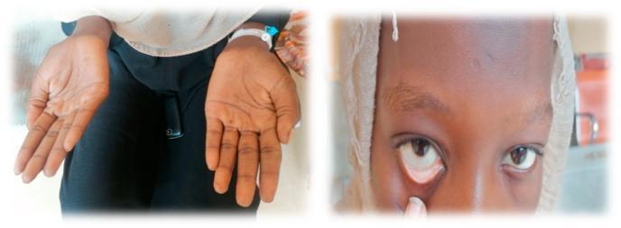
This composite clinical photograph illustrates common physical examination findings associated with severe anemia. The left panel shows the bilateral palms of a patient, demonstrating marked palmar pallor. The skin on the palms and creases appears significantly lighter and lacks the typical pinkish hue of healthy vascularized tissue, suggesting reduced hemoglobin concentration. The right panel is a close-up of the patient's face with the lower eyelid retracted to reveal the palpebral conjunctiva. Instead of the normal vibrant red or pink color, the conjunctiva exhibits conjunctival pallor, appearing whitish or very pale pink. These visual signs are critical diagnostic markers in hematology and general medicine for identifying systemic conditions such as iron-deficiency anemia or other forms of chronic blood loss. The image serves as a teaching tool for medical students and clinicians to recognize mucosal and cutaneous indicators of decreased red blood cell mass or oxygen-carrying capacity.
🩸 Anemia (Khoon ki Kami) - Hindi Lecture
📌 Anemia Kya Hai?
Normal Haemoglobin (Hb) Values:
| Vyakti | Normal Hb |
|---|---|
| Purush (Adult Men) | 13.5 - 17.5 g/dL |
| Mahila (Adult Women) | 12.0 - 15.5 g/dL |
| Garbhawati (Pregnant Women) | 11.0 g/dL se upar |
| Bachche (Children) | Umar ke hisaab se alag |
🔬 Anemia Kaise Hoti Hai? (Pathophysiology)
1. Khoon Banna Kam Ho Jaana (Decreased RBC Production)
- Iron, Vitamin B12, ya Folic Acid ki kami
- Bone marrow (haddi ka marrow) ki bimari
- Chronic bimariyan (kidney failure, cancer)
2. Khoon Ki Zyada Haraat (Increased Blood Loss)
- Andar ya bahar se khoon bahna (ulcer, periods mein zyada khoon, accident)
- Piles (bawaseer) se khoon jana
3. Laal Khoon Ke Kankad Jaldi Toot Jaana (Increased RBC Destruction - Hemolysis)
- Sickle Cell Disease
- Thalassemia
- Autoimmune causes (body apne khoon ke kankad ko khud tod deti hai)
🏷️ Anemia Ke Prakar (Types of Anemia)
A. Iron Deficiency Anemia (Lohe ki Kami)
- Sabse zyada common type
- India mein bahut prevalent - especially mahilaon aur bachon mein
- RBCs chhote aur pale (microcytic, hypochromic) ho jaate hain
- Karan: kam iron wala khana, periods mein zyada khoon, pregnancy
B. Megaloblastic Anemia (Vitamin B12 / Folic Acid ki Kami)
- RBCs bahut bade ho jaate hain lekin kaam nahi kar paate
- Vegetarians mein B12 deficiency zyada hoti hai
- Karan: kam meat/dairy khana, pernicious anemia
C. Anemia of Chronic Disease
- Long-term bimarion mein hoti hai - kidney failure, TB, rheumatoid arthritis
- Kidney ki bimari mein EPO (Erythropoietin) kam banta hai
D. Hemolytic Anemia
- RBCs jaldi toot jaate hain
- Malaria is ka ek aham karan hai (India mein)
- Autoimmune, genetic ya infection se ho sakta hai
E. Aplastic Anemia
- Bone marrow kaam karna band kar deti hai
- Serious condition hai
F. Sickle Cell Anemia / Thalassemia
- Genetic (vanshanugatik) bimariyan hain
- India ke kuch ilaqon mein zyada milti hain
🤒 Anemia Ke Lakshan (Symptoms)
- Kamzori aur thakaan (weakness, fatigue)
- Chehre, haath, aur aankhon ka pila padna (pallor)
- Sans lene mein takleef (breathlessness)
- Dil ki dhadkan tez hona (palpitations)
- Sar dard (headache)
- Chakkar aana (dizziness)
- Thande haath-paon
- Seene mein dard
- Behoshi aa jaana
- Dil pe zyada load padna
🧪 Anemia Ki Jaanch (Diagnosis)
- CBC (Complete Blood Count) - Hb, RBC count, MCV dekha jaata hai
- Peripheral Blood Smear - RBC ka size aur shape
- Serum Iron + Ferritin + TIBC - Iron deficiency ke liye
- Vitamin B12 and Folate levels
- Reticulocyte Count - Kitna naya khoon ban raha hai
- Bone Marrow Biopsy - Gaambhir cases mein
💊 Anemia Ka Ilaaj (Treatment)
| Prakar | Ilaaj |
|---|---|
| Iron Deficiency | Ferrous Sulphate (60-120 mg elemental iron daily), iron-rich foods |
| B12 Deficiency | Vitamin B12 injections ya oral tablets |
| Folic Acid Deficiency | Folic Acid tablets (400-1000 mcg daily) |
| Anemia of CKD | Erythropoietin (EPO) injections + IV iron |
| Severe/Aplastic | Blood transfusion, Bone Marrow Transplant |
| Hemolytic | Steroids, splenectomy, underlying cause ka ilaaj |
🥗 Anemia Mein Kya Khaana Chahiye? (Diet Tips)
- Paalak, methi, sarson
- Rajma, chana, masoor dal
- Jaggery (gud)
- Pomegranate (anaar)
- Non-veg: Liver, egg, red meat
- Nimbu paani, amla, orange saath khaayen
- Chai, coffee (tannins iron ko absorb hone se rokta hai)
📊 Anemia - Quick Summary
Khoon mein Hb kam → Oxygen kam milti hai → Kamzori, pallor, thakaan

Causes:
├── Kam banna (Iron, B12, Folate ki kami)
├── Zyada khoon bahna (periods, ulcer)
└── Zyada tootna (hemolysis, sickle cell)

Diagnosis: CBC + Blood Smear + Iron studies
Treatment: Cause ke hisaab se