
Abnormal uterine bleeding
"abnormal uterine bleeding" AND management
abnormal uterine bleeding PALM-COEIN classification diagram
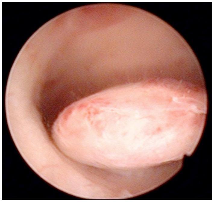
This diagnostic image is an intraoperative hysteroscopic photograph of the uterine cavity. The visual focus is a well-defined, oval-shaped pedunculated endometrial polyp protruding from the posterior uterine wall. The polyp demonstrates a smooth, glistening surface with a pale pink to white color, interspersed with focal areas of erythema and fine vertical striations across its body. Its morphology is slightly lobulated, and it is attached to the uterine wall by a distinct stalk. The surrounding endometrial tissue appears healthy, smooth, and pink, although there is a slight darkening near the base of the pedicle, likely representing minor blood accumulation. This clinical photograph illustrates a common structural cause of abnormal uterine bleeding (AUB), frequently categorized under the 'Polyp' component of the FIGO PALM-COEIN classification system. Such findings are critical in the diagnostic workup of perimenopausal and postmenopausal women to differentiate benign focal abnormalities from endometrial hyperplasia or malignancy.
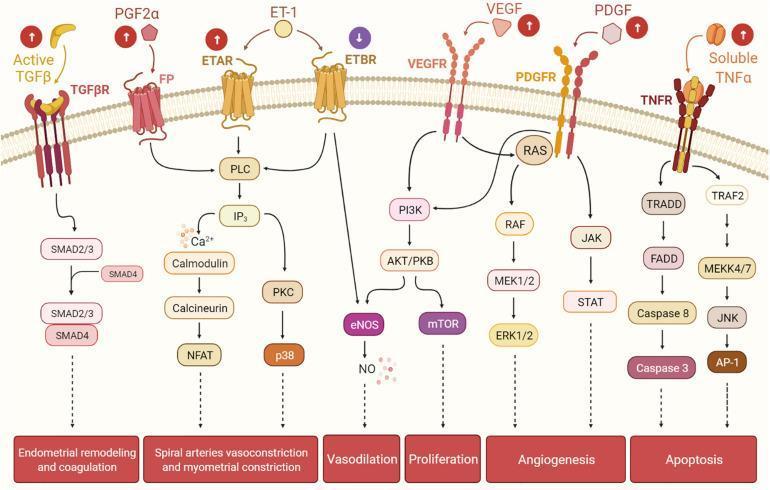
A complex pathophysiology diagram illustrating various cellular signaling pathways in the human endometrium, specifically highlighting mechanisms dysregulated in uterine fibroids. The flow begins with extracellular ligands binding to transmembrane receptors and concludes with functional physiological outcomes. Key pathways include: (1) TGF-β/SMAD signaling leading to endometrial remodeling and coagulation; (2) PGF2α/FP and ET-1/ETAR/ETBR signaling involving PLC, IP3, Calmodulin, and PKC cascades, which result in spiral artery vasoconstriction and myometrial constriction; (3) ETBR/VEGFR/PDGFR cross-talk through PI3K/AKT/mTOR and RAS/RAF/MEK/ERK pathways, driving vasodilation (via NO), proliferation, and angiogenesis; and (4) TNFα/TNFR signaling through Caspase cascades (apoptosis) and JNK/AP-1 pathways. Icons with upward arrows next to ligands like TGF-β, PGF2α, and VEGF indicate molecular upregulation typically seen in the presence of uterine fibroids. This educational diagram is targeted at an advanced level, illustrating how ligand-receptor interactions contribute to heavy menstrual bleeding and abnormal uterine vascular architecture.
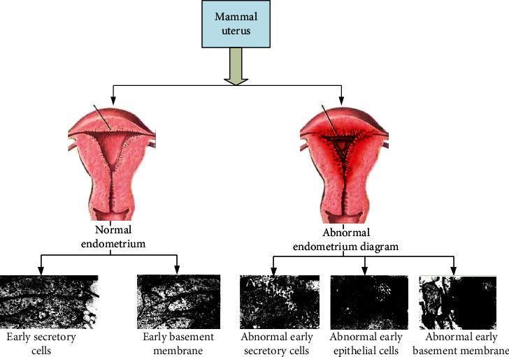
An educational comparison diagram illustrating the differences between normal and abnormal endometrium (specifically endometriosis). The top section displays two anatomical illustrations of a mammal uterus. The 'Normal endometrium' shows a smooth, uniform uterine cavity with an open, Y-shaped internal structure. The 'Abnormal endometrium diagram' depicts a constricted, triangular uterine cavity with thickened, irregular linings and growths, suggestive of ectopic tissue implantation. The bottom section presents five high-magnification grayscale diagnostic images representing cellular pathology. The normal side shows 'Early secretory cells' and 'Early basement membrane' with visible, organized textures and clear cellular arrangements. In contrast, the abnormal side shows 'Abnormal early secretory cells,' 'Abnormal early epithelial cells,' and 'Abnormal early basement membrane,' which exhibit significantly darker, less defined clusters and disrupted structural integrity. This diagram serves as a visual guide to the gross anatomical and microscopic cellular changes associated with endometrial pathology.
Abnormal Uterine Bleeding (AUB)
Definition
- Heavy menstrual bleeding (HMB): excessive flow during regular cycles
- Intermenstrual bleeding (IMB): bleeding between cycles
- Postcoital bleeding (PCB): bleeding after intercourse
- Postmenopausal bleeding (PMB): any bleeding >12 months after last menstrual period
PALM-COEIN Classification (FIGO 2011)
PALM - Structural Causes
| Cause | Notes |
|---|---|
| P - Polyp (AUB-P) | Endometrial/endocervical polyps; may be sessile or pedunculated; up to 5% malignant transformation risk |
| A - Adenomyosis (AUB-A) | Endometrial glands/stroma within the myometrium; diagnosed on ultrasound or MRI |
| L - Leiomyoma (AUB-L) | Most common benign gynecologic tumor; affects up to 70% of women by age 50; Black patients disproportionately affected; classified by FIGO as types 0-8 based on location |
| M - Malignancy/Hyperplasia (AUB-M) | Endometrial carcinoma and hyperplasia; must be excluded, especially in postmenopausal women |
COEIN - Non-Structural Causes
| Cause | Notes |
|---|---|
| C - Coagulopathy (AUB-C) | Up to 20% of women with HMB have an underlying coagulopathy; von Willebrand disease is most common (up to 13% of AUB cases), often first presenting at menarche |
| O - Ovulatory dysfunction (AUB-O) | Most common non-structural cause; due to PCOS, hyperprolactinemia, thyroid disease, hypothalamic dysfunction (anorexia nervosa), perimenopause, or physiologic immaturity in adolescents |
| E - Endometrial (AUB-E) | Primary endometrial disorders - endometritis, impaired local hemostatic mechanisms |
| I - Iatrogenic (AUB-I) | Exogenous hormones, IUCDs, anticoagulants, tamoxifen (induces uterine abnormalities in 10-40% of users), gonadal steroids |
| N - Not yet classified (AUB-N) | No identifiable cause found |
Age-Based Differential Diagnosis
| Age Group | Likely Causes |
|---|---|
| Prepubertal | Infection, trauma/sexual abuse, vaginal foreign body, structural lesion |
| Adolescents | Coagulopathy (von Willebrand disease presenting at menarche), HPO axis immaturity, PCOS |
| Reproductive age | Structural lesions (polyps, leiomyomas), anovulation (PCOS), endometrial hyperplasia |
| Perimenopause (>40 years) | Anovulatory bleeding, leiomyoma, endometrial hyperplasia/carcinoma |
| Postmenopausal | Atrophy (most common), endometrial carcinoma (must be excluded), polyps |
Workup and Investigations
Essential Initial Workup
- Pregnancy test - mandatory in all reproductive-age women
- Full blood count - assess for anemia; hemoglobin/hematocrit if hemodynamically unstable
- TSH - thyroid dysfunction (especially hypothyroidism) associated with AUB
- Prolactin - if anovulatory dysfunction suspected
- Coagulation screen (PT, aPTT, platelet count) - if bleeding since menarche, family history of coagulopathy, or signs of systemic bleeding
- STI screening - Chlamydia linked to AUB via endometrial inflammation
- Cervical cancer screening - if not up to date
Imaging
- Transvaginal ultrasound (TVUS): first-line imaging; identifies fibroids, endometrial thickening, focal masses, adenomyosis features
- In postmenopausal women, endometrial thickness < 4-5 mm reliably excludes endometrial cancer
- Saline infusion sonography / 3D ultrasound: improves detection of intracavitary lesions
- MRI: superior for fibroids (FIGO typing) and adenomyosis
Endometrial Biopsy - Indications
- Age ≥45 years with any AUB, including IMB
- Age < 45 with: obesity/unopposed estrogen exposure, PCOS, persistent AUB refractory to medical treatment, elevated familial cancer risk
- Suspected endometrial pathology at any age
- Hysteroscopy + biopsy has superior sensitivity and specificity vs. blind biopsy alone
Management
Acute/Emergency Management (heavy/unstable bleeding)
- Resuscitation: IV fluids, blood products for hemodynamic instability
- IV conjugated estrogen: 25 mg IV every 4-6 hours for up to 24 hours
- Intrauterine tamponade: 26Fr Foley catheter with 30 mL saline
- Surgical options: urgent dilation and curettage (D&C), uterine artery embolization (UAE), endometrial ablation, or hysterectomy
Medical Management (non-emergent)
| Agent | Indication |
|---|---|
| Combined oral contraceptive pills | Ovulatory dysfunction, cycle regulation, HMB reduction |
| Levonorgestrel-releasing IUD (LNG-IUD) | HMB, endometrial protection; first-line medical option |
| Progestogens | Anovulatory AUB; cyclic or continuous regimens |
| Tranexamic acid | HMB; antifibrinolytic, used during menses only |
| NSAIDs | HMB; reduce prostaglandin-mediated bleeding |
| GnRH agonists | Leiomyoma; induces amenorrhea; short-term use (bone loss) |
Surgical/Procedural Management
| Procedure | Indication |
|---|---|
| Hysteroscopic polypectomy | Uterine/endocervical polyps |
| Myomectomy | Leiomyoma (fertility-sparing) |
| UAE | Leiomyoma, significant bleeding |
| Endometrial ablation | HMB with completed childbearing; contraindicated if future fertility desired |
| Hysterectomy | Definitive treatment for adenomyosis, large fibroids, refractory AUB, endometrial carcinoma |