

Like this generate an image depicting level of lymph nodes of neck, triangle’s of neck , different surgery for neck dissection , different special component in relation to clinical aspect of neck i/v/o ent
A comprehensive, detailed medical education poster titled "CLINICAL ANATOMY OF THE NECK - ENT OVERVIEW" with a clean white background, organized into 4 clearly labeled sections: SECTION 1 - TOP LEFT: "CERVICAL LYMPH NODE LEVELS (AAO-HNS)" - A detailed lateral view anatomical illustration of the neck showing color-coded lymph node levels: Level I (yellow) = Submental & Submandibular, Level II (green) = Upper Jugular (IIA and IIB subdivisions), Level III (blue) = Middle Jugular, Level IV (purple) = Lower Jugular, Level V (orange) = Posterior Triangle (VA and VB), Level VI (red) = Central/Anterior Compartment, Level VII (brown) = Superior Mediastinal (below sternal notch). Each level clearly labeled with Roman numerals on the diagram. SECTION 2 - TOP RIGHT: "TRIANGLES OF THE NECK" - Anatomical anterior and lateral view of the neck showing triangles: Anterior Triangle subdivided into Submental triangle, Digastric/Submandibular triangle, Carotid triangle, Muscular triangle. Posterior Triangle subdivided into Occipital triangle and Subclavian/Supraclavicular triangle. Boundaries labeled: Anterior triangle bounded by midline, mandible, SCM. Posterior triangle bounded by SCM, trapezius, clavicle. Each triangle in a different pastel color with clear boundary lines and labels. SECTION 3 - BOTTOM LEFT: "TYPES OF NECK DISSECTION (AAO-HNS CLASSIFICATION)" - A clean table/diagram showing: 1. Radical Neck Dissection (RND) - removes levels I-V + SCM + IJV + CN XI, 2. Modified Radical Neck Dissection (MRND) - levels I-V preserved one or more: Type I (preserves CN XI), Type II (preserves CN XI + IJV), Type III (preserves CN XI + IJV + SCM) = Functional Neck Dissection, 3. Selective Neck Dissection (SND) - Supraomohyoid (I-III), Lateral (II-IV), Posterolateral (II-V), Central (VI), 4. Extended Neck Dissection - includes additional nodes/structures. Color-coded boxes for each type. SECTION 4 - BOTTOM RIGHT: "CLINICALLY IMPORTANT STRUCTURES IN NECK (ENT)" - Annotated cross-sectional and lateral diagram showing: Carotid sheath (CCA, IJV, Vagus nerve CN X), Accessory nerve CN XI (through posterior triangle - surgical landmark), Hypoglossal nerve CN XII, Lingual nerve, Marginal mandibular branch of facial nerve (VII) - danger zone in Level I dissection, Phrenic nerve (C3,4,5 - on anterior scalene), Thoracic duct (left side - terminates at junction of left IJV + subclavian), Brachial plexus roots, Sympathetic chain (Horner's syndrome risk), SCM muscle, Omohyoid muscle (landmark for levels II/III boundary), Hyoid bone, Thyroid and parathyroid glands (Level VI). Each structure color-coded and labeled with clinical significance notes. Overall style: medical textbook quality, similar to a professional ENT teaching slide, with bold section headers, clean lines, anatomically accurate proportions, color-coded regions with a legend, and clinical pearls in small text boxes.
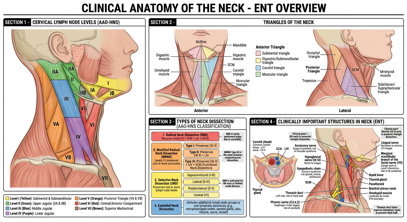
- Color-coded levels I through VII with full nomenclature (submental, submandibular, upper/middle/lower jugular, posterior triangle, central, superior mediastinal)
- IIA/IIB and VA/VB subdivisions marked
- Anterior triangle with all 4 subdivisions: submental, digastric/submandibular, carotid, muscular
- Posterior triangle with occipital and subclavian divisions
- Boundaries clearly labeled (SCM, trapezius, clavicle, mandible, midline)
- Radical Neck Dissection (levels I-V + SCM + IJV + CN XI)
- Modified Radical (MRND Type I, II, III / Functional)
- Selective Neck Dissection (Supraomohyoid, Lateral, Posterolateral, Central)
- Extended Neck Dissection
- CN XI (spinal accessory) - key in posterior triangle surgery
- Marginal mandibular branch of VII - danger in Level I dissection
- Phrenic nerve (C3,4,5) - on anterior scalene
- Thoracic duct - left-sided, at IJV/subclavian junction
- Horner's syndrome risk (sympathetic chain)
- Carotid sheath contents (CCA, IJV, CN X)
- Omohyoid muscle as surgical landmark (Level II/III boundary)