
Are T-inversions benign also?
Reading File
T-wave inversions are not always benign - their significance depends heavily on the lead location, clinical context, and patient symptoms.
Searching sources...
Finding Sources
Searching PubMed
T wave inversion ECG clinical significance
Finding Sources
Finding Sources
Reading File
Finding Sources
Finding Sources
Finding Sources
Reading File
Finding Sources
Finding Sources
Reading File
Reading File
Now I have enough information for a thorough answer. Let me also fetch the ECG image for T-wave inversion in NSTEMI for visual clarity.
Loading Image
Loading Image
No - T-wave inversions are not always benign. Their significance depends entirely on the lead location, morphology, depth, clinical context, and associated symptoms. Here is a structured breakdown:
T-Wave Inversions: Benign vs. Pathological
When T-Wave Inversions ARE Normal (Benign)
| Situation | Detail |
|---|---|
| Lead aVR | T inversion is normal in aVR in virtually everyone |
| Lead V1 | Isolated T inversion in V1 is normal |
| Lead III | T inversion in lead III alone is usually a normal variant |
| Neonates/infants | Flat or inverted T waves in right precordial leads (V1-V3) are normal in the first years of life and may persist into adolescence |
| Children | T-wave changes are common in infants and children and rarely reflect ischemia |
As Tintinalli's Emergency Medicine notes: "T-wave inversion in the right pre-cordial leads is common in the first years of life and may persist into adolescence or revert in early childhood to the typical upright pattern seen in adults. T-wave changes are common in infants and children and rarely reflect ischemia."
When T-Wave Inversions Are PATHOLOGICAL
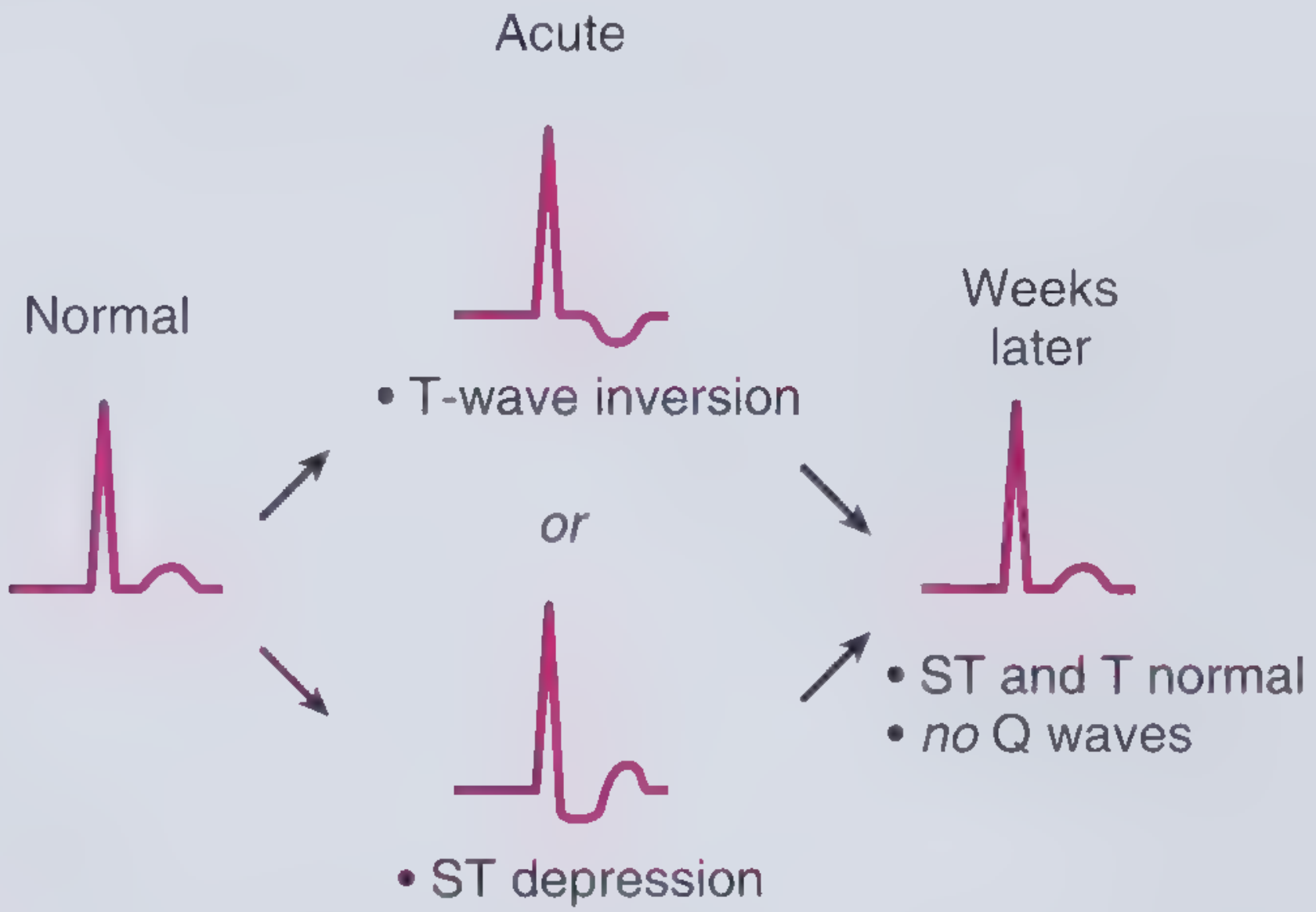
1. Acute Coronary Syndrome (UA/NSTEMI)
T-wave inversion ≥0.1 mV in 2 contiguous leads with a prominent R wave or R/S ratio >1 is a diagnostic criterion for UA/NSTEMI. This is one of the most common and important pathological causes.
2. Wellens' Syndrome (Critical LAD Stenosis)
This is one of the most dangerous patterns - deep, symmetric T-wave inversions in V2-V3 (75% of cases) or biphasic T waves (25%) occurring when a patient is pain-free after an anginal episode. The criteria include:
- History of episodic chest pain consistent with unstable angina
- During pain, ECG may not display abnormal T waves
- When pain-free: deep symmetric T-wave inversion most prominent in V2 and V3, often in V1 and V3, occasionally V3-V6
- No pathologic Q waves or loss of R waves
- Normal or minimally elevated cardiac biomarkers
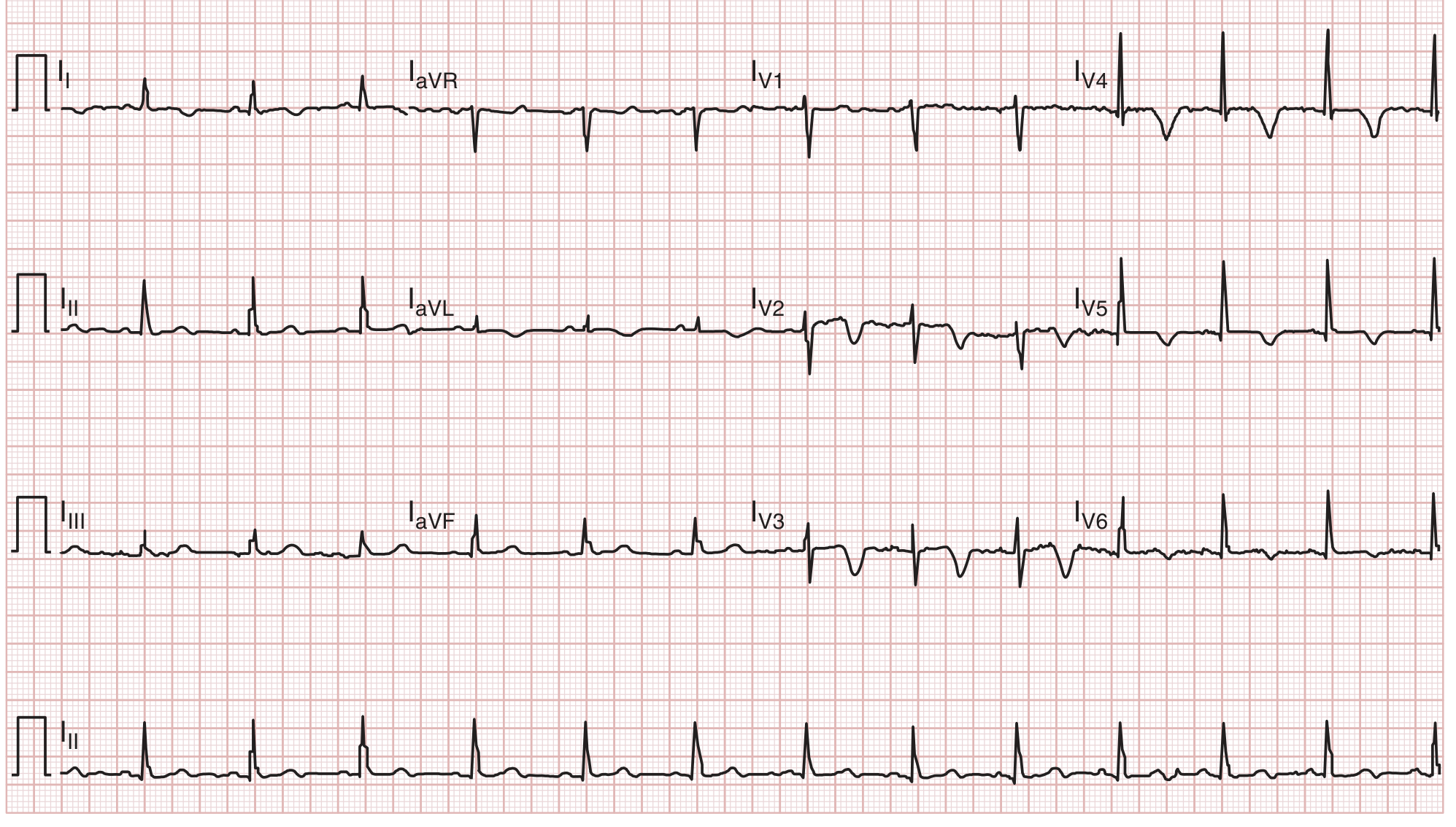
This pattern indicates critical proximal LAD stenosis and is a pre-infarction warning - the patient is at high risk of massive anterior STEMI if not treated urgently.
3. Pulmonary Embolism
T-wave inversion in V1 through V4 is a classic ECG sign of acute pulmonary hypertension from PE. It is part of the PE ECG severity scoring system - T inversion in V1-V4 scores 4 points and isolated V1 inversion >2 mm scores 2 points (Tintinalli's Emergency Medicine, PE chapter).
4. Right Ventricular Strain
Cor pulmonale, pulmonary hypertension, or acute RV overload can produce T inversions across the right precordial leads.
5. Post-STEMI
After ST-elevation resolves following reperfusion, T-wave inversions (sometimes deep and symmetric, similar to Wellens') may follow. These can become permanent in some patients.
6. Structural / Other Causes
- Hypertrophic cardiomyopathy - often dramatic T inversions in lateral leads
- Left bundle branch block and ventricular paced rhythms - discordant T inversions (expected, but mask ischemia assessment)
- Takotsubo (stress) cardiomyopathy - can mimic STEMI/Wellens'
- Hypokalemia - T wave flattening or inversion with prominent U waves
- Digoxin effect - scooped ST/T changes
- Subarachnoid hemorrhage - "cerebral T waves," deeply inverted, widespread, in the context of neurological presentation
Key Clinical Rule
The same morphology means very different things depending on:
-
Which leads: V1 alone = benign; V1-V4 in a dyspneic patient = think PE; V2-V3 deep symmetric in chest pain = Wellens'
-
Symptoms: Asymptomatic isolated findings vs. chest pain/dyspnea dramatically changes the significance
-
Depth and symmetry: Deep, symmetric inversions are more concerning than flat, asymmetric ones
-
Acuity: New inversions on a serial ECG are never benign until proven otherwise
-
Frameworks for Internal Medicine, ACS chapter
-
Tintinalli's Emergency Medicine, Chapters 49 (ACS) and 56 (PE)
Can panic attacks produce T inversions
Yes, panic attacks can produce T-wave changes including inversions, primarily through catecholamine surge and hyperventilation effects on repolarization.
Searching sources...
Finding Sources
Searching PubMed
panic attack T wave inversion ECG changes
Finding Sources
Finding Sources
Finding Sources
Searching the Web
panic attack T wave inversion ECG changes mechanism
Reading File
Reading Web Page
https://pmc.ncbi.nlm.nih.gov/articles/PMC4325305
Finding Sources
Finding Sources
Finding Sources
Reading File
Reading File
This gives a thorough picture. Here is the complete answer:
Can Panic Attacks Produce T-Wave Inversions?
Yes, they can - through several mechanisms, ranging from transient nonspecific changes to a more serious stress-induced syndrome that genuinely mimics ACS.
Mechanism 1: Nonspecific T-Wave Changes from Sympathetic Surge
During a panic attack, there is a marked surge in noradrenergic discharge from the locus coeruleus and elevated circulating catecholamines. This autonomic activation can produce nonspecific ST-segment and T-wave changes, including T-wave flattening or mild inversions. These are generally transient, reversible, and not associated with structural cardiac disease.
Key point from the web literature: anxiety-related T-wave changes tend to be less pronounced than depression-associated inversions, and there is limited evidence that anxiety alone meaningfully changes T-wave amplitude or QTc. Most changes are non-specific and rate-related (tachycardia alters repolarization).
Mechanism 2: Hyperventilation
Panic attacks almost always involve hyperventilation. Hypocapnia from hyperventilation causes:
- Coronary vasospasm
- Alkalosis-driven shifts in electrolytes (particularly calcium and potassium)
- Altered myocardial repolarization
These can produce ST-segment depression and T-wave changes on ECG that mimic ischemia and resolve when breathing normalizes.
Mechanism 3: Takotsubo (Stress) Cardiomyopathy - The Important One
This is the most clinically significant pathway. Takotsubo syndrome (TS) - also called stress cardiomyopathy or "broken heart syndrome" - is triggered by intense emotional or physical stress through a catecholamine storm mechanism. Key facts from Rosen's Emergency Medicine:
- Serum catecholamines in TS are more than twice as high as in myocardial infarction
- Anxiety is explicitly listed as a risk factor for TS
- Emotional stress is one of the recognized triggers
- ECG shows ST-segment elevations or deep T-wave inversions - indistinguishable from ACS
- Serum troponin is mildly elevated
- BNP is elevated in up to 80% of patients
- 85% of US cases occur in women over age 60
- In the ED, TS is usually not distinguishable from AMI and must be treated accordingly
So a severe panic attack in a susceptible individual (older woman, anxiety history, cardiovascular risk factors) can trigger genuine Takotsubo syndrome with real, deep T-wave inversions, not just artifact.
The Clinical Problem: Panic Disorder Mimics ACS (and vice versa)
Harrison's 22nd edition notes a striking statistic: panic disorder was the primary diagnosis in 43% of patients with chest pain who had normal coronary angiograms, and was present in 9% of all outpatients referred for cardiac evaluation. This cuts both ways:
- Panic attacks produce chest pain, palpitations, dyspnea - identical to ACS symptoms
- They can produce ECG changes including T-wave inversions
- But ACS can also present with anxiety and panic-like symptoms
This means you cannot diagnose a panic attack based on T-wave inversions alone - cardiac causes must be ruled out first, especially in first presentations.
Bottom Line
| Mechanism | T Inversions? | Severity |
|---|---|---|
| Sympathetic/catecholamine surge | Mild, nonspecific | Benign, transient |
| Hyperventilation/hypocapnia | Mild ST-T changes | Benign, reversible |
| Takotsubo syndrome triggered by panic | Deep, significant | Serious - mimics STEMI |
The safest approach: treat T-wave inversions in a patient presenting with chest pain and panic-like symptoms as cardiac until proven otherwise, get serial ECGs and troponins, and consider Takotsubo in the differential - particularly in older women after severe emotional stress.
- Rosen's Emergency Medicine, Takotsubo Syndrome chapter
- Harrison's Principles of Internal Medicine 22E, Panic Disorder chapter
- PMC review: Cardiac and non-cardiac causes of T-wave inversion
This is a shared conversation. Sign in to Orris to start your own chat.