
when di u call moon faciesis tere a definition
moon facies definition clinical criteria when to call it
moon facies Cushing syndrome rounded face
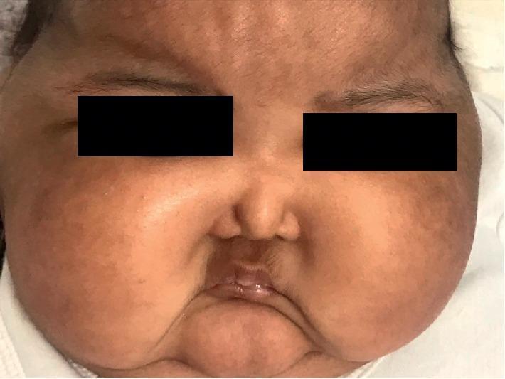
This clinical photograph shows a close-up facial view of an infant demonstrating classic 'moon facies,' a characteristic finding associated with Cushing syndrome or systemic corticosteroid exposure. The face exhibits a markedly rounded, plethoric, and symmetrical appearance due to subcutaneous fat redistribution and edema. Notable features include prominent, puffed cheeks (buccal fat pad hypertrophy) that obscure the jawline and give the face a circular contour. The central facial features, specifically the nose and mouth, appear small and recessed relative to the enlarged, convex cheeks. The eyes are partially obscured by periorbital puffiness, and the forehead appears broad. This visual finding is a key diagnostic indicator for hypercortisolism, which in this clinical context is related to the iatrogenic absorption of potent topical steroids used for diaper dermatitis.
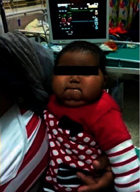
This clinical photograph displays a 15-month-old child exhibiting 'moon facies,' a classic sign of hypercortisolism (Cushing syndrome). The face is notably rounded and plethoric with a circular contour. There is significant soft tissue fullness in the bilateral malar regions (prominent cheeks) and noticeable submental fat accumulation beneath the chin. The child has a medium-to-dark complexion. The image is set in a clinical environment, with a patient monitor displaying vital signs visible in the background. This visual finding is characteristic of exogenous steroid exposure, either through the mother's milk (secondary to maternal prednisolone use) or direct topical application of potent corticosteroids like clobetasol. The photograph serves as an educational example of pediatric iatrogenic Cushing syndrome and its associated physical manifestations.
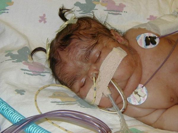
Clinical photograph of a neonate demonstrating characteristic facial features of neonatal Cushing syndrome. The infant displays 'moon facies,' characterized by a rounded face with prominent, full cheeks. Visible facial plethora (reddish discoloration) and hirsutism (excessive hair growth on the face and forehead) are present. The infant is shown in a medical setting, evidenced by intensive care monitoring and support devices. Visible equipment includes nasal prongs or a nasogastric tube secured with tan adhesive tape across the midface, and circular ECG electrodes with lead wires attached to the chest for cardiac monitoring. The clinical presentation is highly suggestive of hypercortisolism, potentially as a component of McCune-Albright Syndrome (MAS), where such signs are often associated with café-au-lait spots and polyostotic fibrous dysplasia.
Moon Facies - Definition and When to Call It
Definition
- Frameworks for Internal Medicine defines it as: "rounded face due to fat deposition" - a clinical manifestation of Cushing's syndrome
- ScienceDirect / Practical Pediatric Endocrinology defines it as: "a rounded red face that is characteristic of Cushing syndrome"
- Cleveland Clinic: fat deposits build up along the sides of the skull, and the face can become so round that the ears are not visible from the front
When Do You Call It Moon Facies?
| Feature | Moon Facies |
|---|---|
| Shape | Uniformly round/circular ("full moon") |
| Cause | Fat redistribution, not fluid |
| Location | Cheeks and jaw bilaterally |
| Onset | Gradual, over weeks to months |
| Associated findings | Buffalo hump, central obesity, striae, plethora |
Main Causes
- Cushing's syndrome - excess endogenous cortisol (adrenal tumor, pituitary adenoma - Cushing's disease, ectopic ACTH)
- Exogenous corticosteroids - the most common cause in clinical practice; develops in slightly more than 10% of patients on steroids, but is uncommon below physiologic replacement doses (Rheumatology, 2-Volume Set, 2022)
- Obesity - can produce a similar appearance but without the other Cushingoid features
Classic Cluster (Cushingoid Appearance)
- Buffalo hump (dorsal fat pad)
- Supraclavicular fat pads
- Central/truncal obesity with thin limbs
- Violaceous abdominal striae
- Skin thinning and easy bruising
- Hypertension
- Hirsutism (in women)
Why is there proximal myropathy in cusging ssepcofcallt explain ans articels
"Cushing syndrome" AND "proximal myopathy" AND glucocorticoid muscle
proximal myopathy Cushing syndrome mechanism glucocorticoid muscle wasting pathophysiology
glucocorticoid myopathy muscle atrophy mechanism
https://pmc.ncbi.nlm.nih.gov/articles/PMC8258338
PMID: 41148376
PMID: 40806744
Proximal Myopathy in Cushing's Syndrome - Mechanism Explained
What Does It Look Like Clinically?
- Weakness is proximal (shoulders, hips, thighs) - distal muscles are largely spared
- Legs affected more than arms
- Onset is typically insidious - patients first notice difficulty rising from a chair, climbing stairs, or lifting arms overhead
- Eventually leads to frank muscle wasting
- Crucially: serum CK (creatine kinase) is usually normal - this is a key distinguishing feature from inflammatory myopathies
- Reported in up to 70% of patients with active primary Cushing's syndrome (Firestein & Kelley's Textbook of Rheumatology, 2022)
Why Proximal? The Core Mechanisms
1. Type II Muscle Fiber Atrophy (The Most Important Mechanism)
- Muscle biopsy shows increased vacuolation and glycogen accumulation in type II fibers (Firestein & Kelley's Rheumatology)
- GCs activate the glucocorticoid receptor (GR) inside muscle cells, which then drives the transcription of genes that destroy muscle protein
2. Increased Muscle Protein Catabolism (Breaking Down Muscle)
| Proteolytic System | What It Does |
|---|---|
| Ubiquitin-Proteasome System (UPS) | Tags muscle proteins with ubiquitin, then degrades them via the proteasome ("atrogenes" MuRF-1 and MAFbx/Atrogin-1 are upregulated) |
| Lysosomal system (cathepsins) | Lysosomal enzyme activation further degrades muscle structural proteins |
| Calcium-dependent system (calpains) | Calcium-activated proteases degrade myofilaments |
3. Suppression of Protein Synthesis (Blocking Muscle Building)
- Suppresses PI3K/AKT/mTORC1 signaling - this pathway is the master regulator of muscle protein synthesis. Glucocorticoids suppress it directly via GR activation, and also by inducing SIRT6, which inhibits AKT/mTORC1
- Reduces IGF-1 (Insulin-like Growth Factor-1) locally in muscle - IGF-1 normally promotes satellite cell proliferation and new muscle fiber synthesis
- Upregulates myostatin - a powerful inhibitor of muscle growth; glucocorticoids increase myostatin expression, further blocking regeneration
- Reduces insulin sensitivity in muscle - less glucose uptake means less energy for muscle synthesis
4. Mitochondrial Dysfunction
5. Additional Contributing Factors
- Hypokalemia - Cortisol has mineralocorticoid-like activity, causing potassium wasting through the kidney. Low potassium directly impairs muscle membrane excitability and contraction
- Hypophosphatemia - Can further reduce muscle contractility
- In endogenous Cushing's (ACTH-dependent causes): excess ACTH itself may impair neuromuscular junction transmission by reducing quantal content of the end-plate potential - an additional myopathic mechanism on top of the cortisol effect
Why Does It Persist Even After Treatment?
Summary Diagram of Mechanisms
Excess Cortisol / Glucocorticoids
         |
         ├── Activates GR in muscle
         |        |
         |        ├── Upregulates ubiquitin-proteasome (UPS) → protein degradation
         |        ├── Activates lysosomal cathepsins → protein degradation
         |        ├── Activates calpains → myofilament degradation
         |        ├── Suppresses PI3K/AKT/mTOR → ↓ protein synthesis
         |        ├── Upregulates myostatin → blocks muscle regeneration
         |        └── Reduces IGF-1 → ↓ satellite cell proliferation
         |
         ├── Mitochondrial dysfunction → ↓ energy supply
         ├── Hypokalemia → ↓ membrane excitability
         └── Excess ACTH (endogenous only) → ↓ neuromuscular transmission
                   ↓
         Preferential TYPE II FIBER ATROPHY
         (Type II fibers dominate PROXIMAL muscles)
                   ↓
         PROXIMAL MYOPATHY
Key Articles
-
Gé fard-Gontier et al., Journal of Neurology, 2025 - "Glucocorticoid-induced myopathy: a comprehensive review" - covers pathophysiology (catabolic + anti-anabolic mechanisms), diagnostic challenges, and emerging therapies including IGF-1, branched-chain amino acids, and exercise. PMID: 41148376
-
Permpoon et al., Int J Mol Sci, 2025 - "Glucocorticoid-Mediated Skeletal Muscle Atrophy: Molecular Mechanisms and Potential Therapeutic Targets" - covers GR transactivation, SIRT6/AKT/mTORC1 suppression, the kynurenine/IDO-1 pathway, LSD1 cooperation with GR, and novel drug targets including 11β-HSD1 inhibitors and myostatin blockers. PMID: 40806744
-
Reincke M., Endocrinol Metab, 2021 - "Cushing Syndrome Associated Myopathy: It Is Time for a Change" - specifically focused on Cushing's-associated myopathy, noting its underdiagnosis and calling for updated diagnostic and management approaches. PMID: 33915607
Type II Muscle Fiber Atrophy (The Most Important Mechanism) Glucocorticoids preferentially attack type II (fast-twitch) muscle fibers - specifically type IIa and IIb fibers. These are the fibers that dominate proximal (limb-girdle) muscles, Increased Muscle Protein Catabolism ddrw flow hart
A detailed medical flowchart diagram titled "Glucocorticoid-Induced Proximal Myopathy: Type II Fiber Atrophy & Muscle Protein Catabolism". The flowchart starts at the top with a box labeled "EXCESS CORTISOL / GLUCOCORTICOIDS (Cushing's Syndrome)". An arrow points down to "Activates Glucocorticoid Receptor (GR) in Muscle Cell". From there, two main branches split left and right with arrows: LEFT BRANCH - titled "INCREASED CATABOLISM (Protein Breakdown)" with three sub-boxes connected by arrows: 1. "Ubiquitin-Proteasome System (UPS) - Upregulates MuRF-1 and Atrogin-1 atrogenes - Tags and degrades muscle proteins" 2. "Lysosomal Cathepsins - Activated lysosomal enzymes - Degrade structural muscle proteins" 3. "Calpains (Ca²⁺-dependent) - Calcium-activated proteases - Break down myofilaments" RIGHT BRANCH - titled "DECREASED ANABOLISM (Protein Synthesis Blocked)" with three sub-boxes connected by arrows: 1. "Suppresses PI3K / AKT / mTORC1 - Master regulator of protein synthesis inhibited - SIRT6 upregulation blocks AKT" 2. "Reduces IGF-1 in muscle - Less satellite cell proliferation - Impaired muscle regeneration" 3. "Upregulates Myostatin - Potent inhibitor of muscle growth - Blocks new fiber formation" Both branches converge with arrows to a central box: "PREFERENTIAL ATROPHY OF TYPE II (FAST-TWITCH) FIBERS - Type IIa and IIb fibers most vulnerable - Dominate proximal limb-girdle muscles" An arrow points down to a final box: "PROXIMAL MYOPATHY - Weakness: hips > shoulders > arms - Legs affected more than arms - Insidious onset, normal CK - Difficulty rising from chair, climbing stairs" Additional small boxes branching off the side showing: "Hypokalemia (mineralocorticoid effect) → ↓ muscle excitability" and "Mitochondrial dysfunction → ↓ energy supply". Clean clinical style with blue and red color coding for catabolism vs anabolism branches.
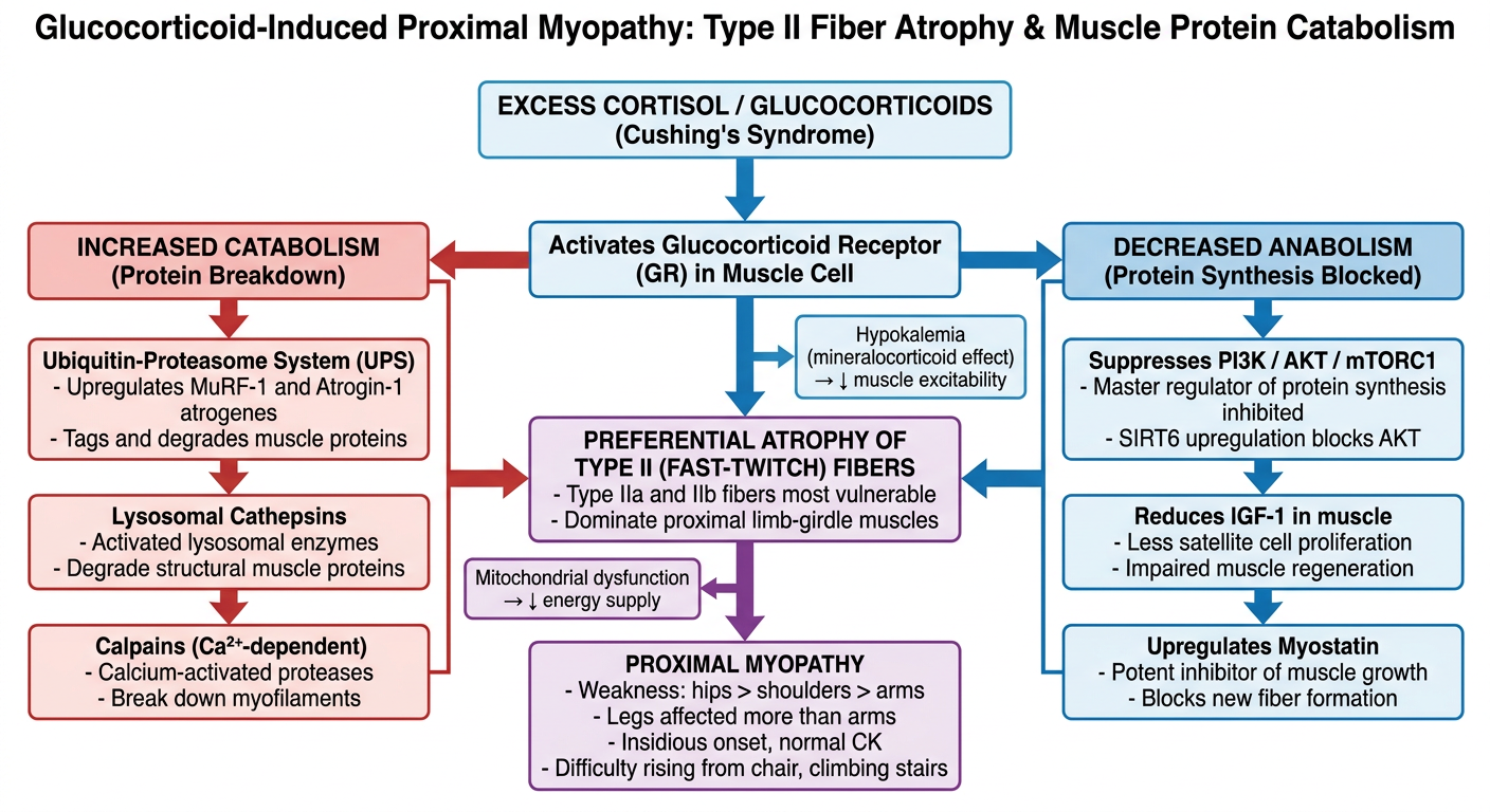
- UPS (ubiquitin-proteasome) upregulates MuRF-1 and Atrogin-1, tagging muscle proteins for destruction
- Lysosomal cathepsins degrade structural muscle proteins
- Calpains (calcium-activated) break down myofilaments
- PI3K/AKT/mTORC1 signaling is suppressed - muscle cannot synthesise new protein
- IGF-1 is reduced - satellite cells cannot regenerate new fibers
- Myostatin is upregulated - a brake on all muscle growth