
Give details about surgery specimens for table viva 1) trichobezoar (2)pckd (3) Splenomegaly (4) teratoma... Possible questions like causes treatment diagnosis etc and answers
Reading File
Now let me search all four topics in parallel.
Finding Sources
Finding Sources
Finding Sources
Finding Sources
Good initial results. Now let me read the key textbook sections in parallel for detailed content.
Reading File
Finding Sources
Reading File
Finding Sources
Good content so far. Let me pull more detailed sections on PCKD and teratoma, plus fetch the trichobezoar image.
Finding Sources
Reading File
Loading Image
Now let me get the PCKD clinical details and the teratoma/dermoid cyst details.
Finding Sources
Reading File
Loading Image
Now let me get the PCKD clinical content and splenomegaly table details.
Finding Sources
Reading File
Let me read the Bailey & Love ADPKD section and get images for splenomegaly and teratoma.
Reading File
Loading Image
I now have comprehensive information from the textbooks on all four specimens. Let me compile the full table viva answer.
Surgery Specimens - Table Viva Guide
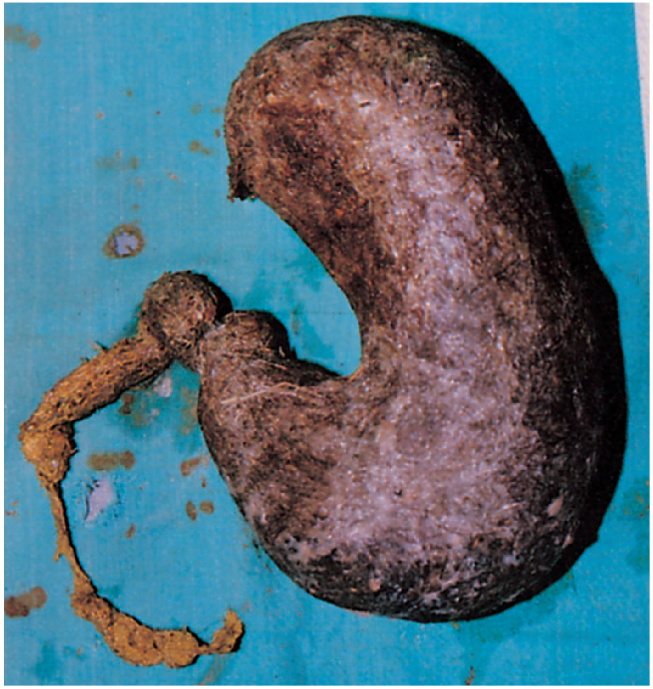
1. TRICHOBEZOAR (Hair Ball)
What is it?
A trichobezoar is a mass formed by the accumulation of swallowed hair in the stomach. Hair is not digested and accumulates over time, taking the shape of the stomach.
Causes
- Trichotillomania (compulsive hair-pulling) followed by trichophagia (eating hair) - an obsessive-compulsive spectrum disorder
- Virtually exclusively found in young female psychiatric patients
- Associated with DSM-5 obsessive-compulsive and related disorders
Types of Bezoars (Know the Full List)
| Type | Composition |
|---|---|
| Trichobezoar | Hair |
| Phytobezoar | Vegetable/plant matter (post-gastric surgery) |
| Pharmacobezoar | Medications |
| Lactobezoar | Inspissated milk (neonates) |
| Diospyrobezoar | Persimmons |
Rapunzel Syndrome
A variant where the trichobezoar extends from the stomach into the small intestine - sometimes reaching the ileocecal valve. This is named after the fairy tale character.
Clinical Features / Complications
- Epigastric mass (palpable)
- Abdominal pain and nausea
- Weight loss, anorexia
- Gastric ulceration and GI bleeding
- Gastric outlet obstruction
- Perforation (rare)
- If Rapunzel syndrome: small bowel obstruction
Diagnosis
- Endoscopy - gold standard, easiest diagnosis
- CT abdomen - shows heterogeneous intraluminal mass with gas pockets (mottled appearance)
- Plain X-ray - may show soft tissue mass with gas lucencies
- USG - echogenic mass with dirty shadowing
Treatment
- Endoscopic removal - first attempt; Coca-Cola dissolution sometimes used for phytobezoars
- Surgical removal (laparotomy/gastrotomy) - for large/complicated trichobezoars; required in most cases
- Psychiatric management and follow-up to prevent recurrence (address trichotillomania/trichophagia)
Possible Viva Questions
- Q: What is Rapunzel syndrome? A: Extension of trichobezoar from stomach into small intestine up to the ileocecal valve.
- Q: Who is typically affected? A: Young female psychiatric patients with trichotillomania/trichophagia.
- Q: What is the diagnosis of choice? A: Upper GI endoscopy.
- Q: What is the treatment? A: Endoscopic removal; if large - open gastrotomy. Must address the underlying psychiatric disorder.
- Q: What complications can occur? A: Ulceration, bleeding, perforation, obstruction, Rapunzel syndrome.
2. POLYCYSTIC KIDNEY DISEASE (PCKD/ADPKD)
What is it?
A heritable cystic disorder where multiple fluid-filled cysts replace normal renal parenchyma bilaterally, eventually leading to end-stage renal disease (ESRD).
Types
| Feature | ADPKD | ARPKD |
|---|---|---|
| Inheritance | Autosomal Dominant | Autosomal Recessive |
| Common name | Adult PKD | Infantile PKD |
| Gene | PKD1 (chr 16) ~78%, PKD2 (chr 4) ~15% | PKHD1 (chr 6) |
| Protein | Polycystin-1 and Polycystin-2 | Fibrocystin/Polyductin |
| Incidence | 1:1000 | 1:20,000 |
| Onset | Usually 3rd-4th decade | In utero/neonatal |
Causes / Genetics
- PKD1 mutation (chromosome 16p13.3) - encodes polycystin-1; causes ~78% of ADPKD cases
- PKD2 mutation (chromosome 4q21) - encodes polycystin-2; milder course
- "Two-hit hypothesis" - germline mutation + somatic second hit causes individual cyst formation
- PKD1 mutations: more severe, ESRD ~10 years earlier than PKD2
Risk Factors for ESRD Progression
- Early age of presentation
- Hypertension
- Male sex
- PKD1 gene type (vs PKD2)
- African ethnic group
Gross Pathology (Specimen Description)
- Hugely enlarged kidneys bilaterally
- Replaced almost entirely by multiple cysts of varying sizes (mm to several cm)
- Cysts contain clear, straw-colored or brown fluid (if bleeding has occurred)
- Some residual compressed normal parenchyma between cysts
- Normal reniform shape may be preserved but grossly distorted
Extrarenal Manifestations
- Liver cysts (most common extrarenal, ~80% of patients)
- Intracranial (berry) aneurysms - 10-30% of patients; risk of subarachnoid hemorrhage - most feared complication
- Pancreatic cysts
- Mitral valve prolapse
- Arachnoid membrane cysts
- Seminal vesicle cysts
Clinical Features
- Usually presents after age 30 (often 4th-5th decade)
- Bilateral flank masses - often first sign
- Haematuria (gross or microscopic)
- Abdominal/flank pain
- Hypertension (most patients >20 years are hypertensive)
- Recurrent UTI, pyelonephritis
- Renal stones
- Progression to ESRD (~50% by age 60 with PKD1)
Diagnosis
- Ultrasound - investigation of choice for screening; bilateral enlarged kidneys with multiple cysts (Unified Ravine/Pei criteria based on age and number of cysts)
- CT scan - more sensitive, can detect smaller cysts
- MRI - most sensitive; used for total kidney volume measurement (TKV) to predict progression
- Genetic testing - PKD1/PKD2 mutation analysis (useful for young family members/potential donors)
- Urinalysis - haematuria, proteinuria
- Renal function tests - rising creatinine
Treatment
- Hypertension control - ACE inhibitors/ARBs (most important; delays progression)
- Tolvaptan (vasopressin V2 receptor antagonist) - slows cyst growth and decline in renal function (FDA approved); monitor for hepatotoxicity
- Somatostatin analogues and mTOR inhibitors - under investigation
- Treat UTI aggressively
- Pain management
- Screen for and treat intracranial aneurysms (if symptomatic/large)
- Renal replacement therapy - dialysis or renal transplantation for ESRD
Possible Viva Questions
- Q: What genes are mutated in ADPKD? A: PKD1 (chromosome 16) and PKD2 (chromosome 4).
- Q: Most common extrarenal manifestation? A: Hepatic cysts.
- Q: Most dangerous extrarenal complication? A: Intracranial berry aneurysm causing subarachnoid haemorrhage.
- Q: Investigation of choice? A: Ultrasound (USG); MRI for TKV monitoring.
- Q: How is progression delayed? A: Control of hypertension (ACE inhibitor/ARB); tolvaptan.
- Q: What is the difference between ADPKD and ARPKD? A: ADPKD is adult onset, dominant, PKD1/PKD2 genes; ARPKD is infantile, recessive, PKHD1 gene.
- Q: What percentage develop ESRD? A: ~50% of affected individuals.
3. SPLENOMEGALY (Enlarged Spleen)
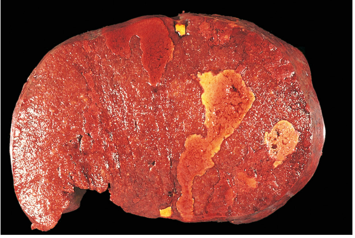
Normal vs Enlarged
- Normal spleen weight: 150-200 g
- Mild splenomegaly: 200-400 g (e.g. acute splenitis)
- Moderate: 500-1000 g
- Massive splenomegaly: >1000 g (up to 5000 g in congestive causes)
Causes (Classified)
Infections
- Bacterial: Infectious endocarditis, tuberculosis, typhoid, brucellosis
- Viral: Infectious mononucleosis (EBV), CMV
- Parasitic: Malaria, visceral leishmaniasis (Kala-azar) - causes massive splenomegaly
Congestive / Portal Hypertension
- Cirrhosis of liver (most common cause of massive congestive splenomegaly)
- Schistosomiasis ("pipestem" fibrosis)
- Portal vein thrombosis
- Splenic vein thrombosis (carcinoma pancreas/stomach)
- Right heart failure (tricuspid valve disease, cor pulmonale)
Haematological
- Haemolytic anaemias (hereditary spherocytosis, thalassemia, sickle cell - early)
- Leukaemias - CML, ALL
- Lymphomas (especially indolent/follicular)
- Myeloproliferative disorders: polycythaemia vera, primary myelofibrosis, essential thrombocythaemia
- Hairy cell leukaemia
Immunological / Inflammatory
- Rheumatoid arthritis (Felty's syndrome)
- SLE
- Sarcoidosis
- Amyloidosis
Storage/Infiltrative
- Gaucher disease (most common lysosomal storage disease causing splenomegaly)
- Niemann-Pick disease
- Glycogen storage diseases
Massive Splenomegaly - Classic Causes (Important for Viva)
Mnemonic: CML KaLa GauLym ThalMy
- CML (Chronic Myeloid Leukaemia)
- Kala-azar (visceral leishmaniasis)
- Gaucher disease
- Lymphoma (indolent/hairy cell)
- Thalassaemia major
- Primary myelofibrosis
- Malaria (chronic hyperreactive malarial splenomegaly)
Gross Pathology (Specimen Description)
- Enlarged, firm organ with thickened fibrous capsule (in chronic congestion)
- Cut surface: congested red pulp, may show infarcts (pale, wedge-shaped, subcapsular)
- In congestive splenomegaly: dilated sinusoids, fibrosis, Gamna-Gandy bodies (hemosiderin deposits)
Clinical Features
- Dragging sensation in left upper quadrant
- Discomfort after eating (pressure on stomach)
- Hypersplenism - triad of: anaemia + leukopenia + thrombocytopenia (due to increased sequestration and phagocytosis)
- Spleen palpable below left costal margin
- Associated features of the underlying cause
Diagnosis
- Clinical examination - splenomegaly detected on palpation/percussion
- Ultrasound - confirms, measures size
- CT scan - defines size and nature; detects infarcts
- Blood counts - CBC (cytopenias in hypersplenism)
- Bone marrow biopsy - for haematological causes
- Liver function tests, viral serology, malarial smear as indicated
- Peripheral blood smear - target cells (thalassemia), smudge cells (CLL), tear-drop cells (myelofibrosis)
Treatment
- Treat the underlying cause - primary goal
- Splenectomy indications:
- Hereditary spherocytosis
- Immune thrombocytopenic purpura (ITP) refractory to medical therapy
- Symptomatic hypersplenism
- Splenic rupture
- Staging/diagnosis (selected cases)
- Post-splenectomy: vaccinate against encapsulated organisms (Pneumococcus, Meningococcus, H. influenzae) - risk of overwhelming post-splenectomy sepsis (OPSS)
- Long-term prophylactic penicillin in asplenic patients
Possible Viva Questions
- Q: What is hypersplenism? A: Cytopenias (anaemia, leukopenia, thrombocytopenia) due to excessive sequestration and destruction of blood cells in an enlarged spleen.
- Q: Causes of massive splenomegaly? A: CML, kala-azar, myelofibrosis, Gaucher disease, malaria, thalassaemia, hairy cell leukaemia, lymphoma.
- Q: What is Felty's syndrome? A: Triad of rheumatoid arthritis + splenomegaly + neutropenia.
- Q: Risk after splenectomy? A: Overwhelming post-splenectomy sepsis (OPSS) due to encapsulated bacteria - prevent with vaccines.
- Q: What is the most common cause of massive congestive splenomegaly? A: Cirrhosis of the liver leading to portal hypertension.
- Q: Gamna-Gandy bodies? A: Foci of hemosiderin, calcium, and fibrous tissue in the spleen - seen in chronic congestive splenomegaly, sickle cell disease; appear as low-signal foci on MRI.
4. TERATOMA
What is it?
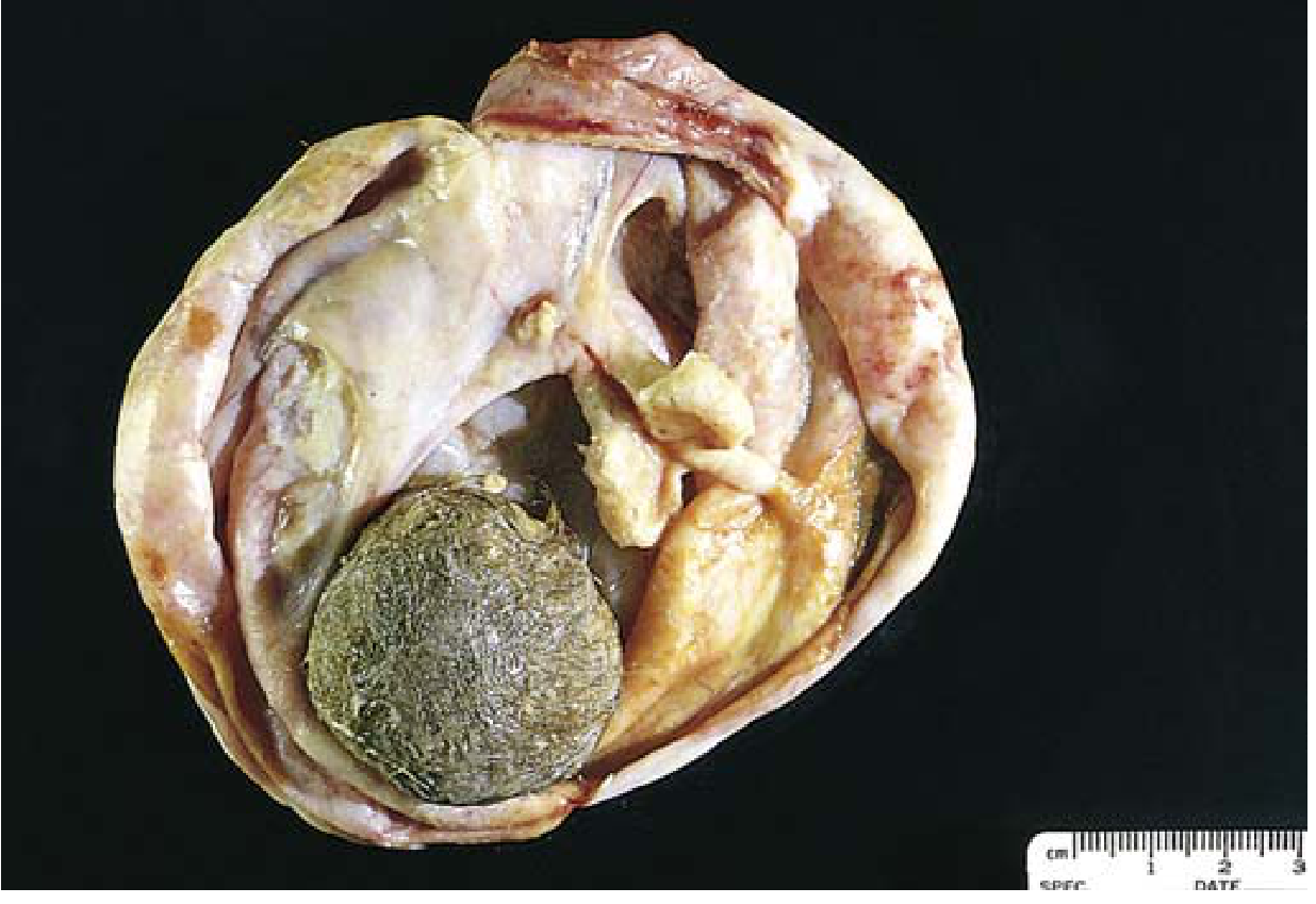
A teratoma is a germ cell tumor containing tissues from all three embryonic germ layers (ectoderm, mesoderm, endoderm). It arises from totipotent germ cells.
Classification
| Type | Features | Behavior |
|---|---|---|
| Mature (benign) cystic teratoma - Dermoid cyst | Most common; contains hair, teeth, sebum, skin; lined by squamous epithelium; karyotype 46,XX | Benign; 1% malignant transformation (usually SCC) |
| Mature solid teratoma | Rare | Benign |
| Immature (malignant) teratoma | Contains immature neuroepithelium, embryonal tissue; mean age 18 years | Malignant; risk correlates with proportion of immature neuroepithelium |
| Monodermal (specialized) | e.g., Struma ovarii (thyroid tissue), ovarian carcinoid | Variable |
Location by Site
- Ovary - most common site in females; mature cystic teratoma (dermoid cyst) is the most common ovarian germ cell tumor
- Testis - teratoma in post-pubertal males is considered malignant; in pre-pubertal boys it is benign
- Sacrococcygeal - most common germ cell tumor in neonates/infants
- Mediastinum - anterior mediastinum; must distinguish from thymoma/lymphoma on imaging
- Retroperitoneum
- Pineal gland/CNS
Gross Pathology (Specimen Description - Ovarian Dermoid)
- Cystic ovarian mass
- Cut section shows: hair, cheesy sebaceous material (yellow/white), teeth (calcifications), skin
- "Rokitansky nodule" or "dermoid plug" - solid protuberance within the cyst wall from which hair tufts emerge
- Usually unilateral (10-15% bilateral)
Histology
- Mature cystic teratoma: cyst lined by stratified squamous epithelium; skin appendages (sebaceous glands, hair follicles); may contain teeth, bone, cartilage, neural tissue, thyroid tissue
- Immature teratoma: primitive neuroepithelium, embryonal/fetal-type tissues
- Origin: parthenogenetic development from a single germ cell (hence 46,XX karyotype)
Tumour Markers
- Mature teratoma - usually negative
- Immature teratoma - may have raised AFP (alpha-fetoprotein)
- Mixed GCT with teratoma component - AFP and/or beta-hCG elevated depending on other elements
Imaging Features
- X-ray / CT - calcification (teeth/bone), fat density, cystic component - classic triad
- Ultrasound - hyperechoic component with posterior acoustic shadowing (calcification/teeth); "tip of iceberg" sign
- MRI - fat signal (high T1, suppresses on fat-sat), cystic (high T2), soft tissue elements (isointense to muscle)
- Dermoid mesh (floating hair strands within cyst) on ultrasound
Complications
- Torsion - most common complication (10-15% of cases); presents as acute abdomen - surgical emergency
- Rupture - can cause chemical peritonitis from spillage of sebaceous material
- Malignant transformation - in ~1% of mature dermoids; usually squamous cell carcinoma
- Infection - rare
- Autoimmune encephalitis - limbic encephalitis with anti-NMDA receptor antibodies in women with mature neural tissue-containing teratomas; often remits after tumor resection
Treatment
- Ovarian cystectomy (cyst removal, preserve ovarian tissue) - for young women with mature dermoid cyst; laparoscopic approach preferred
- Oophorectomy - if large, recurrent, or in postmenopausal women
- Orchidectomy - for testicular teratoma (post-pubertal, as it is malignant)
- Chemotherapy (BEP - Bleomycin, Etoposide, Cisplatin) - for malignant/immature teratoma; note teratoma per se is chemo-resistant
- Surgery for residual mass after chemotherapy - to excise residual teratoma (which is chemo-resistant)
Possible Viva Questions
- Q: What is a dermoid cyst? A: Mature cystic teratoma of the ovary; contains tissues from all 3 germ layers, predominantly ectodermal (hair, skin, sebaceous glands, teeth).
- Q: Most common complication? A: Torsion (10-15%); presents as acute surgical emergency.
- Q: Most common site of teratoma? A: Ovary (females), sacrococcyx (neonates).
- Q: What is the karyotype of mature ovarian teratoma? A: 46,XX - arises by parthenogenesis from a single germ cell.
- Q: Malignant transformation in dermoid? A: ~1%; usually squamous cell carcinoma.
- Q: How does teratoma look on imaging? A: Fat + calcification + cystic components = pathognomonic on CT. Teeth are classical.
- Q: Rokitansky nodule? A: Solid protuberance/mural nodule within dermoid cyst from which hair and teeth arise.
- Q: What is anti-NMDA receptor encephalitis? A: Autoimmune limbic encephalitis associated with ovarian teratoma containing neural tissue; treat by removing the tumor.
- Q: Difference between mature and immature teratoma? A: Mature (benign) - all adult-type tissues, 46XX; Immature (malignant) - embryonal/primitive neuroepithelium, mean age 18 years.
- Q: Why is teratoma resistant to chemotherapy? A: It is histologically benign/differentiated tissue even in a metastatic setting; chemo cannot kill mature differentiated cells.
Sources: Bailey & Love's Short Practice of Surgery 28th Ed.; Robbins, Cotran & Kumar Pathologic Basis of Disease; Robbins & Kumar Basic Pathology; Comprehensive Clinical Nephrology 7th Ed.; Harrison's Principles of Internal Medicine 22nd Ed.; Frameworks for Internal Medicine; Berek & Novak's Gynecology.
This is a shared conversation. Sign in to Orris to start your own chat.