
Molecular and genomic profiling of endometrial cancer with Pathogenesis clinical features morphology md pathology 10 marks answer
"endometrial cancer" AND "molecular classification" AND "TCGA"
PMID: 36791751
endometrial carcinoma histology endometrioid serous morphology
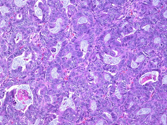
This is a histopathology image (hematoxylin and eosin stained) of an ovarian mass demonstrating a well-differentiated endometrioid carcinoma. The tumor displays tightly packed, back-to-back glands with minimal stroma, lined by neat, pseudostratified columnar epithelium. Nuclei are relatively uniform, with mild to moderate hyperchromasia and rare mitotic figures, consistent with low-grade morphology. The glandular lumina are often crescentic or slit-like; there may be cribriform arrangements in places. Surrounding ovarian stroma shows focal desmoplastic reaction and focal infiltration, supporting invasion by malignant epithelial cells. The tumor shows a predominantly tubular architecture resembling endometrial glands; mucin production is limited. Overall, the morphology fits endometrioid carcinoma of ovarian origin, grade 1. Differential considerations include mucinous or serous type ovarian carcinomas, metastatic endometrial adenocarcinoma, and clear cell carcinoma; however, the glandular pattern, cytologic uniformity, and absence of significant hobnail cells or prominent papillary features favor endometrioid histology. Clinically significant because endometrioid ovarian carcinomas are often associated with endometriosis and may present at an early stage; they may be estrogen-responsive; immunohistochemistry can show PAX8 positivity and variable ER/PR expression. This image is useful for educational teaching, histopathology differential diagnosis, and radiologic-pathologic correlation. Correlation with clinical staging and management is recommended.
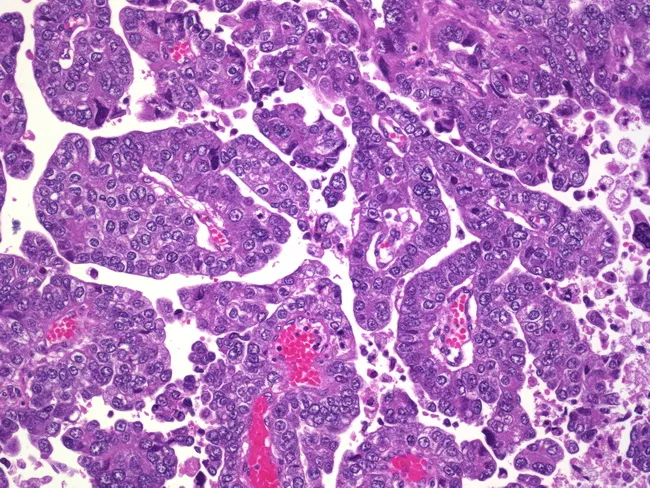
Imaging modality: Histopathology, prepared as a hematoxylin and eosin stained section of uterine endometrium. The slide demonstrates papillary clusters of malignant epithelial cells forming intricate papillary fronds with fibrovascular cores. Cells show high-grade nuclear atypia, marked pleomorphism, conspicuous nucleoli, and brisk mitotic activity with frequent mitotic figures. Necrosis is evident in several regions. Psammoma bodies are present as concentric calcified cores within papillary structures. Notably, there is invasion into the myometrium, indicating an infiltrative growth pattern and depth of invasion that carries prognostic and staging implications (FIGO staging for endometrial carcinoma). The architecture is complex and papillary, rather than glandular, favoring serous-type histology; psammoma bodies and high-grade features strongly support serous endometrial carcinoma over endometrioid types. Inflammatory cells and stromal desmoplasia may be variably present. This histology correlates with aggressive behavior, propensity for lymphovascular invasion, and higher likelihood of extra-uterine spread. Clinically, this morphology aligns with postmenopausal bleeding in many cases and requires staging workup, including surgical intervention with lymph node assessment and consideration of adjuvant therapy (carboplatin/paclitaxel +/- radiation). Differential considerations include clear cell carcinoma and metastatic ovarian serous carcinoma to the endometrium. Accurate pathologic subtyping is critical for prognosis and treatment planning. This description enhances searchability and education.
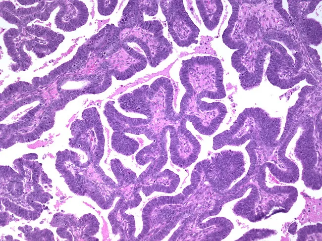
This is a histopathology image (Hematoxylin and Eosin stained) of an ovarian tumor. The specimen is ovarian parenchyma with gland-forming epithelium showing a villoglandular pattern characteristic of endometrioid carcinoma. The glands are lined by stratified columnar epithelium with pale to dark nuclear chromatin, mild to moderate nuclear atypia, and apical cytoplasmic clearing in an overall well-differentiated architecture. The glandular spaces vary in size, with some back-to-back arrangements and occasional cribriform/ villous projections; stromal invasion is suggested by irregular gland termination into surrounding stroma. The histology resembles endometrium (hence endometrioid type) and the tumor constitutes approximately 10-25% of primary ovarian carcinomas, consistent with literature. The differential includes serous, mucinous, clear cell, and metastatic endometrial carcinoma to the ovary; immunohistochemistry and clinical correlation aid in distinguishing these entities. Diagnostic significance: recognition of ovarian endometrioid histology has implications for prognosis and treatment and warrants evaluation for synchronous endometrial pathology. This image is suitable for educational purposes, pathology review, and research on ovarian epithelial tumors, particularly for pattern recognition of villoglandular growth and stratified epithelium. Potential clinical use: differential diagnosis of ovarian neoplasms, surgical planning, grading, and correlation with patient management. Color and morphology of the glands may correlate with imaging features and tumor markers (e.g., CA-125) in clinical practice.
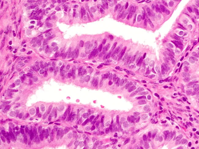
Imaging modality: bright-field light microscopy of a hematoxylin and eosin (H&E) stained endometrial tissue section from a biopsy. The tissue demonstrates nonatypical endometrial hyperplasia with crowding and architectural complexity of glands. The glands are lined predominantly by ciliated columnar epithelial cells with eosinophilic cytoplasm, showing typical tubal-type epithelium within the endometrium. Interspersed among the ciliated cells are round, clear cells consistent with tubal metaplasia. The stromal background is preserved, with no conspicuous cellular atypia or high-grade mitotic activity. The glands exhibit variable diameter and branching, yet maintain orderly polarity without cribriform destruction. Overall histology indicates benign metaplastic transformation rather than malignant invasion, and there is no definite cytologic atypia to suggest endometrial intraepithelial neoplasia or carcinoma. Recognizing tubal metaplasia in this setting is critical to avoid misinterpretation as serous or endometrioid carcinoma and to distinguish it from reactive or hormonal changes. Clinically, the finding may reflect underlying hormonal influences or sampling artifact; management decisions should integrate morphology with patient age and risk factors. This description supports educational use, differential diagnostic discussions, and research on endometrial metaplasia patterns in hyperplastic endometrium. The image thereby serves as a teaching exemplar for distinguishing benign tubal metaplasia from malignant glandular processes in endometrial pathology.
Molecular and Genomic Profiling of Endometrial Cancer: Pathogenesis, Clinical Features, and Morphology
Overview and Classification
I. Traditional Type I vs. Type II Classification
| Feature | Type I (Endometrioid) | Type II (Serous/Non-endometrioid) |
|---|---|---|
| Frequency | ~80-85% | ~15% |
| Estrogen relation | Estrogen-dependent | Estrogen-independent |
| Background | Endometrial hyperplasia | Endometrial atrophy |
| Patient profile | Perimenopausal, obese | Older, postmenopausal, non-obese |
| Grade | Mostly low grade | High grade |
| Prognosis | Favorable (5-yr survival >85%) | Poor (overall survival ~20%) |
| Precursor | Atypical complex hyperplasia | Endometrial intraepithelial carcinoma (EIC) |
II. Pathogenesis
A. Type I - Endometrioid Carcinoma
- Obesity (peripheral conversion of androgens to estrogens in adipose tissue)
- Anovulation / polycystic ovarian syndrome (PCOS)
- Exogenous estrogen use without progestin
- Estrogen-secreting tumors (e.g., granulosa cell tumor)
- Late menopause, nulliparity, tamoxifen use
-
PTEN inactivation - found in 30-83% of cases; one of the earliest molecular events; tends to occur in well-differentiated, minimally invasive tumors. PTEN normally suppresses PI3K/AKT signaling.
-
PIK3CA mutation - activating mutations in ~40% of cases; encodes catalytic subunit of PI3K; directly amplifies PI3K/AKT signaling.
-
KRAS mutation - present in ~10-25% of tumors; stimulates PI3K/AKT signaling; codons 12 or 13 affected; associated with unfavorable prognosis.
-
ARID1A mutation - in ~one-third of tumors; disrupts SWI/SNF chromatin remodeling; loss enhances pro-oncogenic changes mediated by PI3K/AKT.
-
CTNNB1 (β-catenin) mutation - activates WNT/β-catenin signaling; found particularly in endometrioid tumors with squamous differentiation.
-
DNA Mismatch Repair (MMR) defects / Microsatellite Instability (MSI) - occurs in ~20% of sporadic tumors; MLH1 silencing via CpG island promoter hypermethylation (epigenetic silencing) is the mechanism in sporadic cases; in contrast to hereditary Lynch syndrome where germline MMR gene mutations (hMLH1, hMSH2, PMS1, PMS2, hMSH6) are responsible. MSI and MLH1 hypermethylation are early events found in precancerous lesions.
-
POLE mutation - a small subset (<10%) has mutations disrupting the proofreading function of DNA polymerase epsilon (POLE); these tumors have the highest somatic mutation burden of any human cancer ("ultramutated").
The hallmark of endometrioid carcinoma is that a single tumor may harbor multiple mutations all converging on the PI3K/AKT pathway - endometrial carcinoma has the highest frequency of PI3K/AKT pathway mutations of any tumor type studied by TCGA.
B. Type II - Serous Carcinoma
-
TP53 mutation - found in >90% of serous carcinomas; typically missense mutations causing accumulation of p53 protein; present early in precursor EIC lesions; contrast with endometrioid where TP53 mutations are rare.
-
Chromosomal instability (CIN) - extensive copy number alterations (somatic copy number alterations, SCNAs) are characteristic; in sharp contrast to endometrioid tumors which have few SCNAs.
-
HER2/neu amplification - targetable alteration found more frequently in serous tumors.
-
p16 inactivation - found in ~45% of serous tumors.
-
E-cadherin loss - contributes to invasive phenotype.
-
MSI is rare (<5%) in serous carcinoma; PTEN and KRAS mutations are also uncommon.
III. TCGA Four-Group Molecular Classification (2013)
| Group | Key Feature | Mutation Burden | Prognosis |
|---|---|---|---|
| 1. POLE ultramutated | POLE exonuclease domain mutations | Extremely high (>100/Mb) | Excellent |
| 2. MSI hypermutated | MMR deficiency / MSI (usually MLH1 methylation) | High (~18/Mb) | Intermediate-good |
| 3. Copy Number Low (CN-low) | Low SCNAs, PTEN/CTNNB1 mutations | Low | Intermediate |
| 4. Copy Number High (CN-high) / "Serous-like" | High SCNAs, TP53 mutations | Low/moderate | Poor |
- Most endometrioid tumors fall into groups 1, 2, or 3
- 25% of high-grade endometrioid tumors molecularly resemble serous carcinoma (CN-high group) - meaning histological type alone can be misleading
- Group 4 (CN-high) includes uterine serous carcinomas and serous-like endometrioid tumors
- Extent of SCNA correlates with progression-free survival
- Endometrioid tumors share molecular parallels with colorectal carcinoma (high MSI frequency, POLE mutations, WNT/CTNNB1 activation) but have distinct mechanisms
IV. Morphology
Gross Pathology
- Endometrioid carcinoma: localized polypoid mass or diffuse involvement of endometrial lining; spread by myometrial invasion then extension to adjacent organs; broad ligament invasion creates palpable mass; late: lymph node then distant (lung, liver, bone) metastases
- Serous carcinoma: often arises in a small, atrophic uterus; may be a large bulky tumor or deeply invasive despite minimal surface involvement
Histopathology - Endometrioid Carcinoma (Grading)
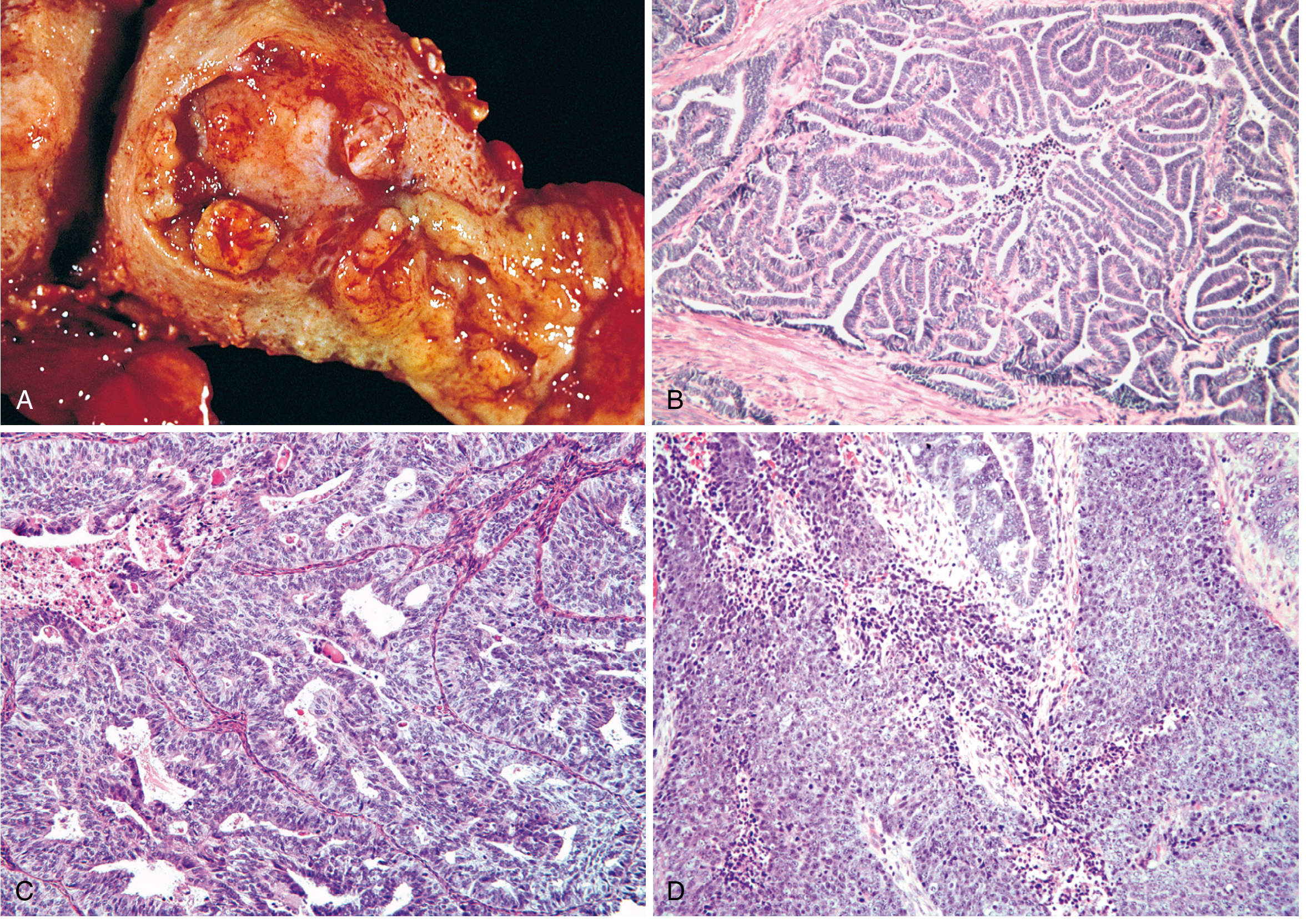
| Grade | Histologic Features |
|---|---|
| Grade 1 (well-differentiated) | >95% glandular; closely packed glands mimicking proliferative endometrium; desmoplastic stroma distinguishes from hyperplasia |
| Grade 2 (moderately differentiated) | Well-formed glands + solid sheets comprising ≤50% of tumor |
| Grade 3 (poorly differentiated) | Solid growth >50%; marked nuclear atypia |
- Up to 20% of endometrioid carcinomas have foci of squamous differentiation (graded on glandular component only)
- Tumors with POLE mutations or MMR deficiency show large numbers of tumor-infiltrating T cells (representing host response to neoantigens)
- A binary system (low grade = G1+G2; high grade = G3) is increasingly preferred
Histopathology - Serous Carcinoma
- Papillary growth pattern with fibrovascular cores; OR predominantly glandular (distinguished from endometrioid by marked cytologic atypia)
- Cells: high nuclear-to-cytoplasmic ratio, hyperchromasia, prominent nucleoli, atypical mitotic figures
- Psammoma bodies may be present
- All serous carcinomas are classified as high-grade regardless of architecture
- The precursor EIC shows identical malignant cells confined to surface epithelium over atrophic endometrium
- Despite superficial endometrial involvement, serous carcinoma may have extensive peritoneal disease (spread via tubal regurgitation / exfoliation) - >1 in 3 patients have extrauterine spread even with minimal myometrial invasion
V. Clinical Features
- Peak incidence: postmenopausal women aged 55-65 years; incidence in younger women is rising
- Usual presentation: irregular or postmenopausal vaginal bleeding (allows early detection)
- Currently no screening test available; diagnosis by endometrial biopsy or curettage
- Transvaginal ultrasound: endometrial stripe ≥5 mm in postmenopausal woman with bleeding warrants sampling
- Lynch syndrome screening: All endometrioid carcinomas are tested for MMR defects at diagnosis; approximately 3-5% of endometrial cancer patients have Lynch syndrome (high risk for colon cancer)
- Stage I - confined to uterine corpus
- Stage II - cervical stromal invasion
- Stage III - local/regional spread (adnexa, vagina, lymph nodes)
- Stage IV - bladder/bowel involvement or distant metastases
- Stage is the major determinant of survival
- Type I: 70% confined to uterus at diagnosis; 5-year survival >85%
- Type II (serous): aggressive course; frequently presents with extrauterine spread; overall survival ~20%
- POLE-mutated tumors: best prognosis even when high-grade
- CN-high (serous-like): worst prognosis
Summary Table: Key Molecular Alterations
| Alteration | Type I (Endometrioid) | Type II (Serous) |
|---|---|---|
| PTEN | 30-83% | Rare |
| PIK3CA | ~40% | Less common |
| KRAS | 10-25% | Rare |
| ARID1A | ~33% | Rare |
| CTNNB1 | Present | Rare |
| TP53 | Rare | >90% |
| MMR defects/MSI | ~20% | <5% |
| POLE | <10% | Very rare |
| Copy number alterations | Few SCNAs | Extensive SCNAs |
| HER2/neu | Less common | More frequent |
- Robbins, Cotran & Kumar, Pathologic Basis of Disease, pp. 933-934
- Robbins & Kumar, Basic Pathology, pp. 718
- Berek & Novak's Gynecology, pp. 2172-2174
- Jamieson & McAlpine, "Molecular Profiling of Endometrial Cancer From TCGA to Clinical Practice," JNCCN 2023 (PMID: 36791751)
- Moving into the modern era of molecular classification for endometrial cancer, J Surg Oncol 2024 (PMID: 38100711)