
Tell me the differential diagnosis of a patient with lung cancer. Also give me some images of type 2 diabetes and what are the recent developments in this field.
type 2 diabetes mellitus pathophysiology
lung cancer types histology CT scan
Differential Diagnosis of Lung Cancer
Primary Malignant Causes
| Category | Conditions |
|---|---|
| Primary lung malignancies | Non-small cell lung cancer (adenocarcinoma, squamous cell carcinoma, large cell carcinoma), Small cell lung cancer (SCLC) |
| Pulmonary carcinoid tumors | Typical and atypical carcinoid (neuroendocrine neoplasms; distinguished from SCLC by Ki-67 immunoreactivity and histology) |
| Rare pulmonary malignancies | Pulmonary sarcoma, pleural mesothelioma, primary pulmonary lymphoma |
Metastatic Disease
Benign Pulmonary Masses / Mimics
| Condition | Key Features |
|---|---|
| Respiratory tract papillomatosis | HPV-related (especially type 11); can undergo malignant transformation to squamous cell carcinoma; difficult to distinguish from lung cancer on imaging; FDG-PET may not be useful due to mild hypermetabolism — Murray & Nadel's Textbook of Respiratory Medicine |
| Rounded atelectasis | CT is particularly useful; differential from lung cancer may require tissue biopsy — Murray & Nadel's Textbook of Respiratory Medicine |
| Pulmonary Langerhans cell histiocytosis (PLCH) | Bronchiolocentric stellate nodules that can cavitate and mimic malignancy; CD1a-positive histiocytes; BRAF mutations; strongly associated with smoking |
| Pulmonary carcinoid | Neuroendocrine origin; low mitotic rate; distinguished from SCLC by neuroendocrine markers and Ki-67 |
| Hamartoma | Most common benign lung tumor; "popcorn" calcification on CT; typically incidental finding |
| Granulomatous disease | Tuberculosis, histoplasmosis, coccidioidomycosis, sarcoidosis — can all produce nodules or masses indistinguishable from cancer on imaging |
| Lung abscess | Cavitary lesion; fever, purulent sputum, leukocytosis |
| Aspergilloma | "Crescent sign" on CT within a pre-existing cavity |
Pleural Disease
- Mesothelioma — asbestos-related; pleural plaques on imaging raise suspicion
- Benign asbestos-related pleural fibrosis — long latency (median ~34 years); can mimic malignancy — Murray & Nadel's Textbook of Respiratory Medicine
Key Distinguishing Investigations
- CT chest/abdomen/pelvis — morphology, nodule characteristics (spiculation, ground glass, calcification)
- PET-CT — metabolic activity; helps stage and identify distant disease
- Bronchoscopy with BAL/biopsy — central lesions
- CT-guided percutaneous biopsy — peripheral lesions
- Immunohistochemistry (IHC) — essential in distinguishing SCLC from carcinoid (TTF-1, Ki-67, synaptophysin, chromogranin). TTF-1 is negative in Merkel cell carcinoma (MCC) and positive in SCLC, making it a critical marker in the differential — Fitzpatrick's Dermatology
Lung Cancer CT Scan Images
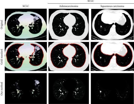
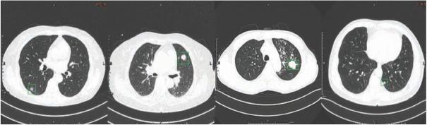
Type 2 Diabetes Mellitus (T2DM)
Images
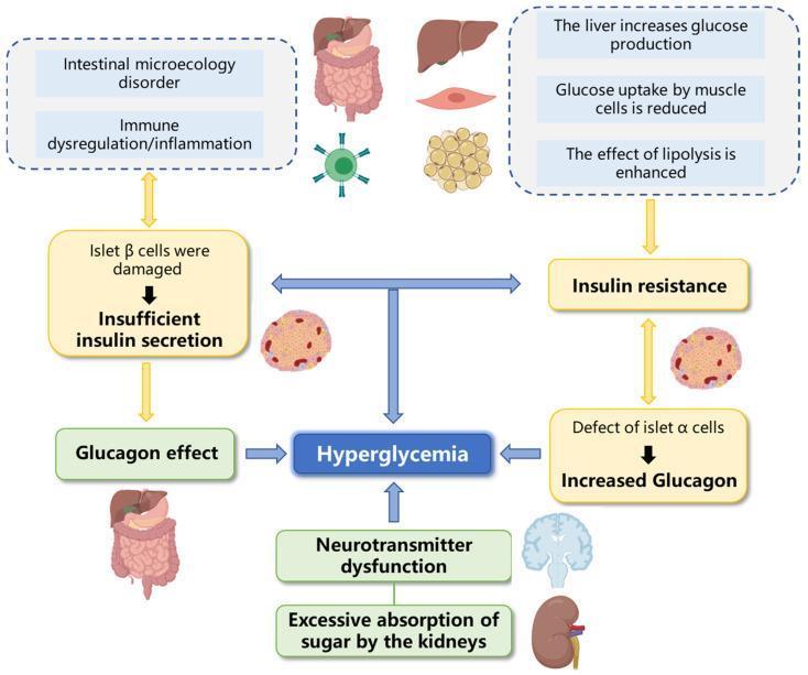
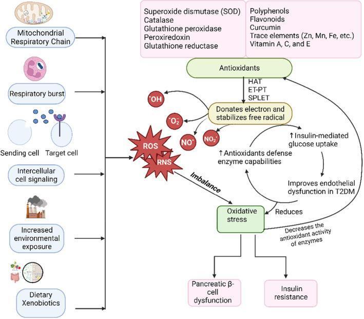
Recent Developments in Type 2 Diabetes
1. Pathogenesis — The "Ominous Octet" Extended
- Gut microbiome dysregulation — intestinal microecology disorder disrupts immune signaling, contributing to β-cell damage
- Incretin deficiency — reduced GLP-1 and GIP secretion/response
- α-cell hypersecretion of glucagon — excess hepatic glucose production
- Renal glucose reabsorption — kidneys reabsorb excess glucose, perpetuating hyperglycemia
- CNS neurotransmitter dysfunction — aberrant dopaminergic and hypothalamic signaling
2. Pharmacological Advances
| Drug Class | Mechanism | Key Benefits |
|---|---|---|
| GLP-1 Receptor Agonists (semaglutide, liraglutide, tirzepatide) | Enhance glucose-dependent insulin secretion, suppress glucagon, delay gastric emptying | Weight loss, CV risk reduction, now approved for obesity independently |
| SGLT-2 Inhibitors (empagliflozin, dapagliflozin, canagliflozin) | Block renal glucose reabsorption → glycosuria | CV and renal protection independent of glucose lowering; now indicated in heart failure and CKD without diabetes |
| Dual GIP/GLP-1 agonists (tirzepatide) | Target both incretin receptors | Superior HbA1c reduction and weight loss vs GLP-1 alone |
| Oral semaglutide | First oral GLP-1 RA | Improved adherence for injection-averse patients |
3. Technology & Monitoring
- Continuous Glucose Monitors (CGMs) — real-time interstitial glucose; now standard of care for insulin-requiring patients; reduces time-in-hypoglycemia
- Closed-loop insulin delivery ("artificial pancreas") — CGM + automated insulin pump; reduces HbA1c without hypoglycemia burden
- Structured SMBG — pairing pre- and post-meal readings (Δ values of 0–50 mg/dL are physiologically normal) guides meal and medication adjustments — Textbook of Family Medicine 9e
4. Lifestyle & Prevention
- Physical activity reduces T2DM risk by 58% in high-risk individuals; GLUT4-mediated glucose uptake in skeletal muscle is impaired at rest but enhanced during muscle contraction
- Reducing caloric intake to 1,100 kcal/day can lower fasting blood glucose in as few as 4 days in obese patients
- ADA nutrition guidelines have replaced the concept of a single "diabetic diet" with individualized healthful eating patterns — Textbook of Family Medicine 9e
5. Cardiovascular & Renal Focus
6. Epigenetic & Genetic Research
make a ppt for this
ls -la /home/daytona/workspace/ 2>/dev/null || echo "workspace not found"
mkdir -p /home/daytona/workspace/medical-ppt && node -e "require('pptxgenjs'); console.log('pptxgenjs OK')"
node /home/daytona/skills/shared/scripts/fetch_images.js \ "https://cdn.orris.care/cdss_images/pmc_clinical_VQA_645087a39ddc8e0c09d514625fb875d0dce13b5e9f6a5d4f7ae812078d3a340b.jpg" \ "https://cdn.orris.care/cdss_images/pmc_clinical_VQA_b8242338e17ddf636825e53ef568fb333eb48cfd0d07dc855cb1c4736c32e1cf.jpg" \ "https://cdn.orris.care/cdss_images/pmc_clinical_VQA_09c4761b1c4dcfc1ed149f3f7dcb1e685afc10b3eb50c0f77460459e58eecf50.jpg" \ "https://cdn.orris.care/cdss_images/pmc_clinical_VQA_ae2e86ab28b9911e764c9e98d550869391806a4db3dc400ac98a871e0d900eb0.jpg" \ 2>&1 | head -5
i dont see any ppt
~/medical-ppt/create_ppt.js
const pptxgen = require("pptxgenjs");
const { execSync } = require("child_process");

// Fetch images
const urls = [
  "https://cdn.orris.care/cdss_images/pmc_clinical_VQA_645087a39ddc8e0c09d514625fb875d0dce13b5e9f6a5d4f7ae812078d3a340b.jpg",
  "https://cdn.orris.care/cdss_images/pmc_clinical_VQA_b8242338e17ddf636825e53ef568fb333eb48cfd0d07dc855cb1c4736c32e1cf.jpg",
  "https://cdn.orris.care/cdss_images/pmc_clinical_VQA_09c4761b1c4dcfc1ed149f3f7dcb1e685afc10b3eb50c0f77460459e58eecf50.jpg",
  "https://cdn.orris.care/cdss_images/pmc_clinical_VQA_ae2e86ab28b9911e764c9e98d550869391806a4db3dc400ac98a871e0d900eb0.jpg"
];

const imgData = JSON.parse(
  execSync(`node /home/daytona/skills/shared/scripts/fetch_images.js ${urls.map(u => `"${u}"`).join(" ")}`).toString()
);

const pres = new pptxgen();
pres.layout = "LAYOUT_16x9";
pres.title = "Lung Cancer & Type 2 Diabetes";

// ─── COLOR PALETTE ─────────────────────────────────────────────────────
const DARK_BG   = "0D1B2A";  // deep navy
const ACCENT1   = "1E88E5";  // bright blue
const ACCENT2   = "00BFA5";  // teal
const ACCENT3   = "E53935";  // red (lung cancer accent)
const ACCENT4   = "F9A825";  // amber (diabetes accent)
const TEXT_LITE = "E3ECFA";  // light text
const TEXT_DIM  = "90A4BC";  // dimmer text
const CARD_BG   = "132238";  // card dark
const CARD_BG2  = "132830";  // card slightly teal-tinted

// ─── HELPER: slide background ─────────────────────────────────────────
function setBg(slide) {
  slide.background = { color: DARK_BG };
}

// ─── HELPER: section header bar ───────────────────────────────────────
function addHeaderBar(slide, label, color) {
  slide.addShape(pres.shapes.RECTANGLE, { x: 0, y: 0, w: "100%", h: 0.75, fill: { color: color }, line: { color: color } });
  slide.addText(label, { x: 0.3, y: 0, w: 9.4, h: 0.75, fontSize: 22, bold: true, color: "FFFFFF", valign: "middle", margin: 0 });
}

// ─── HELPER: page number ──────────────────────────────────────────────
function addSlideNum(slide, n) {
  slide.addText(`${n}`, { x: 9.4, y: 5.3, w: 0.4, h: 0.3, fontSize: 10, color: TEXT_DIM, align: "right" });
}

// ══════════════════════════════════════════════════════════════════════
// SLIDE 1 — TITLE
// ══════════════════════════════════════════════════════════════════════
let s = pres.addSlide();
setBg(s);
// top accent bar
s.addShape(pres.shapes.RECTANGLE, { x: 0, y: 0, w: "100%", h: 0.08, fill: { color: ACCENT1 }, line: { color: ACCENT1 } });
// bottom accent bar
s.addShape(pres.shapes.RECTANGLE, { x: 0, y: 5.54, w: "100%", h: 0.08, fill: { color: ACCENT2 }, line: { color: ACCENT2 } });

// vertical accent stripe
s.addShape(pres.shapes.RECTANGLE, { x: 0.4, y: 1.0, w: 0.06, h: 3.5, fill: { color: ACCENT1 }, line: { color: ACCENT1 } });

s.addText("LUNG CANCER", { x: 0.65, y: 1.05, w: 8.8, h: 0.95, fontSize: 42, bold: true, color: ACCENT3, margin: 0 });
s.addText("& TYPE 2 DIABETES MELLITUS", { x: 0.65, y: 1.9, w: 8.8, h: 0.9, fontSize: 28, bold: false, color: TEXT_LITE, margin: 0 });
s.addShape(pres.shapes.RECTANGLE, { x: 0.65, y: 2.9, w: 4.5, h: 0.04, fill: { color: ACCENT2 }, line: { color: ACCENT2 } });
s.addText("Differential Diagnosis  •  Imaging  •  Pathophysiology  •  Recent Advances", {
  x: 0.65, y: 3.1, w: 8.8, h: 0.5, fontSize: 14, color: TEXT_DIM, italic: true, margin: 0
});
s.addText("Sources: Murray & Nadel's Respiratory Medicine | Textbook of Family Medicine 9e | Fishman's Pulmonary Diseases", {
  x: 0.65, y: 4.8, w: 8.8, h: 0.4, fontSize: 9, color: TEXT_DIM, margin: 0
});

// ══════════════════════════════════════════════════════════════════════
// SLIDE 2 — LUNG CANCER: OVERVIEW
// ══════════════════════════════════════════════════════════════════════
s = pres.addSlide();
setBg(s);
addHeaderBar(s, "Lung Cancer — Overview", ACCENT3);
addSlideNum(s, 2);

const overviewPoints = [
  "Most common cause of a pulmonary mass requiring differential diagnosis workup",
  "Major types: Non-small cell (NSCLC) — Adenocarcinoma, Squamous Cell, Large Cell",
  "Small cell lung cancer (SCLC) — aggressive, central, paraneoplastic syndromes",
  "Key risk factors: smoking, asbestos, radon, HPV (papillomatosis transformation)",
  "Workup: CT chest, PET-CT, bronchoscopy/biopsy, IHC (TTF-1, Ki-67, synaptophysin)",
];

overviewPoints.forEach((pt, i) => {
  s.addShape(pres.shapes.RECTANGLE, { x: 0.3, y: 0.95 + i * 0.87, w: 9.3, h: 0.72, fill: { color: CARD_BG }, line: { color: "1E3A5F", pt: 1 } });
  s.addShape(pres.shapes.RECTANGLE, { x: 0.3, y: 0.95 + i * 0.87, w: 0.06, h: 0.72, fill: { color: ACCENT3 }, line: { color: ACCENT3 } });
  s.addText(pt, { x: 0.55, y: 0.97 + i * 0.87, w: 9.0, h: 0.68, fontSize: 12, color: TEXT_LITE, valign: "middle", margin: 0 });
});

// ══════════════════════════════════════════════════════════════════════
// SLIDE 3 — DIFFERENTIAL DIAGNOSIS: MALIGNANT
// ══════════════════════════════════════════════════════════════════════
s = pres.addSlide();
setBg(s);
addHeaderBar(s, "Differential Diagnosis — Malignant Causes", ACCENT3);
addSlideNum(s, 3);

const malCols = [
  { title: "Primary Lung Malignancies", items: ["Adenocarcinoma (peripheral, GGO)", "Squamous Cell Carcinoma (central, cavitating)", "Large Cell Carcinoma", "Small Cell Lung Cancer (SCLC)"] },
  { title: "Metastatic Disease", items: ["Breast, Colorectal, Renal Cell", "Melanoma, Thyroid", "Multiple nodules → metastatic pattern", "Prior cancer history is key clue"] },
  { title: "Rare Primary Tumors", items: ["Pulmonary Carcinoid (neuroendocrine)", "Typical vs Atypical Carcinoid", "Primary Pulmonary Lymphoma", "Pleural Mesothelioma (asbestos)"] },
];

malCols.forEach((col, ci) => {
  const cx = 0.25 + ci * 3.3;
  s.addShape(pres.shapes.RECTANGLE, { x: cx, y: 0.9, w: 3.1, h: 4.5, fill: { color: CARD_BG }, line: { color: "1E3A5F", pt: 1 } });
  s.addShape(pres.shapes.RECTANGLE, { x: cx, y: 0.9, w: 3.1, h: 0.45, fill: { color: ACCENT3 }, line: { color: ACCENT3 } });
  s.addText(col.title, { x: cx + 0.08, y: 0.9, w: 2.9, h: 0.45, fontSize: 11, bold: true, color: "FFFFFF", valign: "middle", margin: 0 });
  col.items.forEach((item, ii) => {
    s.addText("•  " + item, { x: cx + 0.12, y: 1.45 + ii * 0.72, w: 2.85, h: 0.65, fontSize: 10.5, color: TEXT_LITE, valign: "top", margin: 0 });
  });
});

// ══════════════════════════════════════════════════════════════════════
// SLIDE 4 — DIFFERENTIAL DIAGNOSIS: BENIGN / MIMICS
// ══════════════════════════════════════════════════════════════════════
s = pres.addSlide();
setBg(s);
addHeaderBar(s, "Differential Diagnosis — Benign Mimics", ACCENT1);
addSlideNum(s, 4);

const benignRows = [
  ["Rounded Atelectasis", "CT shows pleural lesion relationship; may need biopsy to exclude cancer"],
  ["Respiratory Papillomatosis", "HPV type 11; malignant transformation to SCC; FDG-PET mildly positive"],
  ["Pulmonary Langerhans Cell Histiocytosis", "Smoking-related; stellate nodules → cavitation; CD1a+, BRAF mutations"],
  ["Hamartoma", "Most common benign tumor; 'popcorn' calcification on CT; incidental"],
  ["Granulomatous Disease", "TB, Histoplasmosis, Sarcoidosis — indistinguishable on imaging alone"],
  ["Lung Abscess", "Cavitary lesion; fever, leukocytosis, purulent sputum"],
  ["Asbestos-related Fibrosis", "Long latency ~34 yrs; pleural plaques; may require biopsy"],
];

benignRows.forEach((row, i) => {
  const y = 0.85 + i * 0.67;
  s.addShape(pres.shapes.RECTANGLE, { x: 0.25, y, w: 9.4, h: 0.6, fill: { color: i % 2 === 0 ? CARD_BG : "0F2030" }, line: { color: "1E3A5F", pt: 0.5 } });
  s.addText(row[0], { x: 0.35, y: y + 0.05, w: 2.9, h: 0.5, fontSize: 10.5, bold: true, color: ACCENT2, valign: "middle", margin: 0 });
  s.addText(row[1], { x: 3.3, y: y + 0.05, w: 6.2, h: 0.5, fontSize: 10, color: TEXT_LITE, valign: "middle", margin: 0 });
});

// ══════════════════════════════════════════════════════════════════════
// SLIDE 5 — LUNG CANCER CT IMAGING (with real images)
// ══════════════════════════════════════════════════════════════════════
s = pres.addSlide();
setBg(s);
addHeaderBar(s, "Lung Cancer — CT Imaging Examples", ACCENT1);
addSlideNum(s, 5);

// Image 1 — 3-type comparison
if (imgData[0] && !imgData[0].error) {
  s.addImage({ data: imgData[0].base64, x: 0.25, y: 0.9, w: 4.5, h: 3.0 });
}
s.addShape(pres.shapes.RECTANGLE, { x: 0.25, y: 3.92, w: 4.5, h: 1.3, fill: { color: CARD_BG }, line: { color: "1E3A5F" } });
s.addText("CT Comparison: SCLC vs Adenocarcinoma vs Squamous Cell Carcinoma", {
  x: 0.35, y: 3.97, w: 4.3, h: 0.45, fontSize: 10, bold: true, color: ACCENT1, margin: 0
});
s.addText("SCLC: large consolidated central mass\nAdenocarcinoma: peripheral rounded nodule\nSquamous: central soft-tissue density near mediastinum", {
  x: 0.35, y: 4.42, w: 4.3, h: 0.75, fontSize: 8.5, color: TEXT_DIM, margin: 0
});

// Image 2 — 4-subtypes
if (imgData[1] && !imgData[1].error) {
  s.addImage({ data: imgData[1].base64, x: 5.1, y: 0.9, w: 4.7, h: 3.0 });
}
s.addShape(pres.shapes.RECTANGLE, { x: 5.1, y: 3.92, w: 4.7, h: 1.3, fill: { color: CARD_BG }, line: { color: "1E3A5F" } });
s.addText("Four Lung Cancer Subtypes on CT (Annotated ROI)", {
  x: 5.2, y: 3.97, w: 4.5, h: 0.45, fontSize: 10, bold: true, color: ACCENT1, margin: 0
});
s.addText("AIS: small well-defined nodule\nSCLC: large homogeneous mass\nSCC: irregular borders\nInvasive Adenocarcinoma: hyperdense irregular nodule", {
  x: 5.2, y: 4.42, w: 4.5, h: 0.75, fontSize: 8.5, color: TEXT_DIM, margin: 0
});

// ══════════════════════════════════════════════════════════════════════
// SLIDE 6 — KEY INVESTIGATIONS
// ══════════════════════════════════════════════════════════════════════
s = pres.addSlide();
setBg(s);
addHeaderBar(s, "Lung Cancer — Key Investigations", ACCENT1);
addSlideNum(s, 6);

const invData = [
  { icon: "CT", label: "CT Chest/Abdomen/Pelvis", desc: "Nodule morphology: spiculation, calcification, ground glass. Guides staging." },
  { icon: "PET", label: "PET-CT Scan", desc: "Metabolic activity (FDG uptake). Identifies distant metastases, guides biopsy." },
  { icon: "BROX", label: "Bronchoscopy + BAL/Biopsy", desc: "Central lesions. Endobronchial ultrasound (EBUS) for mediastinal nodes." },
  { icon: "CTBx", label: "CT-guided Percutaneous Biopsy", desc: "Peripheral lesions. Tissue for histology and molecular markers." },
  { icon: "IHC", label: "Immunohistochemistry (IHC)", desc: "TTF-1 (SCLC+, MCC−), Ki-67, synaptophysin, chromogranin, CK. Distinguishes subtypes." },
  { icon: "MOL", label: "Molecular Testing", desc: "EGFR, ALK, ROS1, KRAS, PD-L1 — guides targeted therapy & immunotherapy." },
];

invData.forEach((inv, i) => {
  const col = i % 2;
  const row = Math.floor(i / 2);
  const x = 0.25 + col * 4.85;
  const y = 0.9 + row * 1.5;
  s.addShape(pres.shapes.RECTANGLE, { x, y, w: 4.6, h: 1.35, fill: { color: CARD_BG }, line: { color: "1E3A5F", pt: 1 } });
  s.addShape(pres.shapes.RECTANGLE, { x, y, w: 0.06, h: 1.35, fill: { color: ACCENT1 }, line: { color: ACCENT1 } });
  s.addText(inv.label, { x: x + 0.18, y: y + 0.1, w: 4.3, h: 0.4, fontSize: 11, bold: true, color: ACCENT2, margin: 0 });
  s.addText(inv.desc, { x: x + 0.18, y: y + 0.52, w: 4.3, h: 0.72, fontSize: 9.5, color: TEXT_LITE, margin: 0 });
});

// ══════════════════════════════════════════════════════════════════════
// SLIDE 7 — T2DM: OVERVIEW & PATHOGENESIS
// ══════════════════════════════════════════════════════════════════════
s = pres.addSlide();
setBg(s);
addHeaderBar(s, "Type 2 Diabetes Mellitus — Overview & Pathogenesis", ACCENT4);
addSlideNum(s, 7);

// Left column — pathogenesis
s.addShape(pres.shapes.RECTANGLE, { x: 0.25, y: 0.9, w: 4.55, h: 4.5, fill: { color: CARD_BG }, line: { color: "2A3A10", pt: 1 } });
s.addShape(pres.shapes.RECTANGLE, { x: 0.25, y: 0.9, w: 4.55, h: 0.45, fill: { color: ACCENT4 }, line: { color: ACCENT4 } });
s.addText("Pathogenesis — The Multi-Organ Failure", { x: 0.35, y: 0.9, w: 4.35, h: 0.45, fontSize: 11, bold: true, color: "000000", valign: "middle", margin: 0 });

const pathItems = [
  "β-cell dysfunction → progressive apoptosis",
  "Insulin resistance (liver, muscle, adipose)",
  "α-cell hypersecretion of glucagon",
  "Gut microbiome dysregulation",
  "Renal glucose reabsorption excess",
  "CNS neurotransmitter dysfunction",
  "18 GWAS polymorphisms identified",
  "GLUT4 impaired at rest, improved with exercise",
];
pathItems.forEach((pt, i) => {
  s.addText("• " + pt, { x: 0.4, y: 1.45 + i * 0.47, w: 4.3, h: 0.42, fontSize: 10, color: TEXT_LITE, margin: 0 });
});

// Right column — diagnostic criteria
s.addShape(pres.shapes.RECTANGLE, { x: 5.05, y: 0.9, w: 4.7, h: 4.5, fill: { color: CARD_BG2 }, line: { color: "1E3A30", pt: 1 } });
s.addShape(pres.shapes.RECTANGLE, { x: 5.05, y: 0.9, w: 4.7, h: 0.45, fill: { color: ACCENT2 }, line: { color: ACCENT2 } });
s.addText("Diagnostic Criteria & Key Facts", { x: 5.15, y: 0.9, w: 4.5, h: 0.45, fontSize: 11, bold: true, color: "000000", valign: "middle", margin: 0 });

const diagItems = [
  ["FPG", "≥ 126 mg/dL (7.0 mmol/L)"],
  ["2-hr OGTT", "≥ 200 mg/dL"],
  ["HbA1c", "≥ 6.5%"],
  ["Prediabetes", "80% β-cell function lost at 2-hr glucose 140–180"],
  ["Retinopathy", "18% of prediabetes patients already affected"],
  ["Normal Δ", "0–50 mg/dL post-prandial rise"],
  ["Lifestyle Rx", "58% risk reduction with diet + exercise"],
  ["Caloric Rx", "1,100 kcal/day → glucose drop in 4 days"],
];
diagItems.forEach((row, i) => {
  const y2 = 1.45 + i * 0.47;
  s.addText(row[0], { x: 5.15, y: y2, w: 1.1, h: 0.42, fontSize: 10, bold: true, color: ACCENT4, margin: 0 });
  s.addText(row[1], { x: 6.3, y: y2, w: 3.35, h: 0.42, fontSize: 10, color: TEXT_LITE, margin: 0 });
});

// ══════════════════════════════════════════════════════════════════════
// SLIDE 8 — T2DM PATHOPHYSIOLOGY DIAGRAMS
// ══════════════════════════════════════════════════════════════════════
s = pres.addSlide();
setBg(s);
addHeaderBar(s, "Type 2 Diabetes — Pathophysiology Diagrams", ACCENT4);
addSlideNum(s, 8);

// Image 3 — multi-organ hyperglycemia
if (imgData[2] && !imgData[2].error) {
  s.addImage({ data: imgData[2].base64, x: 0.25, y: 0.9, w: 4.6, h: 3.2 });
}
s.addShape(pres.shapes.RECTANGLE, { x: 0.25, y: 4.12, w: 4.6, h: 1.1, fill: { color: CARD_BG }, line: { color: "2A3A10" } });
s.addText("Multi-Organ Hyperglycemia", { x: 0.35, y: 4.17, w: 4.4, h: 0.38, fontSize: 10, bold: true, color: ACCENT4, margin: 0 });
s.addText("β-cell dysfunction, insulin resistance (liver/muscle/adipose), elevated glucagon, gut microbiome disorder, renal reabsorption", {
  x: 0.35, y: 4.55, w: 4.4, h: 0.6, fontSize: 8.5, color: TEXT_DIM, margin: 0
});

// Image 4 — endothelial dysfunction + CV risk
if (imgData[3] && !imgData[3].error) {
  s.addImage({ data: imgData[3].base64, x: 5.1, y: 0.9, w: 4.65, h: 3.2 });
}
s.addShape(pres.shapes.RECTANGLE, { x: 5.1, y: 4.12, w: 4.65, h: 1.1, fill: { color: CARD_BG }, line: { color: "2A3A10" } });
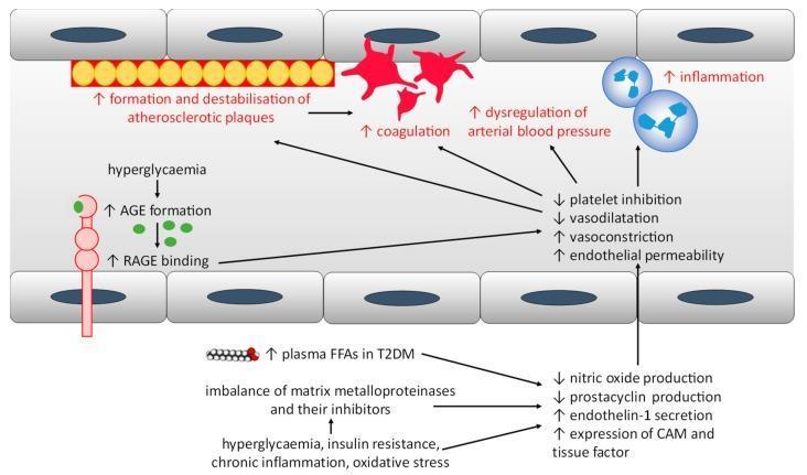
s.addText("Endothelial Dysfunction & CV Risk", { x: 5.2, y: 4.17, w: 4.45, h: 0.38, fontSize: 10, bold: true, color: ACCENT4, margin: 0 });
s.addText("AGE-RAGE signalling, oxidative stress, ↓NO & prostacyclin, ↑ET-1 & CAMs → atherosclerosis, coagulation, hypertension", {
  x: 5.2, y: 4.55, w: 4.45, h: 0.6, fontSize: 8.5, color: TEXT_DIM, margin: 0
});

// ══════════════════════════════════════════════════════════════════════
// SLIDE 9 — T2DM: PHARMACOLOGICAL ADVANCES
// ══════════════════════════════════════════════════════════════════════
s = pres.addSlide();
setBg(s);
addHeaderBar(s, "Type 2 Diabetes — Pharmacological Advances", ACCENT4);
addSlideNum(s, 9);

const drugRows = [
  ["GLP-1 Receptor Agonists", "Semaglutide, Liraglutide", "Glucose-dependent insulin secretion, suppress glucagon, delay gastric emptying", "Weight loss, CV risk ↓, approved for obesity"],
  ["SGLT-2 Inhibitors", "Empagliflozin, Dapagliflozin", "Block renal glucose reabsorption → glycosuria", "CV & renal protection, HF & CKD indication"],
  ["Dual GIP/GLP-1 Agonists", "Tirzepatide", "Targets both incretin receptors simultaneously", "Superior HbA1c ↓ & weight loss vs GLP-1 alone"],
  ["Oral GLP-1 RA", "Oral Semaglutide", "First oral GLP-1 receptor agonist", "Improved adherence for injection-averse patients"],
];

// Header row
const hCols = [1.2, 2.15, 3.5, 2.45];
const hLabels = ["Drug Class", "Agent", "Mechanism", "Key Benefit"];
let hx = 0.25;
hLabels.forEach((hl, hi) => {
  s.addShape(pres.shapes.RECTANGLE, { x: hx, y: 0.85, w: hCols[hi], h: 0.42, fill: { color: ACCENT4 }, line: { color: ACCENT4 } });
  s.addText(hl, { x: hx + 0.05, y: 0.85, w: hCols[hi] - 0.1, h: 0.42, fontSize: 10, bold: true, color: "000000", valign: "middle", margin: 0 });
  hx += hCols[hi] + 0.05;
});

drugRows.forEach((row, ri) => {
  let rx = 0.25;
  const ry = 1.32 + ri * 1.0;
  const rowColor = ri % 2 === 0 ? CARD_BG : "0F2030";
  row.forEach((cell, ci) => {
    s.addShape(pres.shapes.RECTANGLE, { x: rx, y: ry, w: hCols[ci], h: 0.92, fill: { color: rowColor }, line: { color: "1E3A5F", pt: 0.5 } });
    s.addText(cell, { x: rx + 0.07, y: ry + 0.05, w: hCols[ci] - 0.12, h: 0.82, fontSize: 9.5, color: ci === 0 ? ACCENT2 : TEXT_LITE, valign: "middle", margin: 0 });
    rx += hCols[ci] + 0.05;
  });
});

// ══════════════════════════════════════════════════════════════════════
// SLIDE 10 — T2DM: TECHNOLOGY & CARDIORENAL FOCUS
// ══════════════════════════════════════════════════════════════════════
s = pres.addSlide();
setBg(s);
addHeaderBar(s, "Type 2 Diabetes — Technology & Cardiorenal Focus", ACCENT2);
addSlideNum(s, 10);

// Left — Technology
s.addShape(pres.shapes.RECTANGLE, { x: 0.25, y: 0.9, w: 4.55, h: 4.5, fill: { color: CARD_BG2 }, line: { color: "1E3A30", pt: 1 } });
s.addShape(pres.shapes.RECTANGLE, { x: 0.25, y: 0.9, w: 4.55, h: 0.45, fill: { color: ACCENT2 }, line: { color: ACCENT2 } });
s.addText("Technology & Monitoring", { x: 0.35, y: 0.9, w: 4.35, h: 0.45, fontSize: 11, bold: true, color: "000000", valign: "middle", margin: 0 });

const techItems = [
  ["CGM", "Continuous Glucose Monitors — real-time interstitial glucose, reduces hypoglycemia"],
  ["Closed-loop", "'Artificial Pancreas' — CGM + automated insulin pump, reduces HbA1c without hypoglycemia"],
  ["Structured SMBG", "Pre/post-meal paired testing; Δ = 0–50 mg/dL is physiologically normal"],
  ["AI/ML", "AI-assisted dosing algorithms, pattern recognition, personalised management"],
];
techItems.forEach((item, i) => {
  s.addText(item[0], { x: 0.4, y: 1.48 + i * 0.9, w: 1.0, h: 0.38, fontSize: 10, bold: true, color: ACCENT4, margin: 0 });
  s.addText(item[1], { x: 0.4, y: 1.86 + i * 0.9, w: 4.25, h: 0.45, fontSize: 9.5, color: TEXT_LITE, margin: 0 });
});

// Right — Cardiorenal
s.addShape(pres.shapes.RECTANGLE, { x: 5.05, y: 0.9, w: 4.7, h: 4.5, fill: { color: CARD_BG }, line: { color: "1E3A5F", pt: 1 } });
s.addShape(pres.shapes.RECTANGLE, { x: 5.05, y: 0.9, w: 4.7, h: 0.45, fill: { color: ACCENT1 }, line: { color: ACCENT1 } });
s.addText("Cardiorenal Outcomes Trials", { x: 5.15, y: 0.9, w: 4.5, h: 0.45, fontSize: 11, bold: true, color: "FFFFFF", valign: "middle", margin: 0 });

const trialItems = [
  ["EMPA-REG OUTCOME", "Empagliflozin → CV death ↓ 38%, HF hospitalisation ↓ 35%"],
  ["LEADER", "Liraglutide → MACE ↓ 13%, CV death ↓ 22%"],
  ["SUSTAIN-6", "Semaglutide → MACE ↓ 26%"],
  ["DAPA-HF", "Dapagliflozin → HF benefit regardless of T2DM status"],
  ["Guideline Shift", "SGLT-2i or GLP-1 RA first-line in ASCVD, HF, CKD — independent of HbA1c"],
];
trialItems.forEach((item, i) => {
  s.addText(item[0], { x: 5.2, y: 1.48 + i * 0.73, w: 1.9, h: 0.35, fontSize: 9.5, bold: true, color: ACCENT2, margin: 0 });
  s.addText(item[1], { x: 5.2, y: 1.82 + i * 0.73, w: 4.4, h: 0.35, fontSize: 9, color: TEXT_LITE, margin: 0 });
});

// ══════════════════════════════════════════════════════════════════════
// SLIDE 11 — T2DM: GENETIC & EPIGENETIC ADVANCES
// ══════════════════════════════════════════════════════════════════════
s = pres.addSlide();
setBg(s);
addHeaderBar(s, "Type 2 Diabetes — Genetic & Epigenetic Research", ACCENT4);
addSlideNum(s, 11);

const genCards = [
  { title: "GWAS Discoveries", color: ACCENT4, pts: ["18 polymorphisms identified that increase T2DM susceptibility", "Activation of alleles → β-cell apoptosis", "Satiety reduction, increased appetite, reduced energy expenditure"] },
  { title: "Epigenetic Mechanisms", color: ACCENT1, pts: ["DNA methylation changes with aging", "Upregulates chronic inflammation in older individuals", "Circadian misalignment disrupts glucose-insulin metabolism"] },
  { title: "Environmental Triggers", color: ACCENT2, pts: ["Arsenic, dioxins, hexachlorobenzene → β-cell toxic", "PFOA (Teflon) found in 98% of US population's blood", "Second-hand smoke → 18% higher T2DM risk in women"] },
  { title: "Emerging Targets", color: ACCENT3, pts: ["Gut microbiome modulation (prebiotics, FMT)", "Brown adipose tissue activation", "Hepatic gluconeogenesis suppression via novel targets"] },
];

genCards.forEach((card, ci) => {
  const col = ci % 2;
  const row = Math.floor(ci / 2);
  const cx = 0.25 + col * 4.85;
  const cy = 0.9 + row * 2.25;
  s.addShape(pres.shapes.RECTANGLE, { x: cx, y: cy, w: 4.6, h: 2.1, fill: { color: CARD_BG }, line: { color: "1E3A5F", pt: 1 } });
  s.addShape(pres.shapes.RECTANGLE, { x: cx, y: cy, w: 4.6, h: 0.4, fill: { color: card.color }, line: { color: card.color } });
  s.addText(card.title, { x: cx + 0.1, y: cy, w: 4.4, h: 0.4, fontSize: 11, bold: true, color: "000000", valign: "middle", margin: 0 });
  card.pts.forEach((pt, pi) => {
    s.addText("• " + pt, { x: cx + 0.15, y: cy + 0.48 + pi * 0.52, w: 4.3, h: 0.46, fontSize: 9.5, color: TEXT_LITE, margin: 0 });
  });
});

// ══════════════════════════════════════════════════════════════════════
// SLIDE 12 — SUMMARY
// ══════════════════════════════════════════════════════════════════════
s = pres.addSlide();
setBg(s);
// Split header — two colors
s.addShape(pres.shapes.RECTANGLE, { x: 0, y: 0, w: "50%", h: 0.75, fill: { color: ACCENT3 }, line: { color: ACCENT3 } });
s.addShape(pres.shapes.RECTANGLE, { x: "50%", y: 0, w: "50%", h: 0.75, fill: { color: ACCENT4 }, line: { color: ACCENT4 } });
s.addText("Summary", { x: 0.3, y: 0, w: 9.4, h: 0.75, fontSize: 22, bold: true, color: "FFFFFF", valign: "middle", margin: 0 });
addSlideNum(s, 12);

// Left — Lung Cancer summary
s.addShape(pres.shapes.RECTANGLE, { x: 0.25, y: 0.9, w: 4.55, h: 4.5, fill: { color: CARD_BG }, line: { color: "1E3A5F", pt: 1 } });
s.addShape(pres.shapes.RECTANGLE, { x: 0.25, y: 0.9, w: 4.55, h: 0.42, fill: { color: ACCENT3 }, line: { color: ACCENT3 } });
s.addText("🫁  Lung Cancer Differential", { x: 0.35, y: 0.9, w: 4.35, h: 0.42, fontSize: 12, bold: true, color: "FFFFFF", valign: "middle", margin: 0 });
const lcSum = [
  "NSCLC types: Adenocarcinoma, SCC, Large Cell",
  "SCLC: central, aggressive, paraneoplastic",
  "Metastatic disease: always exclude",
  "Benign mimics: TB, hamartoma, papillomatosis, LCH",
  "IHC: TTF-1, Ki-67, synaptophysin key markers",
  "PET-CT for staging + molecular testing for targeted Rx",
];
lcSum.forEach((pt, i) => {
  s.addText("• " + pt, { x: 0.4, y: 1.42 + i * 0.64, w: 4.3, h: 0.58, fontSize: 9.5, color: TEXT_LITE, margin: 0 });
});

// Right — T2DM summary
s.addShape(pres.shapes.RECTANGLE, { x: 5.05, y: 0.9, w: 4.7, h: 4.5, fill: { color: CARD_BG }, line: { color: "1E3A5F", pt: 1 } });
s.addShape(pres.shapes.RECTANGLE, { x: 5.05, y: 0.9, w: 4.7, h: 0.42, fill: { color: ACCENT4 }, line: { color: ACCENT4 } });
s.addText("🩸  Type 2 Diabetes Mellitus", { x: 5.15, y: 0.9, w: 4.5, h: 0.42, fontSize: 12, bold: true, color: "000000", valign: "middle", margin: 0 });
const t2Sum = [
  "Multi-organ pathogenesis: beyond β-cell + insulin resistance",
  "18 GWAS polymorphisms, epigenetic & environmental triggers",
  "GLP-1 RA & SGLT-2i: cardiorenal-protective paradigm shift",
  "Tirzepatide (dual GIP/GLP-1): superior outcomes",
  "CGM + closed-loop: new standard of monitoring",
  "Guideline: SGLT-2i/GLP-1 RA first-line in ASCVD/HF/CKD",
];
t2Sum.forEach((pt, i) => {
  s.addText("• " + pt, { x: 5.2, y: 1.42 + i * 0.64, w: 4.5, h: 0.58, fontSize: 9.5, color: TEXT_LITE, margin: 0 });
});

// Write file
pres.writeFile({ fileName: "/home/daytona/workspace/medical-ppt/Lung_Cancer_T2DM.pptx" })
  .then(() => console.log("SUCCESS: /home/daytona/workspace/medical-ppt/Lung_Cancer_T2DM.pptx"))
  .catch(e => console.error("ERROR:", e));
cd /home/daytona/workspace/medical-ppt && node create_ppt.js
ls -lh /home/daytona/workspace/medical-ppt/Lung_Cancer_T2DM.pptx
Lung Cancer T2DM
Presentation · PPTX
| Slide | Content |
|---|---|
| 1 | Title slide |
| 2 | Lung Cancer — Overview |
| 3 | Differential Diagnosis: Malignant causes (3-column cards) |
| 4 | Differential Diagnosis: Benign mimics (table format) |
| 5 | CT imaging — 2 real annotated scans (SCLC, Adenocarcinoma, SCC, AIS) |
| 6 | Key Investigations (CT, PET, Biopsy, IHC, Molecular testing) |
| 7 | T2DM Overview & Pathogenesis |
| 8 | T2DM Pathophysiology diagrams — multi-organ hyperglycemia + CV risk |
| 9 | Pharmacological Advances (GLP-1, SGLT-2, Tirzepatide, Oral semaglutide) |
| 10 | Technology (CGM, closed-loop) & Cardiorenal outcomes trials |
| 11 | Genetic & Epigenetic Research |
| 12 | Summary slide |