
A 56 yo female presents with history of fainting two times preceded by sweating in last week. H/0 1 episode of TIA 15 years back. HTN on telmi 40. All labs came out normal
Reading File
Now let me search the medical library for syncope evaluation, TIA workup, and relevant content in parallel.
Finding Sources
Finding Sources
Finding Sources
Reading File
Reading File
Reading File
Finding Sources
Finding Sources
Searching PubMed
syncope evaluation management guidelines
Reading File
Finding Sources
Reading File
Finding Sources
Reading File
Reading File
Loading Image
Reading File
Finding Sources
I now have sufficient information to provide a thorough clinical analysis. Let me compile everything.
Clinical Assessment: 56F with Recurrent Syncope + Prior TIA + HTN
Putting the Picture Together
This patient has:
- Two syncopal episodes in one week, both with a diaphoretic prodrome (sweating before LOC)
- Prior TIA 15 years ago (cerebrovascular risk history)
- Hypertension on Telmisartan 40 mg
- Normal labs
The sweating prodrome is a classic feature of neurocardiogenic (vasovagal) syncope. As Braunwald's Heart Disease notes, vasodepressor/cardioinhibitory syncope "can be preceded by manifestations of autonomic hyperactivity such as nausea, abdominal cramping, diarrhea, sweating, or yawning." Fuster & Hurst's The Heart confirms the three stages: prodrome (nausea, sweating, lightheadedness, visual alterations) → abrupt LOC → rapid recovery without postictal state.
However, at age 56 with HTN and a prior TIA, you cannot accept this as a benign diagnosis without ruling out serious etiologies.
Differential Diagnosis (Ranked by Likelihood in Context)
| Category | Specific Entities | Key Features |
|---|---|---|
| Neurocardiogenic / Vasovagal | Reflex syncope | Sweating prodrome, rapid recovery, normal labs |
| Orthostatic hypotension | Drug-induced (telmisartan), dehydration, autonomic dysfunction | Position-related, HTN medication |
| Cardiac arrhythmia | Sick sinus syndrome, AV block, SVT, VT | Age 56, HTN - structural changes possible |
| Structural cardiac | Aortic stenosis, HCM, cardiomyopathy | Must exclude with echo |
| Metabolic | Hypoglycemia, electrolyte (already excluded by labs) | Normal labs make this less likely |
| Neurological | Seizure (postictal state absent), NOT TIA* | TIA does NOT cause LOC per ACC/AHA guidelines |
Critical point about TIA and syncope: Per Braunwald's Heart Disease: "Transient ischemic attacks that result from carotid disease are NOT accompanied by loss of consciousness." The 2017 ACC/AHA/HRS syncope guidelines and textbook sources consistently state that TIA is NOT a cause of true syncope. Her prior TIA history is a cardiovascular risk marker, not a direct cause of her current episodes. Carotid duplex is not indicated in straightforward syncope evaluation.
Red Flags Present in This Patient (Warrant Urgent Evaluation)
Per Washington Manual (Diagnostic Testing section):
- Age >56 with HTN - structural heart disease possible
- Recurrent episodes (2 in one week) - more ominous than a single event
- Prior cerebrovascular disease - high atherosclerotic burden
Investigations: Step-by-Step
Step 1 - Mandatory (do NOW):
- 12-lead ECG - most important initial test; abnormal in ~50% of syncope; diagnoses arrhythmias, conduction disease, ischemia, long QT, Brugada
- Orthostatic BP measurement (supine, sitting, standing) - drop >20 mmHg systolic or >10 mmHg diastolic = orthostatic hypotension
- Detailed history: triggers, position, duration of LOC, recovery time, witnesses, any palpitations, chest pain, tongue bite, incontinence
Step 2 - Strongly Indicated:
- Echocardiogram - exclude structural disease (AS, HCM, LV dysfunction, pulmonary HTN)
- Holter monitor / 24-48 hr ECG monitoring - capture arrhythmias in recurrent syncope
- Blood glucose at time of event (if possible)
Step 3 - Based on Results:
- If echo normal, ECG normal, no structural disease → Tilt-table testing to confirm neurocardiogenic syncope
- If frequent recurrent unexplained syncope → Implantable loop recorder (high diagnostic yield)
- If palpitations or exertional syncope → Electrophysiology study
What NOT to order (per 2017 ACC/AHA/HRS guidelines - Class III):
- Head CT or brain MRI (unless focal neuro signs or head trauma)
- Carotid duplex (not useful in syncope without focal neuro deficits)
- EEG (unless clinical suspicion of epilepsy)
Algorithm for Evaluation
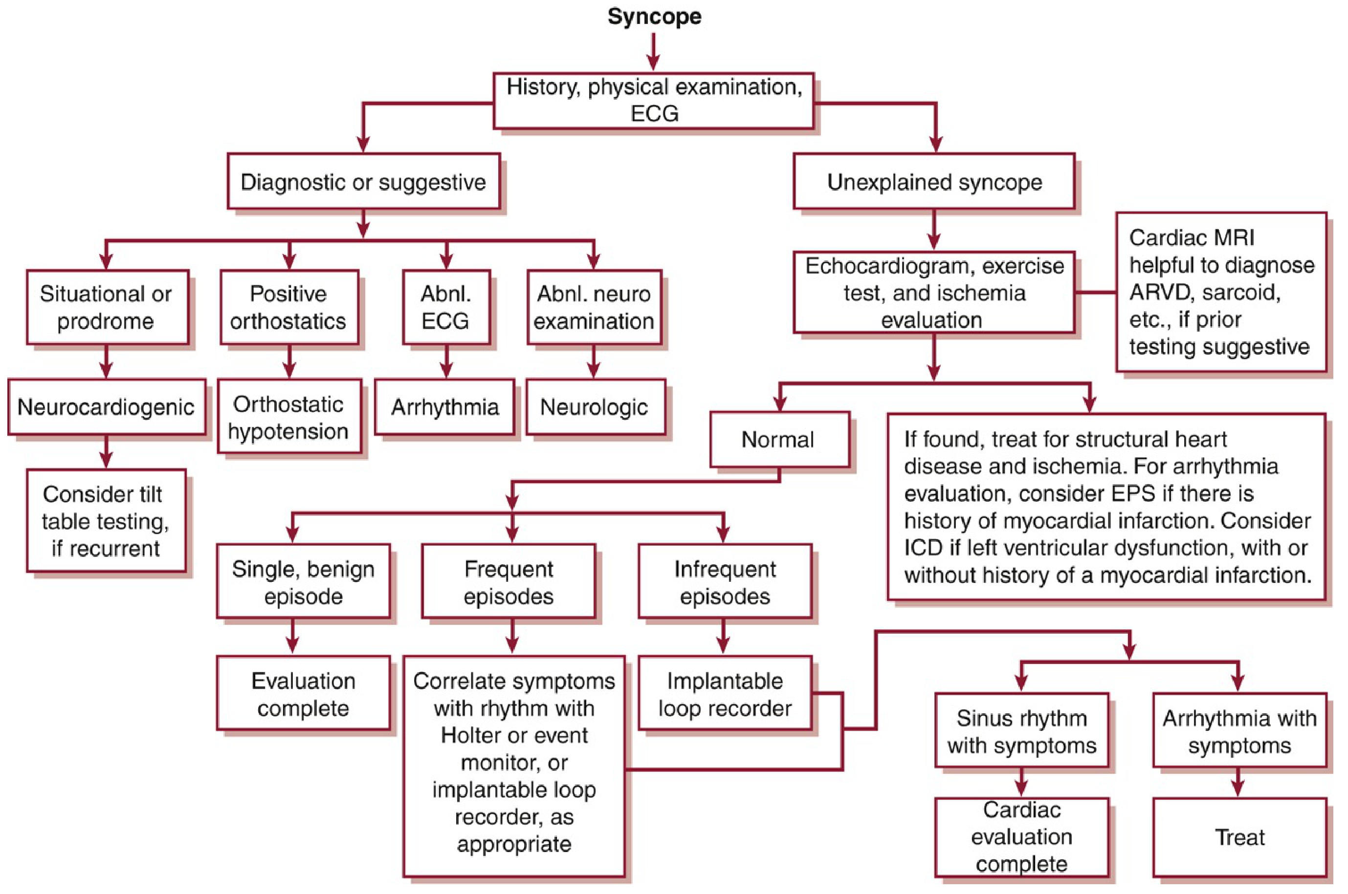
Figure from Washington Manual of Medical Therapeutics - Algorithm for syncope evaluation
Management Direction (Depends on Etiology)
If neurocardiogenic confirmed:
- Educate patient: lie down at prodrome onset, avoid triggers (prolonged standing, hot environments, dehydration)
- Increase fluid/salt intake
- Telmisartan review - ARBs can worsen orthostatic hypotension; consider BP reassessment in the context of syncope
- Physical counterpressure maneuvers (leg crossing, muscle tensing during prodrome)
- Midodrine 5-15 mg TID if recurrent and significant
- Dual-chamber pacemaker only in selected patients with documented cardioinhibitory response
If orthostatic hypotension (telmisartan-related):
- Reduce or time the dose differently; assess need for dosage review
- Compression stockings, adequate hydration
Address cardiovascular risk: Given prior TIA + HTN, ensure antiplatelet therapy, statin, and BP control are optimized to prevent stroke recurrence (independent of the syncope management).
Key Clinical Take-Home Points
- Sweating before syncope = classic vasovagal prodrome - but cannot diagnose by history alone in this age group
- Prior TIA does NOT cause syncope - it is a risk stratifier, not the etiology
- ECG + echo + orthostatic vitals are the non-negotiable first steps
- Telmisartan may be contributing to orthostatic hypotension - review the BP drug
- Two episodes in a week demands expedited cardiac evaluation, not watchful waiting
- Normal labs effectively exclude metabolic causes (hypo/hyperglycemia, electrolyte abnormalities, anemia)
Sources: Braunwald's Heart Disease 2-Vol Set, p. 892-894; Fuster & Hurst's The Heart 15e, p. 1234-1238; Washington Manual of Medical Therapeutics, p. 3117-3155
A 56 yo female presents with history of fainting two times preceded by sweating in last week. H/0 1 episode of TIA 15 years back. HTN on telmi 40. All labs came out normal
Reading File
This is a Clinical Case / Complex Scenario with a Special Population (56F, HTN, prior cerebrovascular disease). Running parallel searches now.
Finding Sources
Finding Sources
Finding Sources
Reading File
Reading File
Finding Sources
Reading File
Reading File
Finding Sources
Reading File
Reading File
Finding Sources
Loading Image
Finding Sources
I now have a rich, well-sourced picture from multiple authoritative textbooks. Here is the full clinical analysis:
56F - Recurrent Syncope with Diaphoretic Prodrome | Clinical Breakdown
Synthesizing the Clinical Picture
| Feature | Significance |
|---|---|
| 2 syncopal episodes in 1 week | Recurrent - cannot dismiss as benign; expedited workup needed |
| Sweating before LOC | Classic prodrome of vasovagal / neurocardiogenic syncope |
| Rapid recovery (implied) | Favors reflex syncope over arrhythmia (arrhythmic recovery is abrupt without prodrome) |
| Prior TIA 15 years ago | Cerebrovascular risk marker - NOT a cause of syncope |
| HTN on Telmisartan 40 | ARBs can cause or worsen orthostatic hypotension |
| Normal labs | Excludes metabolic causes (hypoglycemia, anemia, electrolyte imbalance) |
The Diaphoretic Prodrome - What It Tells You
Per Braunwald's Heart Disease: "Vasodepressor and cardioinhibitory syncope usually unfold more slowly and can be preceded by manifestations of autonomic hyperactivity such as nausea, abdominal cramping, diarrhea, sweating, or yawning." Post-recovery, the patient is typically bradycardic, pale, sweaty, and fatigued - unlike the flushed, tachycardic recovery seen after a Stokes-Adams attack or VT episode.
Per Fuster & Hurst's The Heart (15e): Neurocardiogenic syncope has three classic stages:
- Prodrome - nausea, sweating, lightheadedness, visual changes
- Abrupt LOC
- Rapid recovery without postictal state (fatigue may persist)
However, close to one-third of patients, especially elderly, have little or no prodrome - so the presence of sweating here is actually a reassuring sign pointing toward reflex syncope rather than a dangerous arrhythmia.
Differential Diagnosis
Most Likely:
- Neurocardiogenic (vasovagal) syncope - diaphoretic prodrome, recurrent, normal labs
- Orthostatic hypotension - HTN on Telmisartan, age, possible volume depletion; ARBs known to cause positional BP drops
Must Rule Out (given age + CVD risk):
3. Cardiac arrhythmia - sick sinus syndrome, AV block, SVT; HTN causes structural remodeling
4. Structural cardiac disease - aortic stenosis (syncope in AS is often exertional), HCM, cardiomyopathy
5. Carotid sinus hypersensitivity - more common >50 years; triggered by pressure on neck
Less Likely / Excluded:
- Hypoglycemia / metabolic - ruled out by normal labs
- TIA as cause of syncope - this is a common clinical misconception. Per Braunwald's: "Noncardiac causes of syncope, such as hypoglycemia, transient ischemic attack, and psychogenic causes, can often be excluded by a careful history." Per ACC/AHA/HRS 2017 guidelines: TIA from carotid disease is NOT accompanied by LOC. Her prior TIA is a cardiovascular risk marker, not the current etiology
- Seizure - no postictal confusion mentioned; sweating + diaphoresis not typical of seizure aura (Harrison's differentiates: seizure premonitory symptoms = aura/odd odor; syncope premonitory symptoms = tiredness, nausea, diaphoresis, tunneling of vision)
Investigations - Step by Step
Step 1 - Mandatory First-Line (do immediately):
| Test | Rationale |
|---|---|
| 12-lead ECG | Most important single test; abnormal in ~50% of syncope; detects arrhythmias, conduction disease, ischemia, long QT, Brugada, delta waves |
| Orthostatic BP (supine → sitting → standing at 1 min and 3 min) | Orthostatic hypotension = SBP drop ≥20 mmHg or DBP drop ≥10 mmHg; especially important given Telmisartan use |
| Focused history from patient ± witness | Triggers? Position? Palpitations before? Duration of LOC? Chest pain? Quick recovery? Tongue bite? Incontinence? |
| Cardiovascular + neurological examination | Cardiac murmurs (AS), carotid bruits, pulse character, JVP |
Step 2 - Next Tier:
| Test | Rationale |
|---|---|
| Echocardiogram | Exclude aortic stenosis, HCM, LV dysfunction, pulmonary HTN, cardiomyopathy |
| Ambulatory ECG (Holter 24-48 hrs or event monitor) | Capture rhythm during recurrent episodes |
| Carotid sinus massage (with telemetry, exclude carotid disease first) | Pause ≥3 sec or SBP drop ≥50 mmHg = carotid sinus hypersensitivity |
Step 3 - If Still Unexplained:
| Test | When to Use |
|---|---|
| Tilt-table test | If no structural disease, no ECG abnormality - to confirm neurocardiogenic etiology |
| Implantable loop recorder | High diagnostic yield for recurrent unexplained syncope; captures arrhythmia correlation |
| EPS (Electrophysiology study) | Only if structural heart disease or suspected VT on ECG |
Do NOT Order (Class III - ACC/AHA/HRS 2017, per Braunwald's):
- Head CT or brain MRI (unless focal neuro signs or head trauma - diagnostic yield only 1.1-3.8%)
- Carotid Doppler ultrasound (not useful in syncope without focal neuro deficits)
- EEG (only if seizure is clinically suspected)
Evaluation Algorithm
Management (Guided by Etiology)
If neurocardiogenic confirmed:
- Patient education: recognize prodrome, immediately adopt supine position
- Avoid triggers: prolonged standing, hot environments, dehydration, emotional distress
- Physical counterpressure maneuvers (leg crossing, arm tensing) during prodrome - evidence-based, non-pharmacologic
- Increase salt and fluid intake
- Review Telmisartan - consider whether BP control is tight enough to be causing syncope; check BP in standing position; consider dose adjustment or timing change
- Midodrine 5-15 mg TID - probably helpful in neurocardiogenic syncope (Washington Manual)
- Fludrocortisone - debatable evidence
- Beta-blockers - generally unhelpful in neurocardiogenic syncope
- Permanent dual-chamber pacemaker with hysteresis - only in highly selected patients with documented cardioinhibitory component
If orthostatic hypotension (Telmisartan-related):
- Reassess antihypertensive regimen; consider dose reduction or timing change
- Adequate hydration, salt supplementation
- Compression stockings
- Gradual position changes
Cardiovascular secondary prevention (independent of syncope):
- Given prior TIA + HTN: ensure antiplatelet therapy (aspirin/clopidogrel), statin, and optimal BP control are maintained to prevent stroke recurrence
Key Clinical Points to Remember
- Sweating before syncope = autonomic prodrome - strongly suggests neurocardiogenic (vasovagal) etiology
- TIA does NOT cause syncope - LOC is not a feature of carotid TIA; her TIA history is a cardiovascular risk marker
- Two episodes in one week - this is recurrent syncope and warrants more than just reassurance; expedited ECG + echo + orthostatic vitals are non-negotiable
- Telmisartan may be contributing - ARBs are a recognized drug cause of orthostatic hypotension; check standing BP
- Normal labs effectively remove metabolic causes from the differential
- Arrhythmia must still be ruled out by ECG even when prodrome looks vasovagal - the clinical picture overlaps and the stakes are high in a 56-year-old with HTN
Sources: Braunwald's Heart Disease 2-Vol Set (p. 892-894, 1798); Fuster & Hurst's The Heart 15e (p. 1234-1240); Washington Manual of Medical Therapeutics (p. 3117-3155); Harrison's Principles of Internal Medicine 22e (Table 436-6/436-7); Frameworks for Internal Medicine (p. 6602-6656)
This is a shared conversation. Sign in to Orris to start your own chat.