Make a handrawn easily reproducible diagram of hypertensive retinopathy for my opthalmology theory exam
Hand-drawn medical diagram of hypertensive retinopathy for ophthalmology exam, black ink on white background sketch style, showing a circular fundus view labeled with numbered grades. The diagram shows: - A central optic disc (circle) with emerging retinal vessels (arteries thinner than veins) - A macula/fovea indicated to the side - Grade 1: labeled arrow pointing to generalized arteriolar narrowing (thin artery compared to normal vein) - Grade 2: labeled arrows for AV nicking / arteriovenous nipping where artery crosses over vein, and copper wiring / silver wiring of arterioles - Grade 3: labeled arrows for flame-shaped hemorrhages, cotton-wool spots (fluffy white patches), hard exudates forming a macular star pattern - Grade 4: labeled arrow for papilledema / disc swelling - All labels in clear printed text - Inset small boxes showing close-up of AV nicking, copper wiring, macular star, and cotton-wool spot - Title at top: "Hypertensive Retinopathy - Keith-Wagener-Barker Classification" - Clean easily reproducible black and white hand-drawn illustration style suitable for medical student exam notes
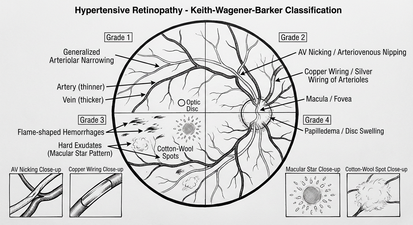
Keith-Wagener-Barker Grading
| Grade | Features |
|---|---|
| 1 | Generalized arteriolar narrowing (A:V ratio < 2:3) |
| 2 | AV nicking/nipping at crossings + copper wiring (sclerotic arterioles) → silver wiring (severe sclerosis) |
| 3 | Flame-shaped hemorrhages, cotton-wool spots (nerve fiber layer infarcts), hard exudates in macular star pattern |
| 4 | All of Grade 3 + papilledema = "malignant hypertension" |
Key features to remember for the exam:
- AV nicking — arteriole compresses the vein at crossing points (shared adventitial sheath)
- Flame-shaped hemorrhages — follow the nerve fiber layer; distinguish from dot-blot hemorrhages of diabetic retinopathy
- Macular star — hard lipid exudates radiating from fovea (due to leaky vessels + Henle's fiber layer anatomy)
- Cotton-wool spots — microinfarcts of the nerve fiber layer, appear fluffy/white
- Copper/silver wiring — progressive arteriolar sclerosis reflecting chronic HTN
- Elschnig spots — focal chorioretinal atrophy from previous choroidal infarcts (sign of prior acute HTN episodes)