

IMG-20260526-WA0293.jpg
What Happened With Both Lungs
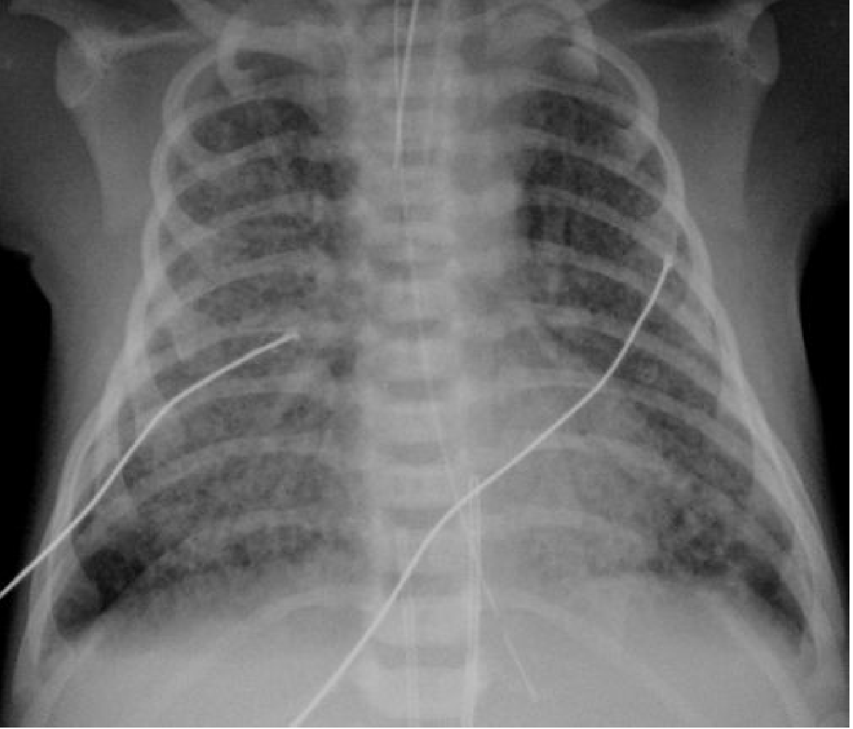
I can see this is a chest X-ray (CXR) of a pediatric patient labeled "B/O AYESHA" (Baby of Ayesha), taken in PA (posteroanterior) view.
Let me read the medical library skill first, then analyze this image carefully.
Searching sources...
Reading File
Finding Sources
Finding Sources
Finding Sources
Finding Sources
Reading File
Now let me fetch the reference CXR image from the textbook for comparison, and also look at the X-ray findings more carefully:
Loading Image
Finding Sources
Now I have a good reference. Let me do one more targeted search on the key findings I see in the X-ray:
Finding Sources
Finding Sources
Reading File
I now have enough information for a thorough analysis. Here is my full interpretation:
Chest X-Ray Analysis - B/O Ayesha (Pediatric/Neonatal CXR, PA View)
Important disclaimer: This is an educational interpretation only. Formal radiological reporting must be done by a qualified radiologist with full clinical context.
Key Radiological Findings
Looking carefully at the X-ray, here is what stands out in both lungs:
| Finding | Description |
|---|---|
| Bilateral hyperinflation | Both lung fields are excessively dark (black), indicating air trapping. The ribs are spread horizontally (barrel chest appearance) and the diaphragm appears flattened |
| Increased bronchial markings | Prominent perihilar markings (peribronchial thickening) visible in both lung fields, giving a "dirty chest" appearance |
| Bilateral interstitial infiltrates | Increased haziness/streakiness throughout both lungs, most visible in the perihilar regions |
| Flattened diaphragm | A hallmark of air trapping - the diaphragm is pushed downward |
| No focal consolidation | No dense lobar opacity to suggest bacterial pneumonia |
| Heart not clearly displaced | Mediastinum appears central |
The Primary Differential for These Bilateral Findings
1. Bronchiolitis (Most Likely)
- The classic CXR picture of bilateral hyperinflation + peribronchial thickening in an infant is bronchiolitis (most commonly RSV - Respiratory Syncytial Virus)
- Presents with bilateral air-trapping due to small airway obstruction
- The lungs look overinflated because air gets trapped behind mucus plugs in inflamed bronchioles
2. Viral Pneumonia / Chlamydial Pneumonia
- Chlamydial pneumonia in infants shows bilateral symmetric interstitial infiltrates with hyperinflation, exactly as seen here
- Per Rosen's Emergency Medicine: "Chest radiograph usually shows hyperinflation with bilateral and symmetric, diffuse, interstitial infiltrates"
3. Transient Tachypnoea of the Newborn (TTN) - if this is a neonate
- Shows mild overinflation, prominent blood vessels, perihilar interstitial shadowing
- Per Grainger & Allison's Diagnostic Radiology: "the most common appearances are mild overinflation, prominent blood vessels, perihilar interstitial shadowing and fluid in the transverse fissure"
- Usually resolves within 24-48 hours
4. Meconium Aspiration Syndrome (MAS) - if post-term neonate
- Shows bilateral hyperinflation + asymmetrical coarse opacification
- Textbook reference image (below) for comparison:
Summary of What's Happening to Both Lungs
The bilateral lung findings in this CXR indicate air trapping with diffuse small airway disease - both lungs are overinflated and show increased interstitial markings. The most likely diagnoses in a young infant (B/O Ayesha = baby of a mother named Ayesha, suggesting a newborn or young infant) are:
- Bronchiolitis (if infant >1 month) - viral inflammation of small airways causing bilateral air trapping
- Viral/Chlamydial pneumonia - bilateral symmetric interstitial disease
- TTN or MAS - if this is a neonate in the first few days of life
The key clinical next steps would be:
-
Clinical history: age of baby, gestational age, onset of symptoms, feeding difficulties, fever
-
O2 saturation and work of breathing assessment
-
Nasopharyngeal swab for RSV/viral panel if bronchiolitis is suspected
-
Supportive care is the mainstay (oxygen supplementation, hydration)
-
Grainger & Allison's Diagnostic Radiology, neonatal chest section
-
Rosen's Emergency Medicine, pediatric respiratory section
This is a shared conversation. Sign in to Orris to start your own chat.