Make a handrawn easily reproducible diagram of diabetic retinopathy for my opthalmology theory exam
Hand-drawn pencil sketch style medical diagram for ophthalmology exam, showing diabetic retinopathy classification. Clean black ink on white background, like a student's notebook drawing. Title at top: "DIABETIC RETINOPATHY" with a subtitle "Classification & Features" Layout: Three columns side by side showing progression: LEFT COLUMN - "MILD NPDR": - Simple circle representing fundus view - A few small dots labeled "Microaneurysms (MA)" only - Arrow pointing down to label "Earliest change" MIDDLE COLUMN - "MODERATE/SEVERE NPDR": - Circle with fundus view showing: - Multiple dots = "Microaneurysms" - Flame shapes = "Dot & blot haemorrhages" - Fluffy white patches = "Cotton wool spots (CWS)" - Wavy line near disc = "Venous beading" - Small branching vessels = "IRMA" - White/yellow deposits = "Hard exudates" - "4-2-1 Rule" box: ">20 haem in 4Q / venous beading 2Q / IRMA 1Q = SEVERE" RIGHT COLUMN - "PDR (Proliferative DR)": - Circle showing new vessels on disc (NVD) drawn as chaotic fine vessels sprouting from optic disc - New vessels elsewhere (NVE) on retina - Wavy line = "Vitreous haemorrhage" - Detachment lines = "Tractional RD" - Label "VEGF-driven neovascularisation" BOTTOM SECTION - horizontal band: "DIABETIC MACULOPATHY (any stage)" - Small circle showing: - Ring of hard exudates around macula = "Circinate exudates" - Thickening = "Macular oedema (DME)" - Dotted area = "Macular ischaemia" - Treatment note: "Rx: Anti-VEGF | Laser | Vitrectomy" Small legend box bottom-right with symbols: dot=MA, flame=haem, cloud=CWS, squiggle=venous beading Overall style: clean hand-drawn sketch, anatomical diagram style, black ink, minimal shading, labels with leader lines
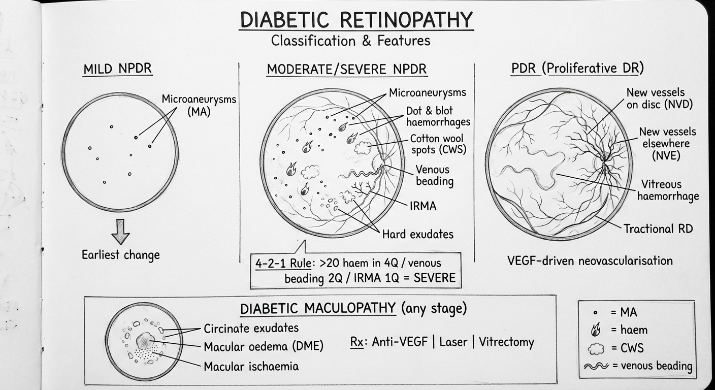
| Stage | Key Features |
|---|---|
| Mild NPDR | Microaneurysms only — earliest change |
| Moderate NPDR | + Dot/blot haemorrhages, hard exudates, cotton wool spots |
| Severe NPDR | 4-2-1 rule: >20 haem in all 4Q / venous beading in 2Q / IRMA in 1Q |
| PDR | NVD / NVE (VEGF-driven) → vitreous haemorrhage, tractional RD |
- Clinically significant macular oedema (CSME)
- Circinate hard exudate rings
- Macular ischaemia
- Rx: Anti-VEGF (first-line), focal laser, vitrectomy
- Draw 3–4 simple circles representing fundus views, left to right
- Add features progressively (more dots, blot haems, CWS, then chaotic vessels)
- Box off the 4-2-1 rule — examiners love that you know the threshold for severe NPDR
- Always add maculopathy as a separate horizontal note at the bottom