
Give me clinical cases with questions and answers on topic of anatomy NEUROANATOMY
neuroanatomy spinal cord cross section tracts Brown-Sequard
Wallenberg lateral medullary syndrome brainstem anatomy
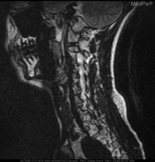
**Imaging Modality:** Sagittal Magnetic Resonance Imaging (MRI), T2-weighted sequence.
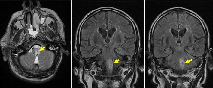
This diagnostic image set consists of brain MRI scans, including an axial T2-weighted sequence and two coronal views, demonstrating a subacute ischemic lesion. The axial view reveals a localized area of hyperintensity (bright signal) within the brainstem, specifically located in the medulla oblongata on the left side, as indicated by the yellow arrow. In the coronal sections, the lesion is shown as an elongated, hyperintense region extending vertically along the longitudinal axis of the medulla, reaching from the pontomedullary junction toward the caudal portion near the cervical spinal cord. The high signal intensity relative to the surrounding brain parenchyma suggests vasogenic edema or subacute infarction. Clinically, this imaging pattern of a medullary infarct extending caudally is associated with Opalski syndrome, a variant of lateral medullary syndrome (Wallenberg syndrome) that involves corticospinal fibers, leading to ipsilateral hemiparesis. The images serve as an educational example of brainstem vascular anatomy and the radiological presentation of specialized stroke syndromes.
**Imaging Modality:** Magnetic Resonance Imaging (MRI), Diffusion-Weighted Imaging (DWI) sequence.
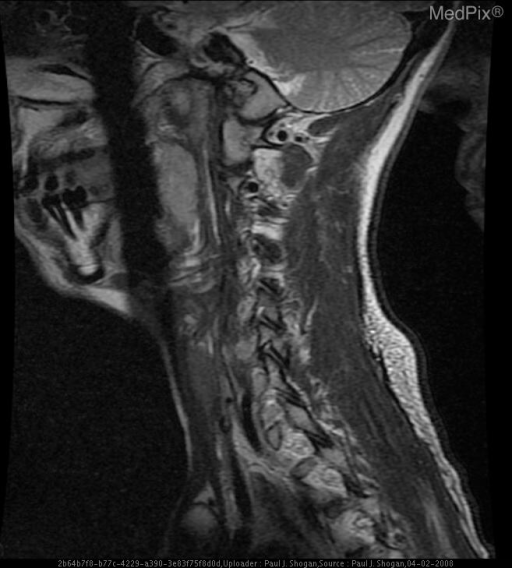
**Imaging Modality:** Magnetic Resonance Imaging (MRI), Sagittal T2-weighted sequence.
cranial nerve anatomy brainstem nuclei diagram
Clinical Cases in Neuroanatomy - Questions & Answers
CASE 1 - The Crossed Syndrome
Clinical Scenario
- Ipsilateral (right) face: Loss of pain and temperature sensation; ptosis, miosis, and anhidrosis (Horner's syndrome); hoarseness; dysphagia; ataxia of the limb
- Contralateral (left) body: Loss of pain and temperature sensation (arm and leg)
- Motor: No limb paralysis
- Nystagmus present (horizontal, towards the lesion side)
Questions
Answers
| Finding | Structure Damaged | Location |
|---|---|---|
| Ipsilateral face - pain/temp loss | Spinal trigeminal nucleus & tract (CN V) | Lateral medulla |
| Contralateral body - pain/temp loss | Spinothalamic tract (already crossed in spinal cord) | Lateral medulla |
| Horner's syndrome (ptosis, miosis, anhidrosis) | Descending sympathetic pathway | Lateral tegmentum of medulla |
| Dysphagia, hoarseness | Nucleus ambiguus (CN IX, X) | Lateral medulla |
| Vertigo, nystagmus | Vestibular nuclei | Lateral medulla |
| Ipsilateral limb ataxia | Inferior cerebellar peduncle | Lateral medulla |
Source: Localization in Clinical Neurology, 8e - "The lateral medullary lesion (Wallenberg syndrome)... combination of a Wallenberg syndrome plus a contralateral hemiplegia" and K.J. Lee's Essential Otolaryngology - "Also called syndrome of the posterior-inferior cerebellar artery thrombosis or lateral medullary syndrome"
CASE 2 - The Knife Wound
Clinical Scenario
- Right lower limb: Complete loss of voluntary movement; loss of vibration and proprioception (position sense)
- Left lower limb: Loss of pain and temperature sensation (beginning 2 segments below T10, i.e., around T12)
- Both limbs: Preserved crude touch
- Bladder and bowel function: intact (incomplete lesion)
Questions
Answers
RIGHT SIDE (ipsilateral to lesion - T10 hemisection):
├── Lateral corticospinal tract → IPSILATERAL motor loss (UMN signs)
└── Posterior columns (fasciculus gracilis/cuneatus) → IPSILATERAL loss of
    vibration, proprioception, fine discriminative touch

LEFT SIDE (contralateral):
└── Lateral spinothalamic tract (already crossed) → CONTRALATERAL loss of
    pain and temperature
- 80-90% regain bowel and bladder function
- 75% regain ambulatory status
- 70% become independent in activities of daily living
Source: ROSEN's Emergency Medicine - "Brown-Séquard syndrome carries the best prognosis of any of the incomplete spinal cord syndromes. Fully 80% to 90% of patients regain bowel and bladder function, 75% regain ambulatory status"
CASE 3 - The Locked-In Patient
Clinical Scenario
Questions
Answers
| Damaged (Ventral Pons) | Spared (Tegmentum) |
|---|---|
| Bilateral corticospinal tracts | Somatosensory pathways |
| Bilateral corticobulbar tracts (face, jaw, tongue) | Brainstem arousal (reticular formation) - consciousness |
| Horizontal gaze pathways (PPRF/CN VI nucleus) | Vertical gaze control (rostral midbrain tectum) |
| CN VI, VII nuclei affected | CN III, IV nuclei spared |
Source: Neuroanatomy through Clinical Cases, 3rd Edition - "Locked-in syndrome often spares vertical eye movements and eye opening. Patients with this syndrome can thus communicate using eye movements."
CASE 4 - The Horner's Triad
Clinical Scenario
Questions
Answers
1st ORDER NEURON (central):
   Hypothalamus → descends through lateral brainstem
   → lateral funiculus of spinal cord → synapses at C8-T2 (ciliospinal center of Budge)

2nd ORDER NEURON (pre-ganglionic):
   C8-T2 → exits ventral root → travels over lung apex
   → over subclavian artery → superior cervical ganglion (synapses here)
   [THIS IS WHERE PANCOAST TUMOR COMPRESSES]

3rd ORDER NEURON (post-ganglionic):
   Superior cervical ganglion → travels along internal carotid artery
   → enters orbit → superior tarsal muscle + iris dilator + sweat glands
- Ptosis (drooping eyelid): The sympathetic system innervates the superior tarsal muscle (of Müller), which contributes to upper lid elevation (~2 mm). Loss → partial ptosis. (Full ptosis = CN III palsy; Horner's ptosis is partial/subtle)
- Miosis (constricted pupil): Iris dilator muscle (dilator pupillae) is sympathetically innervated. Loss of innervation → the parasympathetic sphincter pupillae dominates unopposed → miosis. Pupil fails to dilate in darkness.
- Anhidrosis (lack of sweating): Sweat glands of the face are sympathetically innervated. The 3rd-order fiber from the superior cervical ganglion travels with the external carotid artery to facial sweat glands. Anhidrosis is present if the lesion is at or proximal to the superior cervical ganglion.
Source: Bradley and Daroff's Neurology in Clinical Practice - "The classic Horner syndrome triad of sympathetic dysfunction consists of ipsilateral ptosis, miosis, and facial anhidrosis. The lesion may be anywhere along the three-neuron sympathetic pathway"
CASE 5 - "I Can't Move My Right Side But I'm Awake"
Clinical Scenario
Questions
Answers
- Motor cortex lesion → disproportionate weakness (arm > leg if frontal MCA territory, or leg > arm if parasagittal/ACA territory), often with cortical sensory signs and seizures
- Capsular lesion → proportionate hemiplegia, no cortical signs (no aphasia, no seizure, no hemineglect)
| Feature | Cortical Motor Lesion | Internal Capsule Lesion |
|---|---|---|
| Weakness pattern | Disproportionate | Proportionate |
| Sensory loss | Common | Absent (pure motor) |
| Aphasia/neglect | Yes (if dominant/non-dominant) | Absent |
| Visual field defect | Possible | Absent |
| Seizures | More common | Less common |
| Size of lesion | Larger territory | Small lacune |
Source: Localization in Clinical Neurology, 8e - "With internal capsular lesions, the hemiplegia is often proportionate..." and Adams and Victor's Principles of Neurology - "in the cerebral white matter (corona radiata) and internal capsule, the corticospinal fibers are intermingled"
CASE 6 - "I Can't Feel Anything Below My Waist"
Clinical Scenario
Questions
Answers
| Tract | Location | Function | Status |
|---|---|---|---|
| Lateral corticospinal tract | Lateral funiculus | Voluntary motor | DAMAGED → bilateral weakness |
| Lateral spinothalamic tract | Anterolateral funiculus | Pain, temperature | DAMAGED → bilateral loss |
| Anterior spinothalamic tract | Anterior funiculus | Crude touch | DAMAGED |
| Posterior columns | Posterior funiculus | Vibration, proprioception | SPARED |
| Dorsal root fibers | Posterior horn | Segmental sensation | SPARED |
- Hypertonia (spasticity)
- Hyperreflexia (exaggerated DTRs)
- Positive Babinski's sign (extensor plantar response)
- No muscle atrophy (lower motor neurons intact)
- Clonus may be present
CASE 7 - The Diplopia and Facial Weakness
Clinical Scenario
Questions
Answers
- CN VI fascicles → ipsilateral lateral rectus palsy (medial deviation/esotropia)
- CN VII fascicles → ipsilateral LMN facial palsy
- Corticospinal tract → contralateral hemiplegia (crossed hemiplegia)
- Motor neurons → axons form CN VI nerve → innervate ipsilateral lateral rectus
- Internuclear neurons → axons cross and ascend in the contralateral MLF (medial longitudinal fasciculus) → reach the contralateral CN III nucleus → drive medial rectus for conjugate gaze
| Feature | Central (UMN) palsy | Peripheral (LMN) palsy - Bell's type |
|---|---|---|
| Forehead sparing | YES - forehead spared (bilateral cortical input to upper face) | NO - forehead involved (complete ipsilateral palsy) |
| Tear secretion | Normal | Reduced (if proximal lesion) |
| Taste | Normal | Impaired (if proximal to chorda tympani) |
| Hyperacusis | Absent | Present (if proximal to nerve to stapedius) |
| Site of lesion | Above facial nucleus (cortex/corona radiata/internal capsule) | Facial nucleus or peripheral nerve |
| Cause | Stroke, tumor | Idiopathic, VZV, Lyme disease |
Source: Bradley and Daroff's Neurology in Clinical Practice - "A peripheral seventh cranial nerve palsy usually accompanies a lesion of the sixth cranial nerve nucleus."
CASE 8 - The Vision Field Defect
Clinical Scenario
Questions
Answers
VISUAL PATHWAY (left visual field → right cortex):
Left visual field → falls on NASAL retina of right eye + TEMPORAL retina of left eye
Both → Nasal fibers CROSS at optic chiasm; temporal fibers stay ipsilateral
→ Right optic tract (carries all fibers from left visual field)
→ Right lateral geniculate nucleus (LGN) of thalamus
→ Right optic radiations:
   - Inferior fibers → loop into temporal lobe (Meyer's loop)
   - Superior fibers → through parietal lobe
→ Right primary visual cortex (striate cortex, calcarine fissure, V1)
- Left occipital infarct → left visual cortex destroyed → the patient's left hemisphere cannot receive any visual input (both eyes now only project right visual field information to the right cortex)
- The splenium (posterior corpus callosum) infarct → severs the connection between the right visual cortex and the left angular gyrus (the language/reading center)
- Result: Visual information reaches the intact right visual cortex, but CANNOT cross to the left angular gyrus for reading
- Writing is preserved because the left hemisphere motor system and language areas are intact - the patient can write from internal language representations
- Reading requires visual input to reach the angular gyrus - this pathway is severed
- Contralateral superior homonymous quadrantanopia ("pie in the sky")
- Superior visual field of both eyes is lost
- A parietal lobe lesion would cause the opposite: inferior homonymous quadrantanopia ("pie on the floor")
- The macula (central 5° of vision, area of highest acuity) is represented at the occipital pole, which has dual blood supply from both the PCA and MCA
- In PCA territory infarction, the occipital pole may be spared by MCA collaterals
- This produces homonymous hemianopia with preservation of central vision = macular sparing
- Clinical importance: Patients with macular sparing often retain usable vision and can still read (unlike complete hemianopia)
Source: Localization in Clinical Neurology, 8e - "An intriguing 'disconnection syndrome' in a literate person occurs if there is damage to the left visual cortex and splenium of the corpus callosum... resulting in alexia without agraphia or pure alexia, occasionally associated with right-sided achromatopsia"
Quick Summary Table - Lesion Localization
| Case | Lesion Site | Key Syndrome | Hallmark Finding |
|---|---|---|---|
| 1 | Lateral medulla | Wallenberg's | Crossed facial/body sensory loss + Horner's, NO weakness |
| 2 | Unilateral spinal cord T10 | Brown-Séquard | Ipsilateral motor/proprioception loss + Contralateral pain/temp loss |
| 3 | Ventral pons (bilateral) | Locked-in | Quadriplegia + intact consciousness + preserved vertical gaze |
| 4 | Cervical sympathetic (Pancoast) | Horner's syndrome | Ptosis + miosis + anhidrosis (ipsilateral) |
| 5 | Posterior limb internal capsule | Pure motor hemiplegia | Proportionate face/arm/leg weakness, no sensory loss |
| 6 | Anterior/lateral cord | Anterior cord syndrome | Motor loss + pain/temp loss; vibration/proprioception spared |
| 7 | Dorsal/ventral pons | Millard-Gubler (partial) | CN VI + CN VII palsy + crossed hemiplegia if corticospinal involved |
| 8 | Left occipital + splenium | PCA territory infarct | Homonymous hemianopia + alexia without agraphia |