
Make a hand drawn easily reproducible exam diagram of triad of irvin grass syndrome
- Cystoid Macular Edema (CME) — post-cataract surgery
- Optic disc swelling (optic nerve head leakage on IVFA)
- Vitreous traction to the surgical wound (vitreous incarceration in the corneoscleral wound)
Hand-drawn medical exam diagram of Irvine-Gass Syndrome triad, sketch style on white paper. Title at top: "IRVINE-GASS SYNDROME" with subtitle "(Post-cataract CME)". Three components arranged in a triangle with arrows pointing to a central eye diagram: 1. Top left box labeled "1. CYSTOID MACULAR EDEMA" with a small sketch of retina showing honeycomb cyst pattern at fovea and "petaloid/spoke-wheel pattern on IVFA" note. 2. Top right box labeled "2. OPTIC DISC SWELLING" with a small sketch of swollen optic disc and "leakage on IVFA" note. 3. Bottom center box labeled "3. VITREOUS INCARCERATION in corneoscleral wound" with a small sketch showing vitreous traction to wound. Central cross-section sketch of an eye showing all three features. At the bottom: "Onset: 6-10 weeks post cataract surgery". Additional notes: "Vitreous to wound → traction → CME". Clean black ink sketch style, educational anatomy diagram, easy to reproduce by hand for medical exams.
- Cystoid Macular Edema (CME) — post-cataract surgery
- Optic disc swelling (optic nerve head leakage on IVFA)
- Vitreous traction to the surgical wound (vitreous incarceration in the corneoscleral wound)
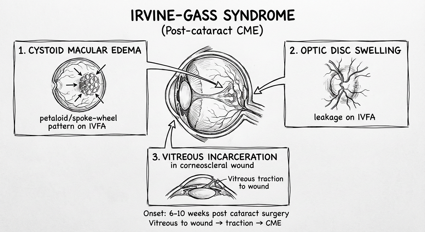
Irvine-Gass Syndrome — The Triad
| # | Component | Key Features |
|---|---|---|
| 1 | Cystoid Macular Edema (CME) | Honeycomb/petaloid cyst pattern at fovea; spoke-wheel leakage on IVFA; loss of foveal reflex |
| 2 | Optic Disc Swelling | Optic nerve head leakage seen on fluorescein angiography |
| 3 | Vitreous Incarceration in wound | Vitreous traction to the corneoscleral wound — the mechanical trigger |
Mnemonic: "VOC" — Vitreous traction → Optic disc swelling → CME
Key exam facts:
- Onset: 6–10 weeks post-cataract extraction (peak incidence)
- Risk factors: Vitreous loss, iris prolapse, uveal incarceration during surgery
- Investigation: IVFA shows petaloid/spoke-wheel leakage; OCT shows intraretinal cysts + macular thickening
- Treatment: Topical NSAIDs (ketorolac) ± topical steroids