

1000061627.jpg
Tell me about the role of calcium in simple language i need to build up my concept first
I can see the image shows diagrams about the role of calcium in muscle contraction. Let me first analyse it properly, then pull up the medical library content for a thorough, simple explanation.
analyse_media
Reading File
Now let me search the medical library for calcium's role in muscle contraction and physiology.
Finding Sources
Finding Sources
Good. Let me now read the key sections from Guyton & Hall and also search for calcium's role in the neuromuscular junction specifically.
Reading File
Finding Sources
Finding Sources
I now have all the content I need to write a thorough, simple explanation. Let me also quickly read the troponin-calcium section from Guyton and read the neuromuscular junction calcium paragraph from Robbins.
Reading File
Reading File
I now have all the information needed. Let me also fetch the diagram image from Guyton to include visually.
Loading Image
Now I have everything I need. Here is a complete, simple, concept-building explanation of the role of calcium:
Role of Calcium - Building Your Concept from Scratch
The Big Picture First
Think of calcium (Ca²⁺) as the "ON switch" for muscle contraction. Without calcium, muscles stay relaxed. When calcium arrives, contraction begins. When calcium is taken away, the muscle relaxes again. That's the core idea.
Now let's follow the full story step by step, exactly matching what your image shows.
Step 1 - The Nerve Sends a Signal (Neuromuscular Junction)
When your brain wants to move a muscle, it sends an electrical signal down a motor nerve. That signal travels to the neuromuscular junction - the meeting point between the nerve and the muscle.
At the nerve terminal:
- The electrical signal (action potential) arrives at the axon terminal
- This opens voltage-gated calcium channels on the nerve ending
- Ca²⁺ flows INTO the nerve terminal from outside
- This calcium entry causes synaptic vesicles to fuse with the membrane and release acetylcholine (ACh) into the synaptic cleft
Calcium's first role: Triggering acetylcholine release at the nerve terminal. Without this Ca²⁺ influx, no ACh would be released, and the muscle would never receive the signal.
As stated in Robbins & Kumar Basic Pathology: "Nerve impulses depolarize the presynaptic membrane, stimulating calcium influx and the release of acetylcholine into the synaptic cleft."
Step 2 - The Muscle Membrane Gets Excited
Once ACh is released:
- ACh diffuses across the synaptic cleft
- It binds to ACh receptors on the muscle membrane (sarcolemma)
- These receptors open, letting Na⁺ rush in and K⁺ flow out
- This creates a new action potential on the muscle membrane (sarcolemma)
- That action potential travels deep into the muscle through the T-tubules (tiny tunnels that carry the electrical signal inward)
Step 3 - The T-Tubule Signal Reaches the Sarcoplasmic Reticulum
The T-tubule is like an electrical wire running deep inside the muscle cell. Here's the clever part:
- The T-tubule wall contains a voltage sensor called the DHPR (Dihydropyridine Receptor)
- Right next to it, the Sarcoplasmic Reticulum (SR) - a calcium storage tank inside the muscle - has a release channel called the Ryanodine Receptor (RyR)
- When the T-tubule depolarizes, DHPR physically pulls open the RyR channel on the SR
Result: Ca²⁺ floods out of the SR into the muscle cell fluid (sarcoplasm) - a 500-fold increase in calcium concentration.
This is exactly what your flowchart shows:
Depolarization of nerve → Depolarization of skeletal muscle → Depolarization of transverse tubular membrane → Charge movement of the slow Ca²⁺ voltage channel (DHPR)...
Step 4 - Calcium "Unlocks" the Contractile Machinery
This is the most important role of calcium. Look at the diagram below:
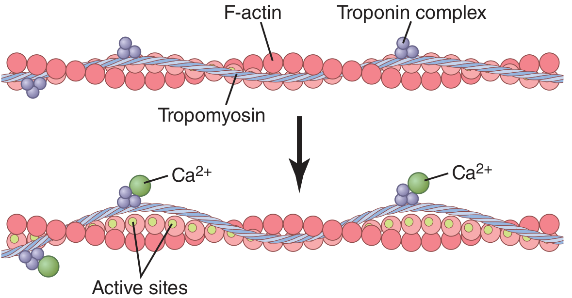
Inside the muscle, the thin filaments (actin) are normally blocked by two proteins:
- Tropomyosin - sits like a lid over the active sites on actin, physically covering them
- Troponin - a complex that holds tropomyosin in the blocking position
In the resting state:
- Ca²⁺ is very low (less than 10⁻⁷ molar)
- Troponin-tropomyosin blocks actin → no contraction possible
When Ca²⁺ rises:
- Ca²⁺ binds to Troponin C (the calcium-sensing subunit)
- This causes a shape change in the troponin complex
- Troponin tugs tropomyosin aside, uncovering the active sites on actin
- Now myosin heads can bind to actin and pull → CONTRACTION
As Guyton & Hall explains: "When the troponin complex binds Ca²⁺, a conformation change occurs that tugs on the tropomyosin and exposes myosin binding sites on the actin, permitting myosin to bind to actin and allowing contraction."
Step 5 - Relaxation: Calcium is Pumped Back
Contraction doesn't last forever. Once the nerve signal stops:
- A pump called SERCA (Sarcoplasmic Reticulum Ca²⁺-ATPase) actively pumps Ca²⁺ back into the SR
- Ca²⁺ levels in the sarcoplasm fall
- Troponin loses its Ca²⁺, tropomyosin slides back to cover actin active sites
- Myosin can no longer bind actin → RELAXATION
Inside the SR, a protein called calsequestrin stores Ca²⁺ (each molecule can bind up to 40 Ca²⁺ ions), helping the SR act as a high-capacity calcium reservoir.
Summary Table
| Step | Where | Calcium's Role |
|---|---|---|
| Neuromuscular junction | Nerve terminal | Ca²⁺ influx triggers ACh vesicle release |
| T-tubule | Muscle membrane | DHPR voltage sensor opens RyR on SR |
| Sarcoplasm | Inside muscle cell | Ca²⁺ released from SR - 500x increase |
| Troponin binding | Thin filament | Ca²⁺ binds TnC → tropomyosin moves → actin sites exposed |
| Cross-bridge cycling | Sarcomere | Myosin-actin interaction → contraction |
| Relaxation | SR membrane | SERCA pump re-sequesters Ca²⁺ → muscle relaxes |
The Simple Way to Remember It
No calcium = blocked actin = no contraction. Calcium arrives = actin unblocked = contraction. Calcium removed = re-blocked = relaxation.
Calcium acts at two places: first at the nerve ending (to release ACh), and second inside the muscle (to unlock the contractile proteins). The SR is the main calcium reservoir, the T-tubule is the electrical messenger, and the troponin-tropomyosin system is the gatekeeper that calcium physically moves to start contraction.
- Guyton and Hall Textbook of Medical Physiology, pp. 109-110
- Robbins & Kumar Basic Pathology, p. 504
- Neuroscience: Exploring the Brain, 5th Edition
Summarise this fonrme as i have to write this concept for 5 marks
Here is a clean 5-mark answer you can write directly:
Role of Calcium in Muscle Contraction
1. At the Neuromuscular Junction
When a nerve impulse arrives at the axon terminal, voltage-gated Ca²⁺ channels open and calcium flows into the nerve ending. This triggers synaptic vesicles to fuse and release acetylcholine (ACh) into the synaptic cleft.
2. Muscle Membrane Depolarization
ACh binds to receptors on the sarcolemma → Na⁺ rushes in → action potential is generated → spreads along the muscle membrane and down the T-tubules.
3. Ca²⁺ Release from Sarcoplasmic Reticulum (SR)
The T-tubule depolarization activates DHPR (voltage sensor), which opens Ryanodine receptors (RyR) on the SR. Ca²⁺ is released into the sarcoplasm - concentration rises 500-fold.
4. Ca²⁺ Triggers Contraction
- Ca²⁺ binds Troponin C on the thin filament
- This shifts tropomyosin away from the active sites on actin
- Myosin heads now bind actin → cross-bridge cycling → contraction
5. Relaxation
Once the signal stops, SERCA pump returns Ca²⁺ back into the SR. Ca²⁺ detaches from troponin → tropomyosin re-covers actin → muscle relaxes.
One-line to end your answer:
Calcium acts as the key messenger that links electrical excitation of the nerve to mechanical contraction of the muscle.
Writing tip: For 5 marks, cover one point per mark - NMJ → depolarization → SR release → troponin unlocking → relaxation. That's exactly 5 points above.
This is a shared conversation. Sign in to Orris to start your own chat.