Now I have comprehensive information. Let me compile a thorough, well-organized answer.
Radiographic Patterns in Dogs and Cats: A Complete Reference
Veterinary radiology uses a systematic pattern-based approach to link radiographic findings with conditions, clinical signs, and treatments. Patterns are broadly divided into thoracic (pulmonary, cardiac, pleural, mediastinal), abdominal, and skeletal categories.
PART 1: THORACIC / PULMONARY PATTERNS
1. Alveolar Pattern
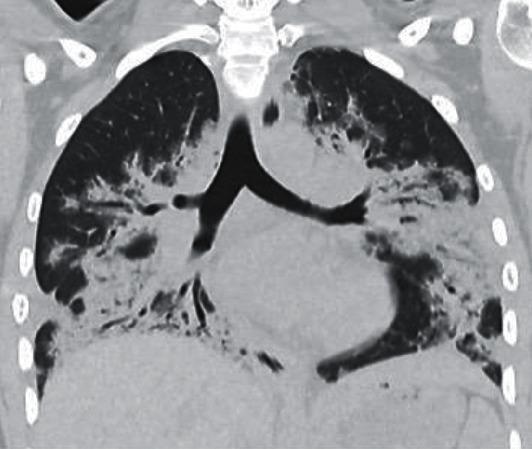
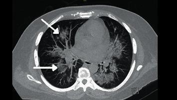
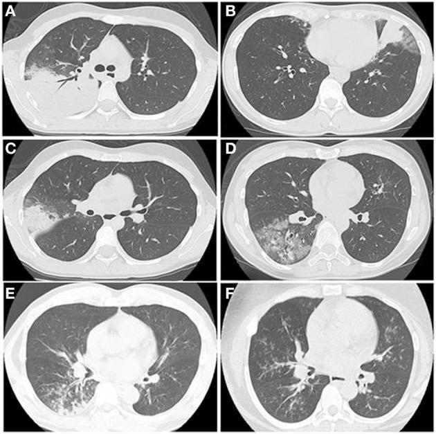
What it looks like: Increased opacity that completely obscures pulmonary vessels. Air bronchograms (air-filled airways visible within opaque tissue) are a hallmark. Border effacement with the heart, vessels, or diaphragm occurs.
Sub-patterns:
- Lobar consolidation - affects one full lobe
- Patchy/multifocal alveolar - scattered coalescent opacities
- Hilar/perihilar distribution - cardiogenic edema in dogs
- Random/diffuse distribution - cardiogenic edema in cats, contusions
| Condition | Species | Clinical Signs | Treatment |
|---|
| Cardiogenic pulmonary edema (mitral endocardiosis, DCM) | Dogs primarily | Dyspnea, cough, exercise intolerance, crackles | Furosemide, pimobendan, ACE inhibitors, oxygen |
| Cardiogenic edema (HCM) | Cats primarily | Open-mouth breathing, labored respiration, lateral recumbency | Furosemide, pleurocentesis if effusion, oxygen |
| Aspiration pneumonia | Dogs > cats | Cough, fever, nasal discharge, crackles ventrally | Broad-spectrum antibiotics (amoxicillin-clavulanate + enrofloxacin), supportive care |
| Bronchopneumonia (bacterial) | Dogs | Cough, pyrexia, mucopurulent nasal discharge | Antibiotics guided by culture/sensitivity |
| Pulmonary contusion (trauma) | Both | Tachypnea, hemoptysis, history of trauma | Supportive: oxygen, rest, analgesics; avoid excessive fluids |
| Atelectasis | Both | Reduced breath sounds, mediastinal shift | Treat underlying cause (remove effusion, bronchodilators) |
| Pulmonary hemorrhage | Both | Hemoptysis, sudden dyspnea | Oxygen, treat coagulopathy (Vit K1 for rodenticide toxicity) |
| Pulmonary neoplasia (primary/metastatic) | Both | Weight loss, chronic cough, dyspnea | Surgery (solitary), chemotherapy, palliative |
2. Interstitial Pattern
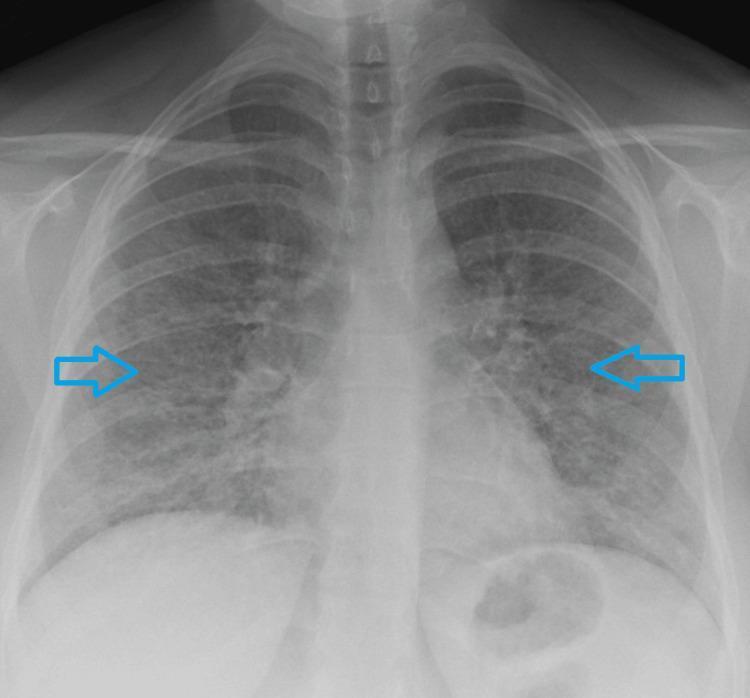
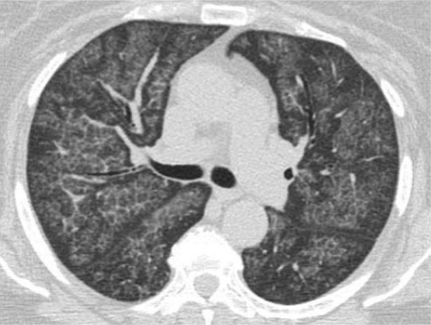
What it looks like: Hazy "ground-glass" opacity - vessels are visible but have fuzzy, indistinct margins ("trees in a fog"). Two sub-types:
- Unstructured interstitial - diffuse haziness
- Structured (nodular) interstitial - discrete soft-tissue nodules (3-30mm)
| Condition | Species | Clinical Signs | Treatment |
|---|
| Early/mild pulmonary edema | Both | Subtle dyspnea, soft cough | Diuretics, cardiac medications |
| Pulmonary fibrosis | Dogs (West Highland White Terriers especially) | Progressive exercise intolerance, dry cough, inspiratory crackles | Sildenafil, bronchodilators (no cure) |
| Viral pneumonia (distemper, FIP) | Dogs/cats | Fever, neurological signs (distemper), FIP signs | Supportive; antivirals (GS-441524 for FIP) |
| Fungal pneumonia (Histoplasma, Blastomyces) | Both | Weight loss, cough, lymphadenopathy | Itraconazole or fluconazole long-term |
| Pulmonary infiltrate with eosinophilia (PIE) | Both | Cough, wheeze, eosinophilia on CBC | Corticosteroids; treat heartworm if present |
| Metastatic neoplasia ("cannonball" nodules) | Both | Weight loss, lethargy | Chemotherapy, palliative care |
| Lymphoma | Both | Dyspnea, weight loss, mediastinal mass | Chemotherapy (CHOP protocol) |
3. Bronchial Pattern
What it looks like:
- "Donut" sign - thickened bronchial walls seen end-on as rings (seen in both dogs and cats)
- "Tram lines" - thickened bronchial walls seen longitudinally as parallel lines (dogs only; airways too small in cats)
- Peribronchial cuffing - indistinct hazy opacity around airways
| Condition | Species | Clinical Signs | Treatment |
|---|
| Feline asthma | Cats | Wheeze, paroxysmal cough, "hunched" breathing posture | Corticosteroids (prednisolone), bronchodilators (salbutamol inhaler via AeroKat) |
| Chronic bronchitis | Dogs | Chronic productive cough >2 months, no fever | Corticosteroids, bronchodilators, coupage; avoid smoke/allergens |
| Heartworm (PIE response) | Both | Cough, dyspnea, eosinophilia; cats: sudden death possible | Dogs: melarsomine + doxycycline; cats: anti-inflammatory steroids, supportive (no adulticidal drug approved for cats) |
| Allergic bronchitis | Both | Cough, wheeze, seasonal | Corticosteroids, antihistamines |
| Lungworm (Aelurostrongylus in cats, Oslerus in dogs) | Both | Cough, dyspnea, weight loss | Fenbendazole or ivermectin |
4. Vascular Pattern
What it looks like: Changes in size of pulmonary arteries and/or veins.
- Enlarged pulmonary arteries (wider than the 9th rib where they cross): Right heart disease, heartworm
- Enlarged pulmonary veins: Left heart failure, pulmonary venous hypertension
- Reduced pulmonary vascularity: Hypovolemia, right-to-left shunts
- Tortuous/blunted arteries: Heartworm disease (especially at caudal lobar arteries)
| Condition | Artery | Vein | Clinical Signs |
|---|
| Heartworm disease | Enlarged, tortuous | Normal | Cough, syncope, exercise intolerance |
| Left heart failure (mitral disease) | Normal | Enlarged (pulm venous congestion) | Dyspnea, cough, orthopnea |
| Pulmonary hypertension | Enlarged | Variable | Syncope, exercise intolerance, right heart enlargement |
| Hypovolemia/shock | Small/thin | Small | Weakness, pale MM, rapid HR |
5. Mixed/Bronchointerstitial Pattern
Very common in practice - features of both bronchial and interstitial patterns simultaneously.
| Condition | Notes |
|---|
| Heartworm PIE | Most common cause |
| Chronic airway disease progressing | Asthma + secondary changes |
| Viral/fungal lower airway disease | Overlapping compartments |
PART 2: CARDIAC RADIOGRAPHIC PATTERNS
Cardiac Size Assessment
- Vertebral Heart Score (VHS): Normal dogs <10.5 vertebrae; normal cats <8 vertebrae
- Cardiomegaly: Generalized or chamber-specific enlargement
| Pattern | Condition | Key Radiographic Sign | Treatment |
|---|
| Left atrial enlargement (dog) | Mitral valve disease | "Bulge" at 2-3 o'clock on VD view; straight left heart border on lateral | Pimobendan, ACE inhibitors, furosemide when CHF develops |
| Generalized cardiomegaly (dog) | Dilated cardiomyopathy (DCM) | Round, enlarged cardiac shadow (VHS >12) | Pimobendan, digoxin, antiarrhythmics |
| Right-sided cardiomegaly (dog) | Pericardial effusion, heartworm, pulm HTN | "Globoid" heart, increased sternal contact | Treat cause: pericardiocentesis, adulticidal therapy |
| "Valentine heart" / sternal contact (cat) | HCM | Increased sternal contact, triangular appearance on VD | Atenolol, furosemide, clopidogrel (anti-thrombotic) |
| Pericardial effusion | Dogs (neoplastic: HSA, chemodectoma) | Globoid, enlarged, rounded cardiac silhouette | Pericardiocentesis; pericardiectomy if recurrent |
PART 3: PLEURAL AND MEDIASTINAL PATTERNS
Pleural Effusion
Radiographic signs: Retraction of lung lobes from chest wall, "scalloped" borders, fissure lines, blunting of costophrenic angles, loss of diaphragmatic/cardiac borders.
| Condition | Species | Fluid Type | Treatment |
|---|
| Pyothorax | Both | Septic exudate | Thoracic lavage + long-term antibiotics |
| Chylothorax | Both | Chylous (milky) | Thoracic duct ligation + pericardectomy |
| Hemothorax | Both | Blood | Treat coagulopathy, drain if compromised |
| Cardiac failure | Both | Modified transudate | Diuretics, cardiac drugs |
| Feline infectious peritonitis (FIP) | Cats | Yellow exudate (high protein) | GS-441524 antivirals |
| Neoplasia (lymphoma, mesothelioma) | Both | Variable | Chemotherapy, palliative drainage |
Pneumothorax
Radiographic signs: Absence of lung markings at periphery, collapsed lung lobes, elevated heart from sternum on lateral, visible pleural line.
| Condition | Clinical Signs | Treatment |
|---|
| Spontaneous (blebs/bullae) | Acute dyspnea | Thoracocentesis; surgery if recurrent |
| Traumatic | History of trauma, dyspnea | Thoracocentesis, chest tube if severe |
Mediastinal Widening/Mass
Cranial mediastinal mass - pushes trachea dorsally on lateral view.
| Condition | Species | Treatment |
|---|
| Lymphoma | Both | CHOP chemotherapy |
| Thymoma | Both | Surgical resection |
| Thymic lymphoma | Cats (young) | Chemotherapy |
| Ectopic thyroid/parathyroid mass | Dogs | Surgery |
PART 4: ABDOMINAL RADIOGRAPHIC PATTERNS
Organ Size/Position Patterns
| Pattern | Condition | Clinical Signs | Treatment |
|---|
| Hepatomegaly (liver caudal to last rib, rounded margins) | Hepatic lipidosis (cats), neoplasia, congestion (right CHF), Cushing's disease | Vomiting, icterus, PU/PD, ascites | Treat underlying cause; nutritional support for lipidosis |
| Microhepatica (liver cranial to last rib, sharp margins) | Portosystemic shunt, cirrhosis, chronic hepatitis | Hepatic encephalopathy, failure to thrive | Surgery (shunt ligation), dietary management |
| Splenomegaly | Splenic mass (HSA, nodular hyperplasia), splenic torsion, lymphoma | Abdominal mass palpable, weakness (if bleeding) | Splenectomy; chemotherapy for lymphoma |
| Renomegaly (bilateral) | Polycystic kidney disease (cats), lymphoma, FIP | PU/PD, weight loss, azotemia | Supportive; antivirals for FIP |
| Small/irregular kidneys | Chronic kidney disease | PU/PD, vomiting, weight loss | CKD management: phosphate binders, diet, fluids |
| Bladder mass/calculi | Transitional cell carcinoma, urolithiasis | Stranguria, hematuria, dysuria | Surgery/piroxicam (TCC); diet/surgery for calculi |
Intestinal Obstruction Patterns
| Pattern | Radiographic Sign | Condition | Treatment |
|---|
| Mechanical obstruction | Dilated small intestinal loops (>2x width of a rib), "stacked" loops, "gravel sign" | Foreign body, intussusception | Emergency surgery |
| Functional ileus | Generalized uniform gas distension | Post-op, electrolyte imbalance, peritonitis | Treat underlying cause, supportive care |
| Gastric dilatation-volvulus (GDV) | "Double bubble" or "C-shape" gas compartmentalization in stomach | Dogs (large/giant breeds), acute abdomen, non-productive retching, shock | Emergency surgery (derotation + gastropexy) |
| Intussusception | "Target sign" on ultrasound; soft-tissue opacity with small bowel dilation on X-ray | Young dogs/cats; vomiting, bloody diarrhea | Surgical reduction or resection |
| Linear foreign body | Plicated (plicating/bunched) small intestine, comma-shaped gas loops | Cats (string/thread); dogs (towels, rope toys) | Emergency surgery |
Free Abdominal Air/Fluid
- Pneumoperitoneum: Free gas under diaphragm on upright/standing lateral - indicates GI perforation (emergency surgery)
- Loss of serosal detail: "Ground glass" or "grey" abdomen indicating peritoneal effusion (peritonitis, hemoabdomen) - abdominocentesis + treat cause
Urinary Calculi (Radiopaque vs. Radiolucent)
| Stone Type | Radiodensity | Breed Predisposition | Treatment |
|---|
| Struvite | Radiopaque | Dogs, cats | Dissolution diet or surgery |
| Calcium oxalate | Very radiopaque | Miniature Schnauzer, Bichon | Surgery/lithotripsy |
| Urate | Radiolucent (need contrast/US) | Dalmatian, Bulldogs | Allopurinol + diet |
| Cystine | Slightly opaque | Dachshund, Newfoundland | Diet + 2-MPG |
PART 5: SKELETAL RADIOGRAPHIC PATTERNS
Bone Lesion Patterns
| Pattern | Description | Conditions | Clinical Signs | Treatment |
|---|
| Lytic (osteolytic) | Bone destruction, "moth-eaten" appearance, cortical loss | Osteosarcoma, fungal osteomyelitis, bacterial osteomyelitis | Lameness, swelling, pain, pathologic fracture | Amputation ± chemotherapy (OSA); antifungals; antibiotics |
| Productive (osteoproliferative) | Increased bone opacity, periosteal reaction, new bone formation | Hypertrophic osteodystrophy (HOD), hypertrophic osteopathy (HO), Panosteitis | Pain, swelling, fever (HOD/HO); shifting limb lameness (Pano) | NSAIDs, corticosteroids; treat underlying cause for HO |
| Mixed lytic and productive | Both destruction and new bone simultaneously | Osteosarcoma, aggressive infection, multilobular osteochondrosarcoma | Severe pain, swelling, firm mass | Surgery ± chemo ± radiation |
| Periosteal reaction | New bone along cortex | Trauma, infection, neoplasia, HO | Pain, swelling | Treat underlying cause |
| Subchondral erosion / joint destruction | Loss of joint space, sclerosis | Rheumatoid/erosive polyarthritis, septic arthritis | Joint swelling, effusion, pain | Immunosuppressives (erosive); antibiotics (septic) |
| Joint space narrowing + osteophytes | Sclerosis, spur formation | Degenerative joint disease (DJD/OA) | Chronic lameness, stiffness | NSAIDs, weight management, physiotherapy, joint supplements |
Specific Skeletal Conditions
| Condition | Radiographic Sign | Species | Treatment |
|---|
| Hip dysplasia | Shallow acetabulum, subluxation, OA changes (Norberg angle <105°) | Dogs (large breeds) | Juvenile: TPO/DPO; adult: THR or FHO; medical: NSAIDs, weight control |
| Intervertebral disc disease (IVDD) | Narrowed disc space, calcified disc, "wedging," spondylosis | Dogs (Dachshund, CKCS); cats (rare) | Conservative: rest + NSAIDs; surgical: hemilaminectomy/ventral slot |
| Osteosarcoma | Aggressive lytic/mixed lesion at metaphysis, "sunburst" periosteal reaction, "Codman's triangle" | Large/giant dogs, distal radius most common | Amputation + chemotherapy (carboplatin/doxorubicin) |
| Hypertrophic osteopathy (HO) | Bilateral palisading periosteal new bone, distal limbs, "corduroy" pattern | Dogs with thoracic mass (primary lung tumor, abscess) | Treat underlying thoracic mass (surgery); HO resolves |
| Panosteitis | Intramedullary opacity/blurring in long bone diaphysis | Young large-breed dogs (German Shepherd) | Self-limiting; NSAIDs for pain |
| Legg-Calve-Perthes (LCPD) | Femoral head deformity, lysis, sclerosis, collapse | Small breed dogs | FHO (femoral head/neck excision) |
| Metaphyseal osteopathy (HOD) | "Double physis" sign - zone of lysis adjacent to physis, periosteal reaction | Young large-breed dogs | NSAIDs, supportive care |
PART 6: SPECIAL RADIOGRAPHIC SIGNS - QUICK REFERENCE
| Sign | Description | Condition |
|---|
| Air bronchogram | Air-filled bronchi visible within opaque lung | Alveolar filling (pneumonia, edema, hemorrhage) |
| Silhouette sign (border effacement) | Loss of border between two same-opacity structures | Alveolar pattern adjacent to heart/diaphragm |
| "Donut" sign | Thickened bronchial wall seen end-on | Bronchial disease (asthma, chronic bronchitis) |
| "Tram lines" | Thickened bronchial walls seen longitudinally | Bronchial disease in dogs |
| Peribronchial cuffing | Hazy opacity around bronchi | Acute airway inflammation |
| "Sunburst" periosteal reaction | Radiating spiculated periosteal new bone | Osteosarcoma |
| "Codman's triangle" | Elevated periosteum at lesion margin | Aggressive bone tumor |
| "Double bubble/C-shape" stomach | Gas compartmentalization | Gastric dilatation-volvulus (GDV) |
| Pleural fissure lines | Sharp linear soft-tissue lines along fissures | Pleural effusion |
| "Cannonball" nodules | Multiple discrete pulmonary nodules | Metastatic neoplasia |
| Vertebral end-plate erosion | Irregular, "fluffy" end-plate | Discospondylitis |
| "Double physis" sign | Radiolucent zone adjacent to growth plate | Hypertrophic osteodystrophy (HOD) |
| "Target sign" (US) | Concentric rings in bowel wall | Intussusception |
| Mediastinal shift | Heart/trachea displaced to one side | Atelectasis (toward), effusion/tension pneumothorax (away) |
Summary by System
| System | Most Common Patterns | Key Differentials |
|---|
| Lung | Alveolar, Bronchial, Interstitial, Vascular, Mixed | Edema, pneumonia, asthma, fibrosis, neoplasia, heartworm |
| Heart | Cardiomegaly (L/R/generalized), pericardial effusion | MVD, DCM, HCM, pericardial neoplasia |
| Pleural | Effusion, pneumothorax | Pyothorax, chylothorax, trauma, neoplasia |
| Abdomen | Organ size changes, obstruction, free gas/fluid | GDV, foreign body, urolithiasis, neoplasia |
| Bone | Lytic, productive, mixed, periosteal | OSA, DJD, IVDD, panosteitis, HO |
Radiographic patterns should always be integrated with signalment (age, breed, sex), clinical signs, physical exam, and ancillary tests (CBC, biochemistry, urinalysis, cytology) for the most accurate diagnosis. Many conditions overlap radiographically, so pattern recognition is a starting point, not an endpoint.