
Development of urinary bladder
I'll start by reading the medical library skill, then search for content on urinary bladder development.
Reading File
Now let me search the library for content on urinary bladder development in parallel:
Finding Sources
Finding Sources
Finding Sources
Excellent! Found dedicated sections in three textbooks. Let me read all of them in parallel:
Reading File
Reading File
Reading File
Now let me read more of The Developing Human content (which has the most detailed embryology) and also fetch the key diagrams:
Reading File
Loading Image
Loading Image
Loading Image
Excellent! Now let me fetch the remaining key diagrams and look for congenital anomalies content:
Loading Image
Loading Image
Finding Sources
Reading File
Now let me fetch the urachal anomalies diagram and read a bit more for bladder exstrophy:
Loading Image
Reading File
I now have comprehensive material from three authoritative textbooks. Let me compile the full answer.
Development of the Urinary Bladder
1. Overview and Embryological Origin
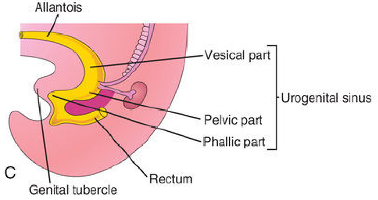
The urinary bladder develops from the endoderm of the urogenital sinus, which itself derives from the primitive cloaca. Between weeks 4 and 7 of gestation, the urorectal septum (a wedge of mesoderm growing caudally between the allantois and hindgut) divides the cloaca into:
- A ventral urogenital sinus (gives rise to the bladder and urethra)
- A dorsal anorectal canal (gives rise to the rectum and anal canal)
The cloacal membrane ruptures to complete this partition. - The Developing Human: Clinically Oriented Embryology, p. 706-707
2. Three Parts of the Urogenital Sinus
For descriptive purposes, the urogenital sinus is divided into three parts:
| Part | Derivatives |
|---|---|
| Vesical part (cranial) | Forms most of the urinary bladder; continuous with the allantois |
| Pelvic part | Becomes the neck of the bladder; prostatic urethra in males; entire urethra in females |
| Phallic part | Grows toward the genital tubercle; forms the spongy urethra in males and vaginal vestibule in females |
3. Development of Bladder Layers
- The entire epithelium (urothelium/transitional epithelium) of the bladder is derived from endoderm of the vesical part of the urogenital sinus
- The detrusor muscle, connective tissue, and serosa develop from the adjacent splanchnic mesenchyme
4. The Allantois and Urachus
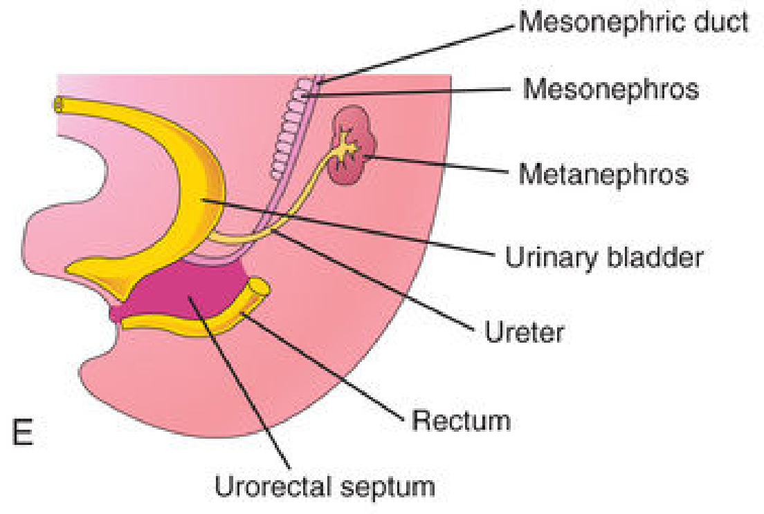
Initially, the superior end of the bladder is continuous with the allantois - an extraembryonic membrane extending toward the umbilicus through the umbilical cord.
- Between weeks 5-7, the allantoic duct constricts and obliterates as the bladder descends into the pelvis
- It becomes a fibrous cord called the urachus, running from the apex of the bladder to the umbilicus
- In adults, the urachus is represented by the median umbilical ligament
5. Formation of the Trigone
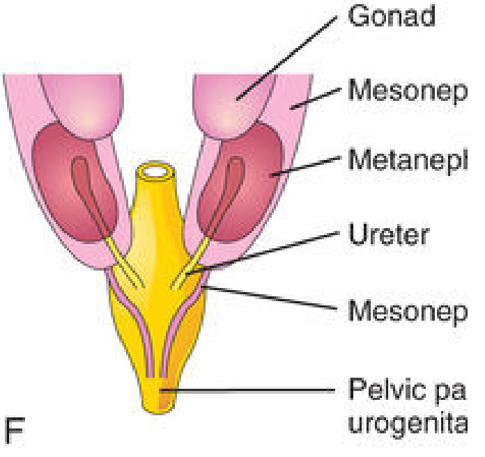
As the bladder enlarges, the distal portions of the mesonephric (Wolffian) ducts are absorbed into the dorsal wall of the bladder. This absorption:
- Contributes connective tissue to the trigone of the bladder
- Causes the ureters to open separately into the bladder wall
- Due to traction from ascending kidneys, ureteric orifices migrate superolaterally, entering the bladder base obliquely
In males, the mesonephric duct orifices move medially to enter the prostatic urethra as the ejaculatory ducts. In females, the distal ends of the mesonephric ducts degenerate. - The Developing Human, p. 708
6. Final Positions by Sex
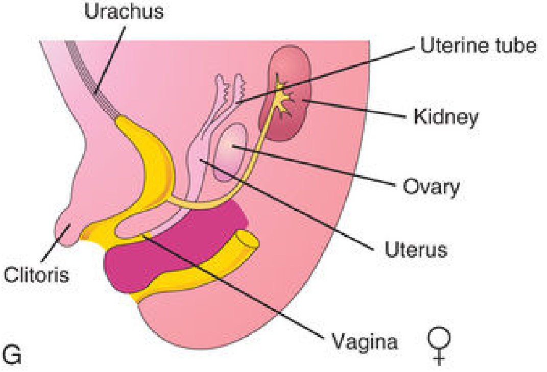
Female (week 12 onward):
Male (week 12 onward):
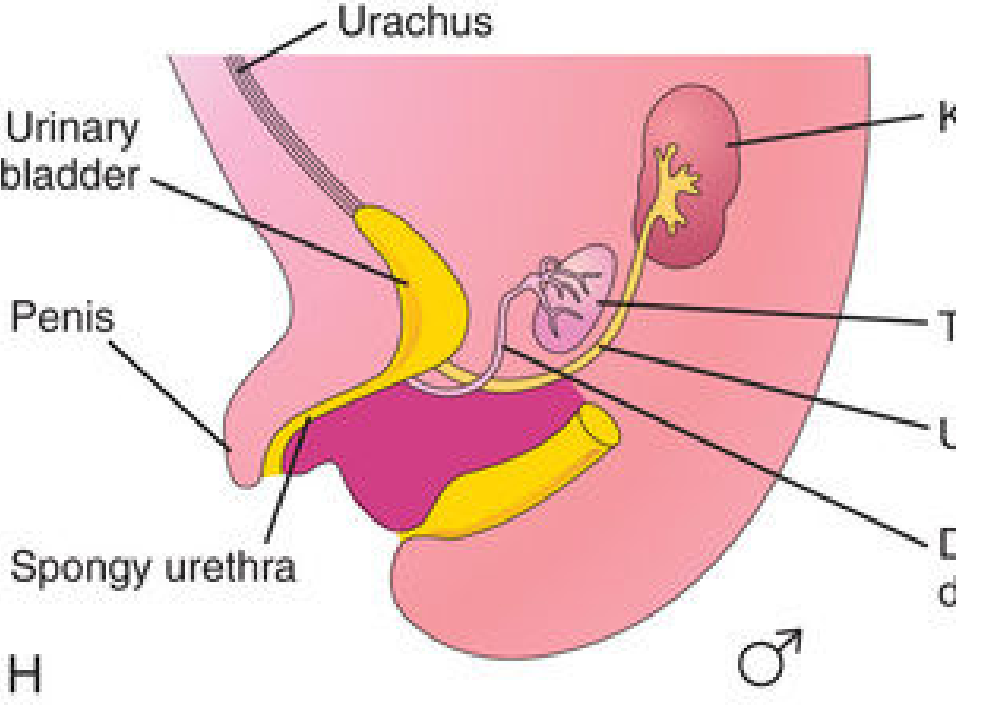
7. Positional Changes with Age
- In infants and children, the urinary bladder is an abdominal organ even when empty
- It begins to enter the greater pelvis at approximately 6 years of age
- It does not become a true pelvic organ until after puberty
8. Onset of Urine Production
Definitive nephrons begin producing urine around the 10th week of gestation. The fetal bladder:
- Can be visualized by ultrasound in ~50% of cases at week 10
- Detection rises to nearly 100% by week 13
- Empties every 15-20 minutes in utero
- Bladder diameter should not exceed 6-8 mm in the first trimester
Campbell-Walsh-Wein Urology, p. 428
9. Congenital Anomalies
A. Urachal Birth Defects
When the urachus fails to fully obliterate, several anomalies result:
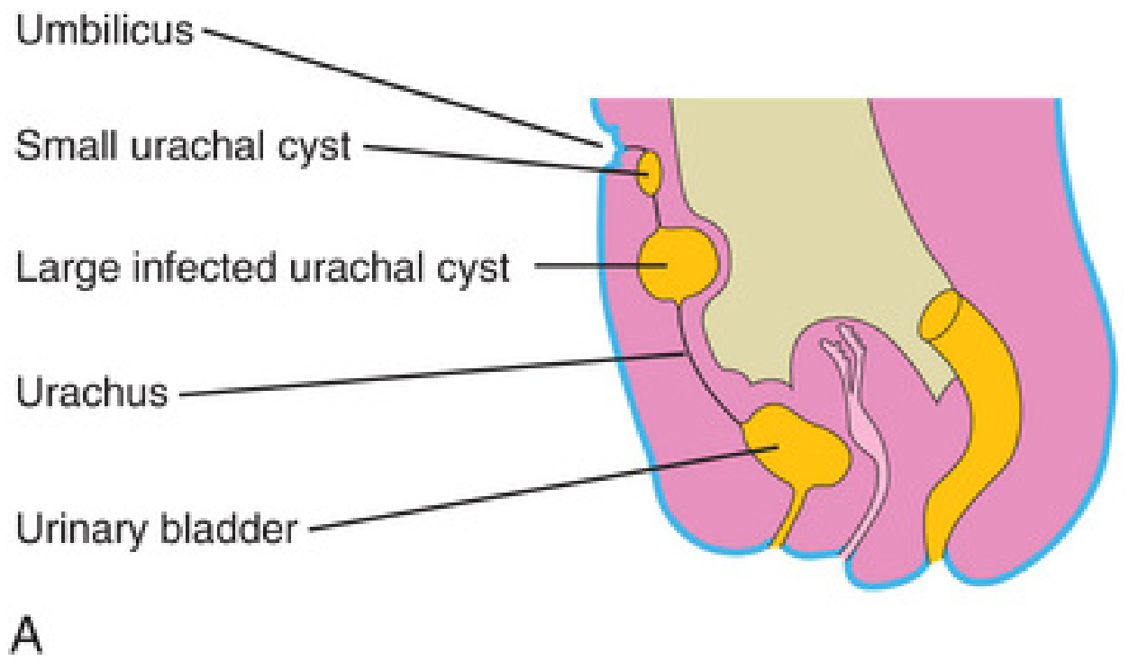
| Anomaly | Description |
|---|---|
| Urachal cyst | Residual epithelium-lined cavity; detected when infected and enlarged |
| Urachal sinus (inferior) | Patent inferior end, opens into the bladder |
| Urachal sinus (superior) | Patent superior end, opens at the umbilicus |
| Patent urachus (urachal fistula) | Entire urachus remains open; urine drips from umbilicus |
B. Congenital Megacystis
A pathologically large bladder caused by:
- Congenital disorder of the ureteric bud (can cause hydronephrosis)
- Posterior urethral valves (most common cause in males)
Many affected infants develop renal failure in early childhood. - The Developing Human, p. 712
C. Exstrophy of the Bladder
- Occurs in approximately 1 in 30,000-50,000 births; more common in males
- Caused by failure of mesoderm to migrate between ectoderm and endoderm of the inferior anterior abdominal wall
- Results in: absent inferior rectus muscles, deficient oblique muscles, exposure of the bladder's posterior mucosal wall
- The trigone and ureteric orifices are exposed; urine dribbles constantly
- Rupture of the cloacal membrane before urorectal septum completion leads to exstrophy of the cloaca (more severe, with hindgut exposure as well)
- Associated with epispadias (urethral opening on the dorsal surface of the penis)
The Developing Human, p. 713-715 | Bailey & Love's Short Practice of Surgery, 28th Ed., p. 1257
Summary Timeline
| Week | Event |
|---|---|
| Week 4 | Cloaca forms; allantois present |
| Weeks 4-7 | Urorectal septum divides cloaca into urogenital sinus + rectum |
| Week 5-7 | Allantois obliterates → urachus forms |
| Week 6-8 | Mesonephric ducts absorbed into bladder wall; trigone forms |
| Week 10 | Kidney begins urine production; bladder detectable on ultrasound |
| Weeks 12+ | Ureters have separate oblique entries; male/female differentiation complete |
| Months 4-5 | Bladder descends into pelvis; urachus elongates → median umbilical ligament |
| Year 6 | Bladder begins entering greater pelvis |
| Puberty | Bladder fully established as pelvic organ |
This is a shared conversation. Sign in to Orris to start your own chat.