
Micro laryngeal surgery and anaesthesia Long note answer in detail also in simple concised and flow chart or images wherever required
Reading File
Now let me search the medical library in parallel for all relevant sections on micro laryngeal surgery and anaesthesia.
Finding Sources
Finding Sources
Finding Sources
Finding Sources
Good. I found key sources. Let me now read the most relevant sections from Morgan & Mikhail (anaesthesia for endoscopy/laryngoscopy), Miller's Anesthesia (laryngeal surgery), and Scott-Brown's (microlaryngoscopy).
Reading File
Finding Sources
Finding Sources
Excellent content. Let me now read the full Morgan & Mikhail section and also pull from Scott-Brown's and Miller's for a complete picture.
Reading File
Reading File
Reading File
Good. Let me now read the core Morgan & Mikhail section on airway management methods (jet ventilation, laser safety), and the Scott-Brown's section on checklist and anesthetic considerations.
Reading File
Reading File
Now let me get the laser precautions section from Morgan & Mikhail and also the Scott-Brown vol2 jet ventilation + MLS setup sections:
Reading File
Reading File
I now have comprehensive material. Let me also fetch the microlaryngoscopy setup image and get the Scott-Brown's chapter on MLS surgical technique/indications.
Loading Image
Reading File
I now have comprehensive material to write a detailed long-note answer. Let me compile it all into a well-structured, flowchart-inclusive response.
Micro Laryngeal Surgery (MLS) and Anaesthesia
1. DEFINITION AND OVERVIEW
Micro Laryngeal Surgery (MLS), also called microlaryngoscopy or phonosurgery, is the endoscopic examination and surgical manipulation of the larynx using a rigid suspension laryngoscope and an operating microscope (or endoscope) for magnification. It provides binocular vision and both hands free for precise, delicate instrumentation.
- Morgan & Mikhail define it as: "laryngoscopy aided by an operating microscope"
- Introduced clinically by Oskar Kleinsasser, who adapted the microscope to direct laryngoscopy to allow fine manipulation of the vocal folds
2. INDICATIONS
| Category | Specific Conditions |
|---|---|
| Benign lesions | Vocal fold polyps, nodules, cysts, Reinke's edema, sulcus vocalis |
| Precancerous | Laryngeal hyperkeratosis (requires biopsy), leukoplakia |
| Neoplastic | Early glottic/supraglottic carcinoma (TLM - transoral laser microsurgery) |
| Infective / Inflammatory | Recurrent respiratory papillomatosis (RRP), arytenoid granuloma |
| Structural | Posterior glottic stenosis, interarytenoid scar, subglottic cysts |
| Diagnostic | Hoarseness workup, assessment of vocal fold mobility, unknown lesions |
| Pediatric | Subglottic cyst, suprastomal granulation, laryngomalacia assessment |
3. SURGICAL ANATOMY - VOCAL FOLD LAYERS
The layered structure of the vocal fold is the foundation for phonosurgical technique:
| Layer | Composition | Depth |
|---|---|---|
| Epithelium | Stratified squamous | ~50 microns |
| Superficial lamina propria (SLP) | Loose fibrous tissue (Reinke's space) | 0.14 mm (F) / 0.30 mm (M) |
| Intermediate lamina propria | Elastic fibers | |
| Deep lamina propria | Collagen fibers | |
| Vocal ligament | Intermediate + deep LP combined | |
| Vocalis muscle (thyroarytenoid) | Deepest layer |
Surgical principle: Preserve the SLP and vocal ligament at all costs. Violation of the vocal ligament leads to scarring and permanent voice impairment.
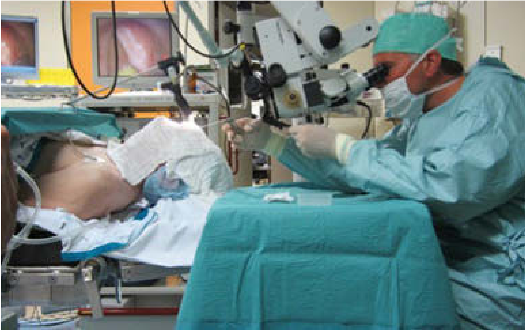
Figure: MLS operating room setup. Patient with cervical flexion and head extension (Boyce-Jackson position), laryngoscope suspended, surgeon using operating microscope.
4. EQUIPMENT AND SETUP
A. Patient Positioning
- Boyce-Jackson position: Flexion at the cervico-thoracic junction + extension at the atlanto-occipital joint ("sniffing position")
- This aligns the oral, pharyngeal, and laryngeal axes to allow direct line-of-sight to the larynx
- A shoulder roll may be used to aid extension
- External laryngeal counter-pressure (by assistant fingers or tape) may improve visualization in difficult cases
B. Laryngoscopes
- Multiple sizes and styles must be available (anterior-lipped, distending types)
- Rigid laryngoscope is suspended using a fulcrum suspension bar attached to the operating table - this leaves both surgeon's hands free for bimanual microsurgery
- Different blade lengths/widths allow access to anterior commissure, posterior glottis, and subglottis
C. Operating Microscope
- Focal length typically 400 mm for adult laryngoscopy
- Provides 6-40x magnification
- CO2 laser may be coupled directly to the microscope (micromanipulator)
D. Instruments
- Bouchayer forceps (atraumatic soft tissue microlaryngeal forceps) - most commonly used
- Curved and straight scissors, microforceps, microelevators, suctions
- Cold steel instruments vs. laser
- Principle: fine, sharp, and well-maintained instruments only - trauma beyond necessary = scarring = worse voice
5. ANAESTHESIA FOR MICRO LARYNGEAL SURGERY
This is a unique anaesthetic challenge: the surgeon and anaesthetist are literally sharing the same airway.
ANAESTHETIC GOALS (Morgan & Mikhail)
┌─────────────────────────────────────────────────────────┐
│           KEY ANAESTHETIC GOALS IN MLS                  │
├─────────────────────────────────────────────────────────┤
│ 1. IMMOBILE SURGICAL FIELD                              │
│    → Profound neuromuscular blockade                    │
│                                                         │
│ 2. MASSETER MUSCLE RELAXATION                           │
│    → For introduction of suspension laryngoscope        │
│                                                         │
│ 3. ADEQUATE OXYGENATION AND VENTILATION                 │
│    → Without obstructing surgical field                 │
│                                                         │
│ 4. CARDIOVASCULAR STABILITY                             │
│    → Despite rapidly varying stimulation                │
│                                                         │
│ 5. RAPID RECOVERY                                       │
│    → Outpatient/day-case procedure typically            │
└─────────────────────────────────────────────────────────┘
5A. PREOPERATIVE ASSESSMENT
History and examination focus:
- Nature of airway problem: voice disorder, stridor, hemoptysis, dysphagia
- Likely diagnosis: papillomas, tracheal stenosis, tumors, vocal cord dysfunction, foreign body
- Can the patient be mask-ventilated? (can we rescue if intubation fails?)
- Can the patient be intubated by conventional/video laryngoscopy?
Investigations:
- Flow-volume loop (especially in subglottic/tracheal stenosis)
- CT/MRI of neck and chest (tumor extent, tracheal narrowing)
- Review of indirect laryngoscopy or flexible nasopharyngoscopy findings
Airway grading:
- Mallampati score, mouth opening, neck extension, thyromental distance
- Previous anaesthetic history, especially any difficult intubation records
Premedication:
- AVOID sedatives in patients with threatening upper airway obstruction
- Glycopyrrolate 0.2-0.3 mg IM (1 hour pre-op): reduces secretions, improves airway visualization, more effective than IV route
5B. INDUCTION OF ANAESTHESIA
FLOW: Preoperative Assessment → Plan Airway Strategy → Induce
                                        ↓
                    Is airway potentially difficult?
                         ↙              ↘
                       YES               NO
                        ↓                ↓
          Awake FOB / Tracheostomy    Standard IV induction
          under local anaesthesia     Propofol + Fentanyl/Remifentanil
                                      + NMB (succinylcholine or rocuronium)
Standard induction agents:
- Propofol 1.5-2.5 mg/kg IV (smooth, antiemetic, good for TIVA)
- Fentanyl or Remifentanil (short-acting opioids preferred for rapid recovery)
- Succinylcholine 1-2 mg/kg (rapid onset, short duration - useful for initial laryngoscopy check)
- OR Rocuronium 1.2 mg/kg with sugammadex reversal (reversed even from profound block with 16 mg/kg)
5C. AIRWAY MANAGEMENT - METHODS OF VENTILATION
This is the most critical and unique aspect of MLS anaesthesia. Multiple techniques exist:
METHOD 1: MICROLARYNGEAL ENDOTRACHEAL TUBE (MLT) - Most Common
┌─────────────────────────────────────────────────────────────┐
│              MICROLARYNGEAL TUBE (MLT)                      │
├────────────────────────────┬────────────────────────────────┤
│ Size: 4.0 / 5.0 / 6.0 mm  │ Same length as adult ETT       │
│ Cuff: HIGH-VOLUME,         │ Disproportionately large cuff  │
│       LOW-PRESSURE         │ Stiff, kink-resistant          │
├────────────────────────────┴────────────────────────────────┤
│ ADVANTAGES:                                                 │
│  ✓ Protects against aspiration                              │
│  ✓ Allows inhalational agents + ETCO2 monitoring            │
│  ✓ Conventional positive-pressure ventilation               │
│  ✓ Secure airway                                            │
├─────────────────────────────────────────────────────────────┤
│ DISADVANTAGES:                                              │
│  ✗ Occupies posterior glottis and vocal cords               │
│  ✗ Obstructs surgical access to posterior commissure        │
│  ✗ Cannot assess true cord mobility                         │
└─────────────────────────────────────────────────────────────┘
Use when: Most elective cases, especially anterior/mid-cord lesions (polyps, nodules, Reinke's edema)
METHOD 2: JET VENTILATION - Field-Clear Technique
Jet ventilation eliminates the tube from the surgical field entirely, providing a completely unobstructed view.
Two approaches:
| Supraglottic Jet | Subglottic / Transtracheal Jet | |
|---|---|---|
| Route | Via laryngoscope side-port above glottis | Via small cannula below glottis |
| Frequency | Low (manual, ~10-15/min) | Low or High-frequency (80-300/min) |
| Mechanism | High-pressure O2 jet (30-50 psi) - entrains air via Venturi effect | Direct subglottic delivery |
| Use in adults | Common for MLS | Hunsaker tube / transtracheal |
| Use in children | Supraglottic described, few centers | Subglottic impractical, barotrauma risk |
Physiology of jet ventilation:
- Inspiration: 1-2 seconds of high-pressure O2 jet
- Expiration: 4-6 seconds - passive, must be allowed to complete
- Chest wall movement must be monitored to prevent air trapping
- ETCO2 is unreliable (constant dilution of alveolar gases) - arterial blood gas may be needed
HFJV (High-Frequency Jet Ventilation):
- 80-300 cycles/minute
- Very small tidal volumes
- Minimal movement of vocal cords - ideal for precise microsurgery
- Requires TIVA (total intravenous anaesthesia) - no inhalational agents possible
METHOD 3: INTERMITTENT APNEA TECHNIQUE
CYCLE:
Pre-oxygenate → Ventilate via mask or ETT
        ↓
   Remove tube / withdraw
        ↓
   Surgeon operates (2-3 min apnea window)
        ↓
  SpO2 monitored continuously
        ↓
   Desaturation → Re-ventilate
        ↓
   Repeat cycle
- Duration of apnea guided by pulse oximetry (usually 2-3 min)
- Risks: Hypoventilation, hypercarbia, failure to re-establish airway, aspiration
- Suitable for short procedures and diagnostic laryngoscopy
METHOD 4: SPONTANEOUS VENTILATION WITH VOLATILE AGENT
- Used mainly in pediatric MLS (diagnostic MLB - microlaryngoscopy and bronchoscopy)
- Sevoflurane via nasopharyngeal tube or oropharyngeal tube at corner of mouth
- Advantage: Dynamic assessment of laryngeal movements possible (cord palsy, malacia)
- Disadvantage: Less control, slower deepening, OR pollution
- Cannot assess dynamic function if paralyzed
SUMMARY TABLE - VENTILATION METHODS
| Method | Tube in Field | ETCO2 | NMB | Inhalational | Best For |
|---|---|---|---|---|---|
| MLT | Yes (small) | Reliable | Needed | Yes/No | Most MLS cases |
| Jet ventilation | No | Unreliable | Needed | TIVA only | Posterior glottis, laser, full exposure |
| Intermittent apnea | No | No | Needed | Possible | Short procedures |
| Spontaneous ventilation | No/minimal | No | Avoided | Yes (sevo) | Pediatric, dynamic assessment |
5D. MAINTENANCE OF ANAESTHESIA
TIVA (Total Intravenous Anaesthesia) - preferred when:
- Jet ventilation is used (no circuit connection)
- Laser surgery (reduces risk of airway fire)
- Day-case requirement
Agents:
- Propofol infusion (TCI or weight-based, 4-12 mg/kg/hr): smooth, antiemetic
- Remifentanil infusion (0.05-0.5 mcg/kg/min): preferred in MLS because:
- Procedure is intensely stimulating during laryngoscopy
- But causes minimal postoperative discomfort (short procedure)
- Titration of longer-acting opioids is difficult in this context
- Dexmedetomidine: useful adjunct for hemodynamic stability
Neuromuscular blockade:
- Profound NMB maintained throughout (immobile field essential)
- Options:
- Rocuronium (1.2 mg/kg) + sugammadex reversal at end - has largely replaced succinylcholine infusion
- Succinylcholine infusion (historical): still an option but largely obsolete now
- Intermittent intermediate-acting NMBs (cisatracurium, vecuronium): for longer procedures
Monitoring:
- Standard: SpO2, ETCO2, ECG, NIBP
- Train-of-four (TOF) monitor to guide NMB depth
- Arterial line during jet ventilation (for ABG if needed)
5E. CARDIOVASCULAR STABILITY
Blood pressure and heart rate fluctuate markedly during MLS because:
- Patients often have significant cardiovascular comorbidities (elderly, smokers, alcohol users)
- The procedure alternates between intense laryngoscopic stimulation and periods of minimal stimulation
Management strategy:
- Modest baseline anaesthesia (don't try to keep constant deep anaesthesia)
- Supplement with short-acting agents during intense stimulation:
- IV bolus propofol or remifentanil
- Esmolol for sudden hypertension/tachycardia
- Nerve blocks (less commonly): glossopharyngeal nerve + superior laryngeal nerve block to blunt cardiovascular response
6. LASER SURGERY CONSIDERATIONS
Laser is commonly coupled to MLS, especially CO2 laser for laryngeal microsurgery. This introduces specific anaesthetic hazards.
Types of Lasers Used
| Laser | Wavelength | Tissue Effect | Use |
|---|---|---|---|
| CO2 | 10,600 nm | High water absorption, superficial, precise | Most common for larynx |
| KTP (532 nm) | 532 nm | Medium penetration, vascular | Vocal fold lesions, papilloma |
| Nd:YAG | 1064 nm | Deep penetration, poor precision | Tracheal/subglottic lesions |
| Diode | Variable | Similar to KTP |
Laser Hazards and Precautions
┌───────────────────────────────────────────────────────────────┐
│                    LASER SAFETY CHECKLIST                     │
├───────────────────────────────────────────────────────────────┤
│  PERSONNEL PROTECTION                                         │
│  ✓ Wavelength-specific laser eye protection for ALL staff     │
│  ✓ Patient's eyes taped shut + moist eye pads                 │
│  ✓ OSHA-compliant respiratory filter masks (laser plume)      │
│  ✓ OR windows covered, laser-in-use signage on door           │
│                                                               │
│  AIRWAY FIRE PREVENTION (Most Critical!)                      │
│  ✓ FiO2 < 30% (if tolerated) - N2 or air as diluent          │
│  ✓ Avoid N2O (supports combustion)                            │
│  ✓ Use laser-resistant ETT (if tube in field)                 │
│  ✓ Cuff inflated with SALINE (not air) - double-cuff tubes    │
│  ✓ No dry cotton or flammable materials near laser            │
│  ✓ Jet ventilation eliminates tube fire risk entirely         │
└───────────────────────────────────────────────────────────────┘
Laser-Resistant ETT Options
| Tube Type | Advantage | Disadvantage |
|---|---|---|
| Polyvinyl chloride (PVC) | Inexpensive, non-reflective | Highly combustible |
| Red rubber | Puncture-resistant | Highly combustible |
| Silicone rubber | Non-reflective | Combustible, toxic ash |
| Metal (stainless steel, flexible) | Combustion-resistant, kink-resistant | Thick flammable cuff, reflects laser |
- Double-cuffed metal tubes (Hunsaker, Mallinckrodt, Laserguard): distal cuff provides backup if proximal cuff fails
- Metallic tape wrapping of standard tubes is suboptimal - no FDA approval, no cuff protection
Airway Fire Protocol
AIRWAY FIRE RESPONSE:
  1. Stop ventilation IMMEDIATELY
  2. Remove endotracheal tube from airway
  3. Disconnect circuit from anaesthesia machine; turn OFF O2
  4. Submerge burning tube in water
  5. Ventilate with face mask; re-intubate
  6. Bronchoscopy to assess airway damage
  7. Serial chest X-rays + ABG
  8. Consider bronchial lavage + IV steroids
7. POSTOPERATIVE CARE
| Issue | Management |
|---|---|
| Edema/stridor | Nebulized adrenaline (1:1000), IV dexamethasone |
| Post-op pain | Usually mild - simple analgesics (paracetamol, NSAID) |
| Nausea/vomiting | Ondansetron + propofol TIVA reduces incidence |
| Voice rest | 5-7 days absolute voice rest after phonosurgery |
| Aspiration risk | Nil-by-mouth until fully awake, head-up position |
| Respiratory depression | Monitor SpO2, have reversal agents ready (sugammadex, naloxone) |
| Day-case discharge | When fully awake, can swallow, stable vitals, pain controlled |
Extubation:
- Deep extubation is controversial - risk of laryngospasm
- Awake extubation preferred: ensure NMB fully reversed (TOF ratio >0.9), spontaneous breathing, responsive
- Have difficult airway trolley immediately available
8. SPECIAL CONSIDERATIONS
A. Difficult Airway / Cannot Intubate Scenario
CANNOT INTUBATE / DIFFICULT AIRWAY ALGORITHM IN MLS:
                    ↓
    Pre-op: Identify risk factors
         (stridor, tumor, prior RT, obesity)
                    ↓
    Awake Flexible Fibreoptic Intubation (AFOI)
         under topical anaesthesia + sedation
                    ↓
    If AFOI fails OR expected severe obstruction:
         Awake tracheostomy under local anaesthesia
                    ↓
    Note: Even tracheostomy does NOT prevent
    intra-op obstruction from surgical manipulation
    or hemorrhage
B. Recurrent Respiratory Papillomatosis (RRP)
- Multiple surgeries required (debulking, not curative)
- Microdebrider or KTP laser via MLS
- Anaesthetic challenge: papillomas can shed and obstruct airway
- Prefer jet ventilation or smallest possible MLT
- Antiviral adjuvants: intralesional cidofovir, bevacizumab
C. Pediatric MLS
- Diagnostic MLB (microlaryngoscopy + bronchoscopy): spontaneous ventilation with sevoflurane preferred for dynamic assessment
- Subglottic jet ventilation impractical and risky for children (barotrauma, air trapping)
- LMA useful if flexible fibreoptic tracheobronchoscopy planned and child difficult to intubate (e.g., mandibular hypoplasia)
- Smaller instruments, greater care with positioning
D. Transoral Laser Microsurgery (TLM) for Laryngeal Cancer
- CO2 laser via suspension MLS for T1/T2 glottic carcinoma
- Excellent oncologic outcomes, voice preservation
- TIVA + jet ventilation is ideal (no flammable tube in field)
- Close coordination between anaesthetist and surgeon for "laser on/off" periods
9. COMPLICATIONS OF MLS
| Complication | Cause | Prevention |
|---|---|---|
| Dental/lip injury | Laryngoscope leverage | Tooth guard, gum protection |
| Nerve injury (glossopharyngeal) | Suspension compression | Careful technique |
| Post-op edema/stridor | Mucosal trauma, RRP | Steroids, adrenaline nebs |
| Airway fire | Laser + O2 + combustible tube | Low FiO2, laser-safe tube, saline cuff |
| Laryngospasm | Post-extubation | Awake extubation, lidocaine |
| Voice worse post-op | Excess mucosal removal, scarring | Conservative phonosurgery, preserve SLP |
| Barotrauma | Jet ventilation | Monitor chest excursion, adequate expiration time |
| Subcutaneous emphysema | Jet ventilation leak | Careful cannula placement |
10. COMPLETE FLOW CHART: ANAESTHETIC MANAGEMENT OF MLS
PATIENT REFERRED FOR MICROLARYNGEAL SURGERY
              ↓
┌─────────────────────────────────┐
│   PRE-OPERATIVE ASSESSMENT      │
│  • Airway: MP, TMD, extension   │
│  • Pathology: stridor? stenosis?│
│  • Imaging: CT, flow-volume     │
│  • Comorbidities, medications   │
│  • Previous anaesthesia records │
└──────────────┬──────────────────┘
               ↓
    CAN AIRWAY BE SECURED SAFELY?
         ↙              ↘
       YES               NO / UNCERTAIN
        ↓                     ↓
IV Induction         AWAKE FOB / Tracheostomy
Propofol +           under local anaesthesia
Remifentanil +
Rocuronium 1.2 mg/kg
        ↓
AIRWAY SECURED - SELECT VENTILATION METHOD:
         ↓
  ┌──────────────────────────────────┐
  │ ANTERIOR/MID CORD LESION?        │
  │  → Microlaryngeal Tube (MLT)     │
  │    4-6 mm, high-vol/low-P cuff   │
  │    Conventional IPPV             │
  └──────────────────────────────────┘
  ┌──────────────────────────────────┐
  │ POSTERIOR GLOTTIS/CORDS/         │
  │ LASER SURGERY?                   │
  │  → Jet Ventilation               │
  │    (Supraglottic via scope port  │
  │     or Subglottic via cannula)   │
  │    TIVA MANDATORY                │
  │    ETCO2 unreliable - ABG PRN    │
  └──────────────────────────────────┘
  ┌──────────────────────────────────┐
  │ SHORT DIAGNOSTIC PROCEDURE?      │
  │  → Intermittent Apnea            │
  │    SpO2 guided, 2-3 min windows  │
  └──────────────────────────────────┘
  ┌──────────────────────────────────┐
  │ PEDIATRIC / DYNAMIC ASSESSMENT?  │
  │  → Spontaneous Ventilation       │
  │    Sevoflurane via NP tube       │
  └──────────────────────────────────┘
               ↓
     MAINTENANCE (TIVA preferred)
     Propofol + Remifentanil infusion
     Profound NMB maintained
     TOF monitoring
               ↓
   IS LASER BEING USED?
         ↙           ↘
       YES             NO
        ↓               ↓
  FiO2 <30%       Standard monitoring
  Avoid N2O        continues
  Laser-safe ETT
  Saline-filled cuff
  Eye protection all
  Laser plume suction
               ↓
     CARDIOVASCULAR STABILITY
     Baseline moderate anaesthesia
     Bolus propofol/remifentanil during
     intense stimulation
     Esmolol for hypertension/tachycardia
               ↓
  END OF PROCEDURE
  Reverse NMB (Sugammadex 16 mg/kg
  from deep block if rocuronium)
  TOF ratio > 0.9
  Awake extubation preferred
               ↓
  POST-OPERATIVE CARE
  SpO2 monitoring, voice rest
  Steroids if edema expected
  Day-case discharge criteria met
QUICK REFERENCE SUMMARY
| Aspect | Key Point |
|---|---|
| Procedure | Suspension laryngoscopy + operating microscope = bimanual microsurgery |
| Position | Boyce-Jackson (cervical flex + head extend) |
| Shared airway | Anaesthetist and surgeon share the same airway |
| Best NMB | Rocuronium + sugammadex (or suxamethonium infusion) |
| Preferred maintenance | TIVA (propofol + remifentanil) |
| Ventilation choice | MLT for most; jet ventilation when full field access or laser needed |
| CO2 laser key risk | Airway fire - FiO2 <30%, no N2O, laser-safe tube with saline cuff |
| Cardiovascular | Fluctuating - use short-acting agents; esmolol for spikes |
| Post-op | Voice rest, steroids for edema, rapid recovery (day case) |
| Difficult airway | Awake FOB or tracheostomy under LA before induction |
Sources:
- Morgan & Mikhail's Clinical Anesthesiology, 7e, Chapter 37 (ENT Anaesthesia, Endoscopy section) - comprehensive anaesthetic management
- Scott-Brown's Otorhinolaryngology Head & Neck Surgery, Chapter 67 (Phonosurgery/MLS technique and anatomy)
- Scott-Brown's Vol 2 (Pediatric MLS, jet ventilation)
- Cummings Otolaryngology HNS (remifentanil in suspension laryngoscopy)
- KJ Lee's Essential Otolaryngology (indications - vocal fold polyps, cysts, subglottic cysts)
This is a shared conversation. Sign in to Orris to start your own chat.