
Postmenopausal bleeding
"postmenopausal bleeding" AND management
postmenopausal bleeding endometrial evaluation algorithm diagram
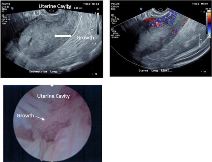
This composite medical image displays three panels illustrating diagnostic findings for endometrial adenocarcinoma in a postmenopausal patient. The top-left panel is a longitudinal grayscale transvaginal ultrasound (TVUS) showing a significantly thickened endometrium (measured at approximately 5 cm) with a heterogeneous echotexture; a white arrow indicates an irregular intraluminal growth. The top-right panel shows a color Doppler ultrasound of the same uterine area, revealing hypervascularity with prominent red and blue signals representing disorganized arterial and venous blood flow within the thickened endometrial tissue. The bottom panel is an endoscopic hysteroscopy image of the uterine cavity, providing a direct view of the lesion. It shows an irregular, raised, pinkish-tan growth with a textured, friable-appearing surface. These images collectively demonstrate the correlation between non-invasive imaging (ultrasound echogenicity and vascular mapping) and direct visualization (hysteroscopy) in the evaluation of postmenopausal bleeding and endometrial malignancy.
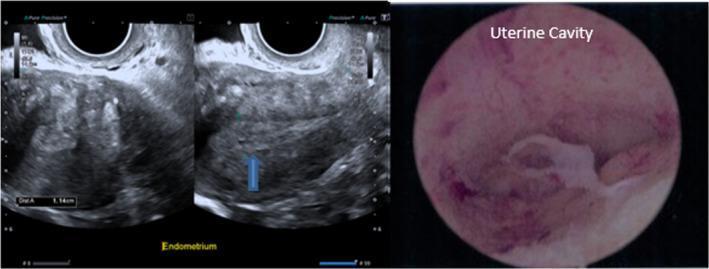
A multi-panel medical image featuring diagnostic imaging of the uterus in a 52-year-old postmenopausal patient. The left panel shows grayscale transvaginal ultrasound (TVUS) images of the uterus in a longitudinal plane. The endometrium is clearly identified, appearing thickened with a heterogeneous echotexture. Electronic calipers indicate an endometrial thickness measurement of 1.14 cm (11.4 mm). A blue arrow highlights a focal hypoechoic area within the thickened endometrial stripe, suggestive of a potential polyp or cystic change. The right panel displays a hysteroscopic view of the uterine cavity. The endoscopic perspective shows a pink, vascular endometrial lining with a prominent, irregular, white-to-tan protruding structure, characteristic of an endometrial polyp. These combined imaging modalities illustrate the evaluation of postmenopausal bleeding and endometrial thickening, which in this clinical case was histologically confirmed as a benign active endometrium.
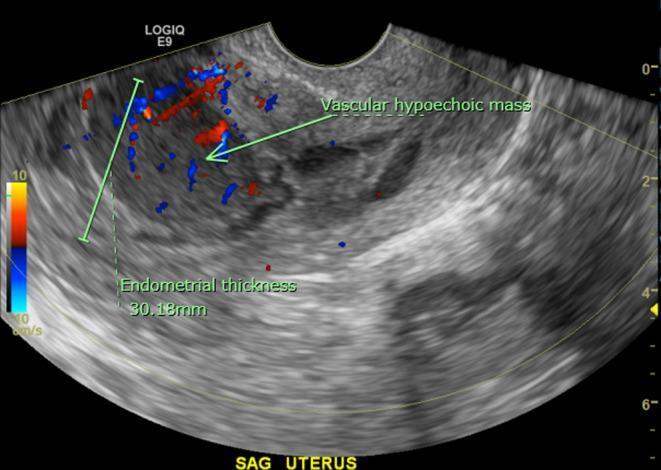
This diagnostic image is a transvaginal ultrasound of the uterus in the sagittal plane, featuring both gray-scale and color Doppler imaging. The uterine view reveals a markedly thickened endometrial stripe measuring approximately 30.18 mm, which is significantly above the normal postmenopausal range. The endometrial contour is irregular and is partially delineated by a trace amount of intrauterine fluid. Within the anterior endometrium, there is a distinct ovoid, homogeneous, hypoechoic mass measuring roughly 3.2 x 2.2 cm. Color Doppler overlay demonstrates internal vascularity within this mass, indicated by red and blue flow signals. Such findings are clinically significant in the context of postmenopausal bleeding, as they necessitate differentiation between endometrial carcinoma, polyps, or rarer entities like primary uterine lymphoma. The image illustrates key sonographic markers for endometrial pathology evaluation including thickness measurement, echogenicity of focal lesions, and assessment of vascular flow patterns.
Postmenopausal Bleeding (PMB)
Definition
Causes and Their Frequency
| Cause | Approximate Frequency |
|---|---|
| Endometrial atrophy | 60-80% |
| Estrogen replacement therapy | 15-25% |
| Endometrial polyps | 2-12% |
| Endometrial hyperplasia | 5-10% |
| Endometrial cancer | ~10% |
- Atrophic vaginitis / endometrial atrophy: Most common cause. Thinning of the endometrial lining and vaginal tissue due to estrogen deficiency. Women with atrophy have typically been menopausal for ~10 years. Endometrial biopsy often yields only blood and mucus, and bleeding usually ceases after biopsy.
- Endometrial polyps: Account for 2-12% of PMB. These are overgrowths of endometrial glands and stroma. The majority are benign, but up to 5% can undergo malignant transformation. Risk factors include increasing age, menopausal status, tamoxifen use, obesity, hypertension, and PCOS.
- Exogenous estrogen: A well-established risk factor. Unopposed estrogen therapy increases endometrial cancer risk 4-8x. Adding a progestin significantly reduces this risk.
- Endometrial hyperplasia: Occurs in 5-10% of PMB cases. Sources of excess estrogen include obesity, exogenous estrogen, or an estrogen-secreting ovarian tumor.
- Endometrial cancer: Present in approximately 10% of PMB cases. Postmenopausal bleeding is the most common presentation of endometrial cancer, and it permits early-stage diagnosis in many cases.
- Uterine leiomyomas (fibroids): Should never be accepted as a cause of PMB - another source must always be excluded.
Non-uterine causes to consider
- Urinary tract sources (hematuria)
- Gastrointestinal bleeding
- Cervical, vaginal, or vulvar lesions (usually evident on exam and require biopsy)
- Traumatic bleeding from an atrophic vagina (accounts for up to 15% of PMB, but uterine cause must first be excluded)
- Functional ovarian tumors producing estrogen
Evaluation
History and Examination
- Thorough history: bleeding pattern, duration, hormone use, medications (especially tamoxifen, anticoagulants), family history
- Pelvic examination: detect local lesions (cervix, vagina, vulva)
- Pap smear (cervical cytology)
Transvaginal Ultrasound (TVUS) - First-line imaging
- ≤4 mm: Low risk for endometrial cancer; endometrial biopsy may be avoided
- >4 mm: Endometrial biopsy is indicated
- Some sources cite a cutoff of <5 mm (Berek & Novak) or even 3 mm as more accurate
Important equity caveat: Recent data show that using a 4 mm cutoff alone may lead to under-diagnosing endometrial cancer in Black patients, and endometrial biopsy should have a lower threshold in this population. (Sabiston Textbook of Surgery, 11th ed.)
Endometrial Biopsy (EMB)
- Office endometrial biopsy is an excellent diagnostic test
- Indicated when:
- Endometrial thickness >4 mm
- Suspected focal lesion on imaging
- Difficult visualization of endometrium
- Persistent bleeding despite normal ultrasound
- Cervical stenosis preventing ultrasound assessment
- Limitation: Can miss focal lesions (polyps); if sampling is inadequate, proceed to hysteroscopy
Hysteroscopy + D&C
- Gold standard for visualizing the endometrial cavity
- Particularly useful when polyps are suspected (difficult to detect with biopsy alone)
- Indicated when office biopsy is non-diagnostic or cervical stenosis precludes biopsy
Sonohysterography (Saline Infusion Sonography)
- Useful adjunct for identifying focal lesions (polyps, submucous fibroids)
Pathology: Endometrial Hyperplasia Classification
| Class | Nomenclature | Treatment |
|---|---|---|
| Benign | Benign endometrial hyperplasia | Medical (progestins) |
| Premalignant | Endometrial intraepithelial neoplasia (EIN) - replaces "atypical hyperplasia" | Surgical (preferred) vs. medical if fertility desired or poor surgical candidate |
| Malignant | Endometrial adenocarcinoma, endometrioid type, well differentiated | Surgical + staging |
Management
Benign causes
- Atrophic vaginitis: Topical or systemic estrogen therapy after malignancy excluded. Vaginal routes (cream, tablet, ring) produce lower systemic levels.
- Cervical polyps: Removed in office
- Endometrial polyps: Hysteroscopic removal
- HRT-related: Regimen adjustment (add or increase progestin)
Endometrial Hyperplasia (EIN / Premalignant)
- Surgical (hysterectomy) is preferred
- Progestin therapy (oral, parenteral, or levonorgestrel IUD) for women who are poor operative candidates or desire fertility
- Follow-up endometrial biopsy every 3 months; recurrence risk approaches 50% with medical management
Endometrial Cancer
- Surgical staging (total hysterectomy + bilateral salpingo-oophorectomy + lymph node assessment) is the primary treatment
- Multidisciplinary review for adjuvant therapy planning
- Progestin therapy for women who are not surgical candidates
Acute Heavy Bleeding
- Unstable women with acute heavy vaginal bleeding: admit for IV estrogen therapy or surgical intervention
Risk Factors for Endometrial Cancer in PMB
- Obesity (unopposed peripheral estrogen from aromatization)
- Unopposed exogenous estrogen
- Late menopause, nulliparity
- Diabetes, hypertension
- Tamoxifen use
- Lynch syndrome / hereditary colorectal cancer syndromes
- Estrogen-secreting ovarian tumor
Key Take-Home Points
- PMB = bleeding >12 months after last menstrual period. Always abnormal - always investigate.
- Most common cause is atrophy (60-80%), but cancer must be excluded in every case.
- ~10% of PMB is endometrial cancer; 90% of endometrial cancer presents with PMB.
- Evaluation begins with TVUS: endometrial thickness ≤4 mm is reassuring; >4 mm warrants biopsy.
- Endometrial biopsy remains the diagnostic cornerstone. Persistent bleeding with a normal ultrasound still warrants biopsy.
- The 4 mm ultrasound threshold may miss cancer in Black patients - maintain a lower threshold for biopsy.
- Uterine fibroids should never be accepted as the cause of PMB.
- Polyps are often missed on blind biopsy - use hysteroscopy when bleeding persists.
Endometrial cancer
"endometrial cancer" AND "treatment"
PMID: 40763788
endometrial cancer staging FIGO histology microscopy adenocarcinoma
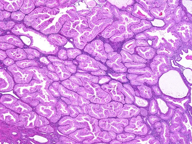
This histology image demonstrates an endometrial glandular neoplasm with well-differentiated, elongated, angulated, and branching glands embedded in a scant stroma. The glandular architecture is predominantly back-to-back, with occasional cribriform-like contours and minimal intervening cytoplasm, consistent with endometrioid histology. Cytologic atypia is mild, featuring uniform nuclei and rare mitotic figures, indicating a low-grade phenotype. The lesion contains a small solid component, described as less than or equal to 5% of tumor volume, which supports a FIGO Grade 1 classification for endometrial adenocarcinoma. The specimen originates from a formalin-fixed paraffin-embedded tissue section, stained with Hematoxylin and Eosin for routine morphologic assessment, and evaluated under bright-field microscopy. Clinically, FIGO Grade 1 endometrioid adenocarcinoma is associated with a relatively favorable prognosis compared with higher-grade counterparts, though invasion depth and myometrial involvement critically influence staging and treatment planning. Diagnostic significance centers on accurate grading, as it guides surgical management and adjuvant therapy decisions. Differential considerations include benign proliferative endometrium, endometrial hyperplasia with atypia, higher-grade endometrioid carcinomas, and serous-endometrial carcinoma. This image is valuable for gynecologic oncology education, pathology training, and research in endometrial carcinogenesis, pattern recognition, and grading criteria. This descriptive caption supports digital teaching libraries, case reviews, and AI-assisted pattern analysis in oncology education.
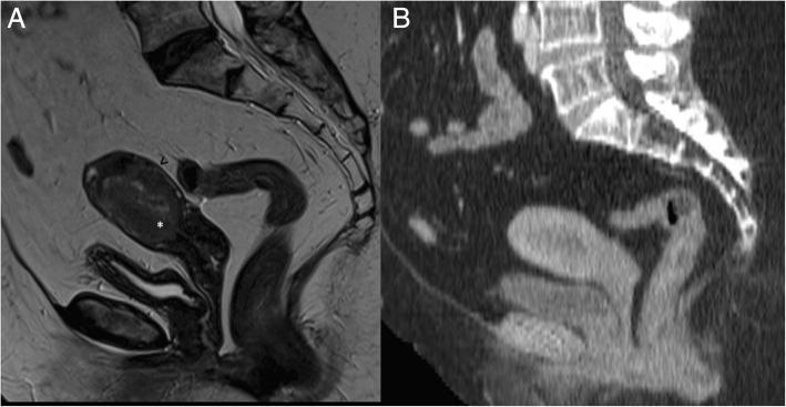
This diagnostic comparison highlights the roles of different imaging modalities in staging endometrial cancer. Image A is a sagittal T2-weighted MRI of the female pelvis showing a 66-year-old patient with grade 3 endometrioid adenocarcinoma. The tumor exhibits intermediate signal intensity and demonstrates deep myometrial invasion (>50% thickness). Key anatomical landmarks include the disruption of the low T2 signal junctional zone (marked by a white asterisk) and the preservation of a thin, low-signal outer myometrial band (indicated by a black arrowhead), consistent with FIGO stage IB. Image B provides a sagittal post-contrast CT scan of the same patient. While CT is utilized for detecting distant metastases, it lacks the soft-tissue contrast resolution seen in MRI, making it difficult to distinguish the specific layers of the uterine wall or the precise depth of myometrial infiltration. This comparison emphasizes MRI's superior sensitivity for local tumor staging and assessing surgical margins in gynecologic oncology.
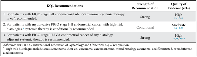
<table> <tr> <th>KQ3 Recommendations</th> <th>Strength of Recommendation</th> <th>Quality of Evidence (refs)</th> </tr> <tr> <td>1. For patients with FIGO stage I-II endometrioid adenocarcinoma, systemic therapy is <i>not</i> recommended.</td> <td>Strong</td> <td>High<br><a href="#">16,17,55</a></td> </tr> <tr> <td>2. For patients with myoinvasive FIGO stage I-II endometrial cancer with high-risk histologies,<sup>*</sup> systemic therapy is conditionally recommended.</td> <td>Conditional</td> <td>Moderate<br><a href="#">16,19,20</a></td> </tr> <tr> <td>3. For patients with FIGO stage III-IVA endometrial cancer of any histology, adjuvant systemic therapy is recommended.</td> <td>Strong</td> <td>High<br><a href="#">16,19,20,56</a></td> </tr> <tr> <td colspan="3"><i>Abbreviations:</i> FIGO = International Federation of Gynecology and Obstetrics; KQ = key question.<br> <sup>*</sup> High-risk histologies include serous carcinoma, clear cell carcinoma, carcinosarcoma, mixed histology carcinoma, dedifferentiated, or undifferentiated carcinoma.</td> </tr> </table>
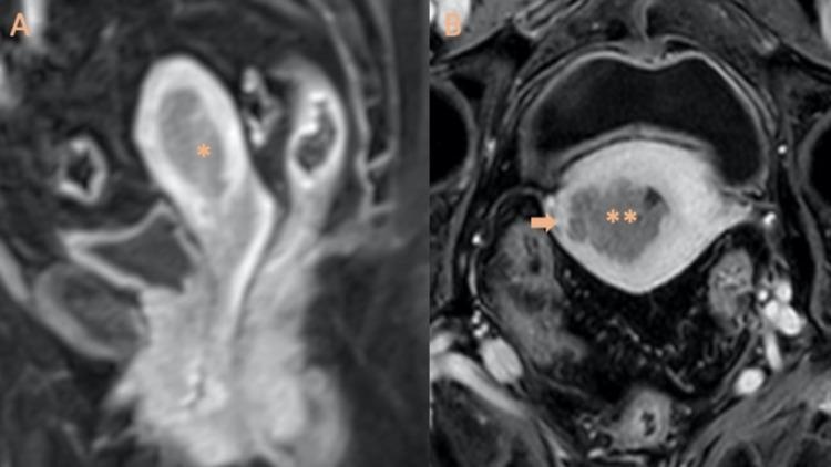
This Comparison Chart consists of two contrast-enhanced T1-weighted MRI images demonstrating the staging of endometrioid adenocarcinoma. Image A (left) displays a Sagittal T1-weighted MRI with intravenous gadolinium, showing a Stage IA grade 2 endometrial tumor (marked with a single asterisk) confined within the uterine cavity without significant myometrial invasion. Image B (right) is a contrast-enhanced MRI showing Stage IB grade 3 endometrioid adenocarcinoma (marked with double asterisks). In this advanced stage, the tumor visibly infiltrates into the outer one-half of the myometrium, with the depth of invasion clearly indicated by an arrow. The use of gadolinium contrast facilitates the differentiation between the enhancing tumor tissue and the surrounding myometrium. These diagnostic images are essential for FIGO staging of endometrial cancer, where Stage IA represents less than 50% myometrial invasion and Stage IB represents 50% or greater invasion. The content is designed for intermediate to advanced medical education in gynecology-oncology and radiology.
Endometrial Cancer
Epidemiology
- ~67,000 new cases and ~13,000 deaths annually in the US
- Fourth most common cancer in women (after breast, lung, colorectal)
- ~3% of women will develop it in their lifetime
- Average age at diagnosis: 60 years; rare before age 45 (only 5% of cases are in women under 40)
- 75% of cases occur in women over 50
- More common in White women but has a higher mortality rate in Black women
Pathogenesis: Type I vs. Type II
| Feature | Type I | Type II |
|---|---|---|
| Frequency | 75-85% | 15-20% |
| Estrogen dependence | Estrogen-dependent | Estrogen-independent |
| Histology | Endometrioid adenocarcinoma | Serous, clear cell, carcinosarcoma |
| Patient profile | Younger, obese, perimenopausal | Older, thin, postmenopausal |
| Background | Arises from hyperplasia | Arises from atrophic endometrium |
| Molecular | PTEN loss, MSI, ER/PR-positive, TP53 wild-type | TP53 mutation, aneuploidy, ERBB2 overexpression |
| Prognosis | Lower grade, earlier stage, favorable | More aggressive, higher mortality |
Critical note: Serous and clear cell carcinomas make up less than 10% of endometrial cancers, yet account for more than half of all endometrial cancer deaths. - Berek & Novak's Gynecology
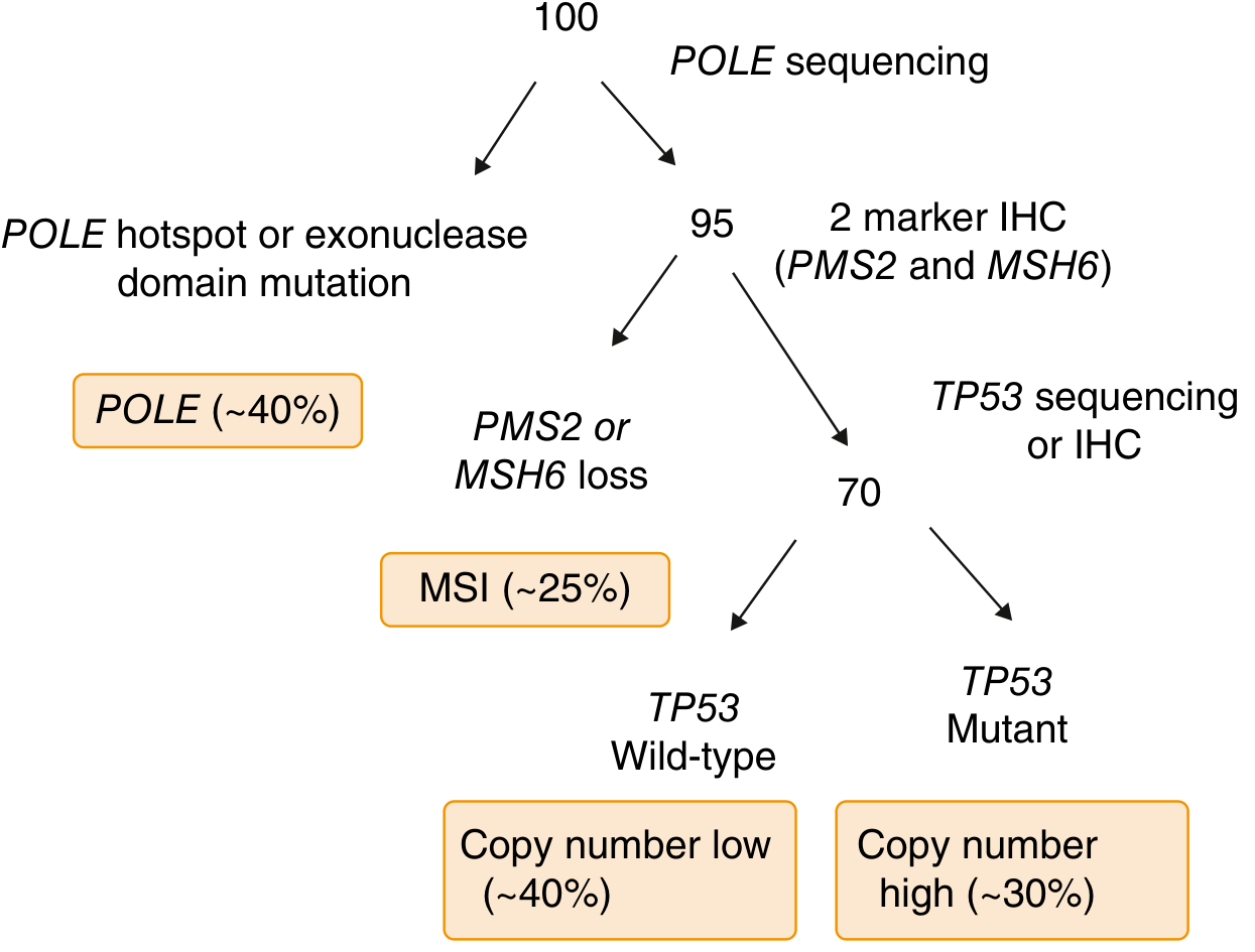
TCGA Molecular Subtypes (Modern Classification)
- POLE ultra-mutated - best prognosis
- Microsatellite instability (MSI) hypermutated - intermediate prognosis; ~30-40% of endometrioid cancers; responds to immunotherapy
- Copy number low (TP53 wild-type)
- Copy number high (TP53 mutated) - worst prognosis, behaves like serous tumors
Risk Factors
| Risk Factor | Relative Risk |
|---|---|
| Complex atypical hyperplasia (EIN) | ~30-fold |
| Unopposed exogenous estrogen | 4-8x |
| Obesity | 1.5-2.5x |
| Late menopause (>52 years) | 2.4x |
| Nulliparity | 2-3x |
| Diabetes mellitus | Elevated |
| Early menarche | Elevated |
| Tamoxifen use | Elevated |
| Lynch syndrome | Up to 60% lifetime risk |
| Cowden syndrome (PTEN mutation) | 13-20% lifetime risk |
| Family history | 1.5-2x |
Genetic Syndromes
- Lynch syndrome (HNPCC): Most common hereditary cause (3-5% of endometrial cancers). Caused by germline mutations in DNA mismatch repair genes: MLH1, MSH2, MSH6, PMS2, or EPCAM. Up to 60% lifetime risk of endometrial cancer; average onset <45 years. All women diagnosed with endometrial cancer should undergo Lynch syndrome screening.
- Cowden syndrome: PTEN tumor suppressor gene mutation. 13-20% lifetime risk.
Clinical Features
Symptoms
- Abnormal vaginal bleeding or discharge in ~90% of cases - the primary presenting symptom
- Most women recognize the significance and seek consultation within 3 months
- Pelvic pressure / discomfort - suggests uterine enlargement or extrauterine spread
- Cervical stenosis can cause hematometra or pyometra (purulent discharge) - associated with poor prognosis
- Less than 5% are asymptomatic at diagnosis
Signs
- Uterine enlargement on pelvic exam (may be absent in early disease)
- Physical exam is often unremarkable in early-stage disease
Histological Subtypes
- Endometrioid adenocarcinoma: Most common (~80%). Well-differentiated glandular pattern
- Mucinous carcinoma: Similar behavior to endometrioid
- Serous carcinoma: Aggressive, spreads early even without deep myometrial invasion
- Clear cell carcinoma: Aggressive; characterized by clear and eosinophilic cells
- Carcinosarcoma (MMMT): Biphasic, contains malignant epithelial AND mesenchymal elements; TP53 mutations common; ~1/3 present with disease beyond uterus
Diagnosis and Pre-treatment Evaluation
Step-by-step workup
- Pelvic examination - assess uterine size, adnexal masses, cervical involvement
- Endometrial biopsy (office aspiration) - first and essential step; sensitivity ~90%
- Transvaginal ultrasound (TVUS): Endometrial thickness >4-5 mm is suspicious; assess depth of myometrial invasion
- Hysteroscopy + D&C: If office biopsy inadequate or non-diagnostic
- MRI pelvis: Best modality for assessing depth of myometrial invasion and cervical involvement (pre-surgical staging)
- CT chest/abdomen/pelvis: Evaluate for lymph node metastasis and distant spread
- Pap smear: Sometimes shows abnormal endometrial cells
Screening
FIGO Staging and Treatment
| Stage | Definition | Standard Treatment |
|---|---|---|
| IA | <50% myometrial invasion | TH + BSO ± vaginal brachytherapy |
| IB | ≥50% myometrial invasion | TH + BSO ± vaginal brachytherapy |
| II | Cervical stromal involvement | TH + BSO + EBRT ± vaginal brachytherapy |
| IIIA | Uterine serosa/adnexa involvement | TH + BSO + systemic therapy ± EBRT |
| IIIB | Vaginal/parametrial involvement | TH + BSO + systemic therapy ± EBRT |
| IIIC1 | Pelvic lymph node involvement | TH + BSO + systemic therapy ± EBRT |
| IIIC2 | Para-aortic lymph node involvement | TH + BSO + systemic therapy ± EBRT |
| IVA | Bladder/bowel invasion | TH + BSO + systemic therapy ± EBRT |
| IVB | Distant metastases / inguinal nodes | TH + BSO + systemic therapy ± EBRT |
Surgical Treatment
- Total hysterectomy + bilateral salpingo-oophorectomy (TH + BSO) is the cornerstone of treatment
- Minimally invasive surgery (laparoscopic or robotic) has largely replaced open surgery for uterine-confined disease
- Peritoneal washings and lymph node assessment are standard
- Sentinel lymph node biopsy is an acceptable alternative to full lymphadenectomy at experienced centers
- Visual inspection of upper abdomen to exclude extrauterine spread
- Full pathologic assessment: histologic subtype, grade, tumor size, depth of invasion, cervical involvement, lymphovascular space invasion (LVSI)
Fertility-Preserving Management
- Continuous progestin-based therapy (oral megestrol or levonorgestrel IUD)
- Repeat endometrial sampling every 3-6 months
- A 2024 systematic review (PMID 39032722) confirms this is an option for carefully selected Stage IA patients
Adjuvant Therapy
Radiotherapy
- Vaginal brachytherapy (VBT): Reduces vaginal recurrence in intermediate-risk Stage I disease
- External beam radiotherapy (EBRT): For cervical involvement (Stage II) and Stage III disease
- Isolated vaginal recurrences can be salvaged by radiotherapy in up to 80% of patients
Chemotherapy
- Used for advanced-stage (III-IV) and high-risk histologies
- Standard regimen: carboplatin + paclitaxel
- Systemic therapy is strongly recommended for FIGO Stage III-IVA disease of any histology
Immunotherapy (emerging / current standard for MMR-deficient tumors)
- Pembrolizumab (PD-1 inhibitor): FDA-approved for MSI-high/MMR-deficient endometrial cancer
- Dostarlimab: Also active in MMR-deficient tumors
- MSI/MMR-deficient tumors respond exceptionally well to checkpoint inhibitors due to high mutational burden
- The NCCN Guidelines Version 3.2025 (PMID 40763788) incorporate updates on the FIGO 2023 staging and new systemic therapy recommendations
Hormonal Therapy
- Progestins: For recurrent disease or fertility-sparing cases; response rates ~25% in advanced disease
- Best response in well-differentiated, ER/PR-positive tumors
Prognostic Variables
- Advancing patient age
- Non-endometrioid or Grade 3 histology
- Deep myometrial invasion (≥50%)
- Lymphovascular space invasion (LVSI)
- Large tumor size
- Cervical extension
- Lymph node metastasis
- Intraperitoneal spread
- Positive peritoneal cytology
Surveillance After Treatment
- Gynecologic exam every 3-6 months for 2-3 years, then annually
- CA-125 if elevated at diagnosis
- CT/PET for symptomatic recurrence
- Vaginal recurrences (most common site of recurrence) are treated with radiotherapy with good salvage rates
Key Take-Home Points
- Endometrial cancer is the most common gynecologic malignancy in developed countries
- Postmenopausal bleeding is the hallmark symptom; 90% present this way, enabling early diagnosis
- Type I (endometrioid, estrogen-related) has a favorable prognosis; Type II (serous/clear cell) is aggressive
- Lynch syndrome is the most important hereditary association - screen all patients
- Surgery (TH + BSO) is the primary treatment; minimally invasive approach preferred
- MMR deficiency / MSI-high status guides immunotherapy eligibility - test all patients
- Adjuvant therapy is guided by surgical stage and histological risk factors
- Isolated vaginal recurrences are potentially curable with radiotherapy (~80% salvage)
Differential diagnosis of post menopausal bleeding
differential diagnosis postmenopausal bleeding causes diagram
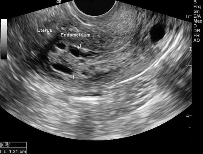
Imaging modality: Transvaginal ultrasound (TVUS) using two-dimensional grayscale ultrasound (B-mode) technique in sagittal (longitudinal) view of the uterus. Precise anatomical localization: uterus with a thickened endometrial stripe measuring approximately 12 mm, consistent with endometrial thickening in a postmenopausal patient. Visual features include a bright, echogenic endometrial lining interrupted by hypoechoic or anechoic cystic spaces within the endometrium, indicating architectural alteration and polypoid or cystic change. In the clinical history provided, endometrial biopsy demonstrated nonatypical endometrial hyperplasia (EH), underscoring nonmalignant proliferative activity but with potential risk for progression if atypia develops. The imaging finding of thickened endometrium (>5 mm in symptomatic postmenopausal women) is non-specific and requires histopathologic confirmation for diagnosis of EH or other pathology such as simple hyperplasia, atypical hyperplasia, or carcinoma. The diagnostic significance lies in risk stratification for uterine neoplasia and guides management decisions including biopsy, surveillance, or therapeutic intervention. Differential considerations include endometrial carcinoma, endometrial polyps, glandular hyperplasia without atypia, chronic endometritis, and atrophic uterus with incidental cystic changes. Clinical correlation emphasizes postmenopausal bleeding history, obesity or polycystic ovarian syndrome risk factors, and the need for biopsy to establish definitive diagnosis. This case (rID 30100) illustrates how TVUS endometrial thickness measurement informs the sequence of workup and confirms pathology via sampling.

Summary : This diagram outlines the process for ruling out other causes of jaundice, listing five main categories and their respective diagnostic steps or criteria. flowchart: # Nodes : • Mechanical obstruction (rectangle): "Rule out HCC/biliary obstruction/Budd-Chiari", "Perform doppler abdominal US and if indicated, MRI" • Drug-induced liver injury (rectangle): "Review detailed history of medication, supplements, pharmacy records", "http://livertox.nih.gov" • Viral hepatitis (rectangle): "Rule out acute Hepatitis A, B, C or E, especially if first episode, or high clinical suspicion" • Auto-immune hepatitis (rectangle): "Rule out severe autoimmune hepatitis if first episode and/or clinical suspicion (ANA, ASMA, IgG)" • Ischemic hepatitis (rectangle): "Presence of hypotension, septic shock, massive bleeding or recent cocaine use" # Connectors : • No explicit arrows shown; nodes are arranged horizontally as parallel diagnostic considerations. # Layout : • Five rectangular nodes arranged in a single horizontal row, each representing a distinct cause to rule out. • Each node contains bullet points listing specific diagnostic actions or criteria. # Analysis : • The diagram provides a systematic checklist for clinicians to exclude major alternative causes of jaundice before proceeding with further workup. Each category is clearly separated, emphasizing the need for a comprehensive differential diagnosis. No decision branches or feedback loops are depicted; the structure is a parallel list rather than a sequential flow.
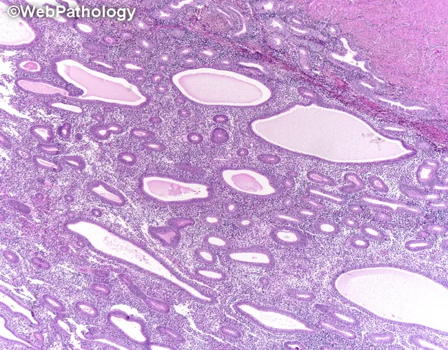
This hematoxylin and eosin stained section presents endometrial tissue from a perimenopausal patient with abnormal uterine bleeding. The architectural pattern shows cystically dilated glands with marked crowding and a back-to-back arrangement within fibromuscular stroma, yielding an overall increased gland-to-stroma ratio. Gland lumina are variably tortuous and irregular in contour, but the epithelial lining demonstrates only minimal cytologic atypia in this field. There is no evident stromal invasion or myometrial extension. The morphology is compatible with endometrial hyperplasia; features may reflect non-atypical hyperplasia or, if nuclear atypia is present in broader sections, atypical hyperplasia/EIN. In the clinical context provided, abnormal non-cyclical bleeding—postmenopausal bleeding, menorrhagia, dysmenorrhea, or intermenstrual bleeding—raises concern for progression to endometrial neoplasia. Differential considerations include complex hyperplasia with atypia, endometrioid intraepithelial neoplasia, and early endometrial carcinoma, particularly if atypia is evident on sampling. Pathologic significance lies in stratifying risk of progression and guiding management, which ranges from surveillance and hormonal therapy to surgical intervention depending on age, fertility wishes, and the presence of cytologic atypia. This image thus supports a radiologic-pathologic correlation for endometrial proliferative disorders in women presenting with abnormal uterine bleeding. Correlation with imaging and clinical data strengthens diagnostic confidence. These findings guide patient-centered treatment planning. decisions.
Differential Diagnosis of Postmenopausal Bleeding
Overview by Frequency
| Cause | Approximate Frequency |
|---|---|
| Endometrial atrophy | 60-80% |
| Exogenous hormone use (HRT) | 15-30% |
| Endometrial polyps | 2-12% |
| Endometrial hyperplasia (EIN) | 5-10% |
| Endometrial cancer | ~10% |
| Other tumors (vulvar, vaginal, cervical) | ~10% |
| Atrophic vaginitis (traumatic) | up to 15% |
Category 1: Non-Genital Tract Sources
Urinary Tract
- Hematuria - cystitis, urethritis, bladder cancer, urethral caruncle
- Urethral caruncle: a benign red, fleshy protrusion at the urethral meatus, common in postmenopausal women due to estrogen deficiency; often mistaken for vaginal bleeding
- Testing: urinalysis, urine culture, cystoscopy if indicated
Gastrointestinal Tract
- Rectal bleeding - hemorrhoids, colorectal cancer, diverticular disease, anal fissure
- Elderly patients may not accurately localize the source between the bladder, uterus, and rectum
- Testing: stool occult blood, proctoscopy, colonoscopy
Category 2: Vulvar and Vaginal Causes
Atrophic Vaginitis (Genitourinary Syndrome of Menopause)
- Most common non-uterine genital cause - accounts for up to 15% of all PMB
- Mechanism: estrogen deficiency causes thinning, dryness, and friability of the vaginal and vulvar epithelium; minor trauma causes bleeding
- Examination: thin, pale, friable vaginal mucosa with loss of rugae; reduced vaginal caliber
- Diagnosis of exclusion: a uterine source must be excluded before attributing bleeding to atrophy
- Treatment: topical or systemic estrogen
Vaginal Lesions
- Vaginal carcinoma (primary or metastatic): usually squamous cell; associated with HPV; presents as a visible vaginal wall lesion - requires biopsy
- Vaginal intraepithelial neoplasia (VAIN): premalignant; detected on colposcopy
- Vaginal trauma: from pessary use, douching, or intercourse in an atrophic vagina
- Vaginal polyps: benign, but must be distinguished from malignant lesions
Vulvar Lesions
- Vulvar carcinoma: squamous cell most common; visible ulcer, plaque, or mass on exam; biopsy required
- Lichen sclerosus: chronic inflammatory condition causing white plaques, thinning, and architectural destruction; may bleed with minor trauma; pre-malignant potential
- Lichen planus / contact dermatitis: inflammatory causes of vulvar bleeding
Category 3: Cervical Causes
Benign
- Cervical polyps: benign pedunculated outgrowths from the endocervical canal; can bleed with minimal contact; easily removed in office; endometrial biopsy still needed to exclude uterine source
- Cervical ectropion: uncommon postmenopausally (usually regresses)
- Cervicitis: uncommon postmenopausally but possible with atrophic changes or retained pessary
Malignant / Premalignant
- Cervical carcinoma: squamous cell (~80%) or adenocarcinoma (~20%); grossly visible lesion (friable, bleeding mass or ulcer) on speculum exam in most cases; diagnosed by biopsy; Pap smear may be abnormal
- Cervical intraepithelial neoplasia (CIN 3): premalignant; identified on colposcopy and biopsy
Category 4: Uterine / Endometrial Causes
Atrophic Endometrium (Most Common)
- Endometrial atrophy: accounts for 60-80% of all PMB; the endometrium thins due to estrogen withdrawal
- Mechanism: fragile, attenuated endometrium bleeds spontaneously or with minor stimulation
- TVUS: very thin endometrial stripe (<4 mm); biopsy yields scant tissue or blood and mucus
- Bleeding usually ceases after biopsy
- Women typically menopausal for ~10 years before presenting
Endometrial Polyps
- Account for 2-12% of PMB
- Benign overgrowths of endometrial glands and stroma (sessile or pedunculated)
- Up to 5% may undergo malignant transformation
- Often missed on blind endometrial biopsy - diagnosed best by hysteroscopy or sonohysterography
- Risk factors: increasing age, menopause, tamoxifen use, obesity, hypertension, PCOS
- Undetected polyps are a common cause of recurrent/persistent PMB
Endometrial Hyperplasia / EIN
- Accounts for 5-10% of PMB
- Benign endometrial hyperplasia: excess estrogen stimulation; treated medically with progestins
- Endometrial intraepithelial neoplasia (EIN): premalignant; ~30-fold risk of progressing to carcinoma; treated surgically (hysterectomy preferred) or with progestins if surgery is contraindicated
- Sources of excess estrogen: obesity (peripheral aromatization), unopposed HRT, estrogen-secreting ovarian tumor
Endometrial Cancer
- Accounts for ~10% of PMB - present in every case until excluded
- The most important diagnosis not to miss
- Type I (endometrioid): estrogen-related, better prognosis
- Type II (serous, clear cell, carcinosarcoma): estrogen-independent, aggressive
- Risk factors: obesity, nulliparity, unopposed estrogen, late menopause, DM, Lynch syndrome, tamoxifen
Uterine Leiomyoma (Fibroids)
- Important: fibroids should NEVER be accepted as a cause of PMB - they typically regress after menopause and another cause must always be excluded
- Rarely, submucosal fibroids may contribute to bleeding if still present and growing
- Rapid growth or growth after menopause raises suspicion for leiomyosarcoma
Uterine Sarcoma
- Rare but aggressive
- Leiomyosarcoma, endometrial stromal sarcoma, carcinosarcoma (MMMT)
- Suspect when fibroids appear to grow rapidly after menopause
Endometritis
- Chronic endometritis can cause irregular bleeding; may be associated with retained tissue or instrumentation
- Pyometra (pus in the uterus) may be associated with cervical stenosis; purulent discharge + bleeding; poor prognostic sign when associated with endometrial cancer
Intrauterine Adhesions / Hematometra
- Cervical stenosis can obstruct blood, causing hematometra
- Usually presents as cyclic pelvic pain without external bleeding, but can present with purulent discharge
Category 5: Ovarian / Adnexal Causes
- Estrogen-secreting ovarian tumors (granulosa cell tumor, thecoma): rare but important; produce excess estrogen leading to endometrial stimulation, hyperplasia, and bleeding
- Bilateral ovarian enlargement or pelvic mass on examination / TVUS
- Evaluate: pelvic TVUS, serum estradiol, inhibin levels (granulosa cell tumor marker)
Category 6: Iatrogenic / Drug-Related Causes
| Drug / Agent | Mechanism |
|---|---|
| Unopposed estrogen HRT | Directly stimulates endometrium; 4-8x risk of endometrial cancer |
| Combined HRT (estrogen + progestin) | Scheduled or unscheduled "breakthrough" bleeding; especially in continuous regimens |
| Tamoxifen (selective estrogen receptor modulator) | Estrogenic effect on endometrium; increases risk of polyps, hyperplasia, endometrial cancer |
| Anticoagulants (warfarin, DOACs) | Impair hemostasis; amplify bleeding from any structural lesion |
| Antiplatelet agents (aspirin, clopidogrel) | Impair platelet function |
| Antipsychotics / antiepileptics | Can affect hormonal axes (less common post-menopause) |
Summary Framework: Anatomical Approach
NON-GENITAL
├── Urinary: hematuria, urethral caruncle
└── GI: rectal bleeding

VULVA / VAGINA
├── Atrophic vaginitis ← most common non-uterine cause
├── Vaginal / vulvar carcinoma
├── Lichen sclerosus
└── Trauma (pessary, intercourse)

CERVIX
├── Cervical polyp
└── Cervical carcinoma

UTERUS ← most clinically critical zone
├── Endometrial atrophy ← most common overall (60-80%)
├── Endometrial polyp (2-12%)
├── Endometrial hyperplasia / EIN (5-10%)
├── Endometrial cancer (~10%) ← must exclude in ALL cases
├── Uterine sarcoma (rare)
└── Endometritis / pyometra

OVARY
└── Estrogen-secreting tumor (granulosa cell, thecoma)

IATROGENIC
├── HRT (unopposed estrogen or breakthrough on combined)
└── Tamoxifen, anticoagulants
Key Diagnostic Principles
- Any PMB = abnormal until proven otherwise. Severity does not correlate with risk of malignancy.
- Atrophy is the most common cause but is a diagnosis of exclusion - endometrial cancer must first be ruled out.
- Uterine fibroids should never be accepted as the explanation for PMB.
- Cervical and vaginal/vulvar lesions are usually visible on speculum exam - examine carefully before attributing bleeding to a uterine source.
- Elderly patients may not accurately localize pain or bleeding between the bladder, uterus, and rectum - always perform a full pelvic exam.
- Tamoxifen is a particularly important iatrogenic cause, as it thickens the endometrium paradoxically and increases polyp/hyperplasia/cancer risk despite being an anti-estrogen at the breast.
- Estrogen-secreting ovarian tumors must be considered, especially when TVUS shows a thickened endometrium without an obvious intrauterine lesion.