
Explained esr and pcv physiology mbbs 1 year
Reading File
Finding Sources
Finding Sources
Searching PubMed
erythrocyte sedimentation rate AND hematocrit
Searching the Web
ESR test hematocrit PCV physiology Merck Manual
Finding Sources
Reading File
Reading File
Searching the Web
erythrocyte sedimentation rate physiology factors increased decreased official clinical laboratory manual
ESR and PCV Physiology
1. ESR: Erythrocyte Sedimentation Rate
Definition:
ESR is the distance that red blood cells (RBCs) fall in a vertical column of anticoagulated blood in 1 hour. It is expressed in mm/hour.
ESR is the distance that red blood cells (RBCs) fall in a vertical column of anticoagulated blood in 1 hour. It is expressed in mm/hour.
It is a nonspecific marker of inflammation. It does not diagnose a particular disease by itself.
Principle of ESR
Normally, RBCs carry a negative surface charge called zeta potential. This causes the cells to repel each other and remain dispersed in plasma.
During inflammation, plasma concentrations of acute-phase proteins, especially fibrinogen, increase.
- Fibrinogen reduces the repulsion between RBCs.
- RBCs stick together in stacks called rouleaux.
- Rouleaux are heavier and settle more rapidly.
- Therefore, the ESR rises.
Key line:
Increased plasma fibrinogen -> rouleaux formation -> faster sedimentation -> raised ESR.
The ESR occurs in three stages:
| Stage | Approximate time | What happens |
|---|---|---|
| Aggregation phase | First 10 min | RBCs form rouleaux, little settling occurs |
| Sedimentation phase | Next 40 min | RBCs settle at a fairly constant rate |
| Packing phase | Last 10 min | RBCs accumulate at bottom, settling slows |
Henry's Clinical Diagnosis and Management by Laboratory Methods, p. 562.
ESR methods
- Westergren method: Standard and commonly used method. Uses a 200 mm vertical tube.
- Wintrobe method: Uses a shorter tube and can also be used for PCV.
Typical reference ranges
Ranges differ among laboratories and rise with age. Common approximate adult values are:
- Male: 0-15 mm in first hour
- Female: 0-20 mm in first hour
Causes of increased ESR
Physiological
- Female sex
- Increasing age
- Pregnancy
- Menstruation
Pathological
- Acute and chronic infections, especially tuberculosis
- Inflammatory and autoimmune disorders, such as rheumatoid arthritis
- Tissue injury or necrosis
- Malignancy
- Chronic kidney disease
- Anemia
- Multiple myeloma and other conditions with increased immunoglobulins
Causes of decreased ESR
- Polycythemia or increased RBC count
- Sickle-cell disease and marked poikilocytosis, because abnormal-shaped RBCs do not form rouleaux well
- Severe leukocytosis
- Low fibrinogen level, for example in hypofibrinogenemia
Factors influencing ESR
| Factor | Effect on ESR | Reason |
|---|---|---|
| Fibrinogen and globulins increase | Increases | Promotes rouleaux |
| Albumin increase | Decreases | Retards sedimentation |
| Anemia / low PCV | Increases | More plasma relative to RBCs, easier settling |
| Polycythemia / high PCV | Decreases | More RBC crowding, slower settling |
| Macrocytes | Increases | Larger cells settle faster |
| Microcytes | Decreases | Smaller cells settle slowly |
| High temperature | Increases | Faster sedimentation |
| Tilted ESR tube | Falsely increases | Alters settling path |
ESR must always be interpreted along with history, examination, and tests such as CRP. It may remain elevated longer than CRP after inflammation improves. The MedlinePlus ESR overview also emphasizes that it is a nonspecific inflammation test.
2. PCV: Packed Cell Volume
Other names: Hematocrit, HCT.
Definition:
PCV is the percentage of total blood volume occupied by packed red blood cells after centrifuging anticoagulated blood.
PCV is the percentage of total blood volume occupied by packed red blood cells after centrifuging anticoagulated blood.
[
\text{PCV (%)} =
\frac{\text{Height of packed red-cell column}}{\text{Total height of blood column}}
\times 100
]
Principle
When anticoagulated blood is centrifuged, its components separate according to density:
- Bottom: Packed RBCs, the heaviest component
- Middle: Buffy coat, made of WBCs and platelets
- Top: Plasma
PCV measures the RBC layer as a percentage of the whole blood column.
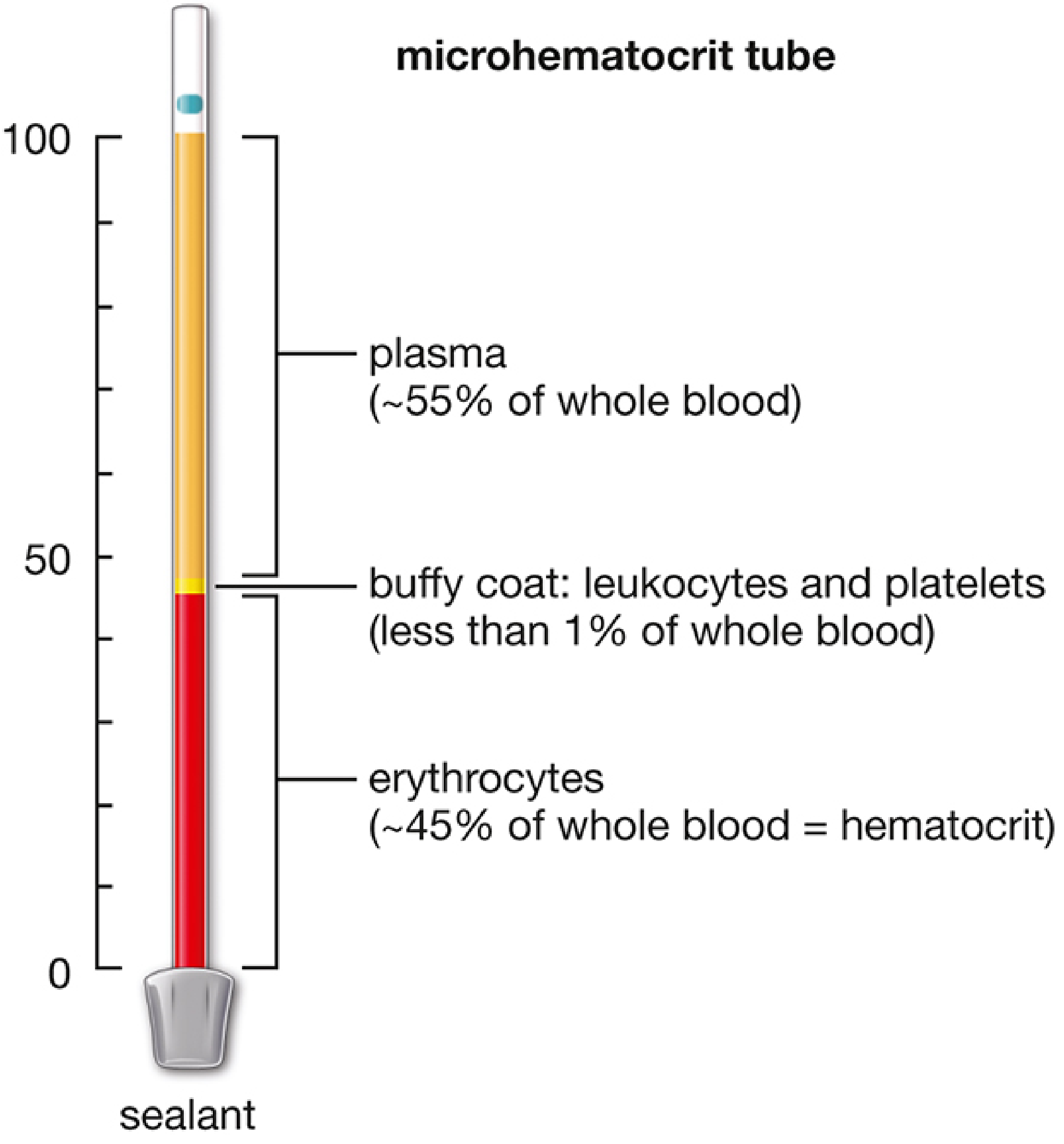
Methods of measuring PCV
-
Microhematocrit method
- Blood is filled in a heparinized capillary tube.
- One end is sealed.
- It is centrifuged at high speed.
- PCV is read using a hematocrit reader.
- Commonly used in practical physiology.
-
Wintrobe method
- Blood is placed in a Wintrobe tube and centrifuged.
- The same tube can help determine ESR and PCV.
-
Automated analyzer
- Modern laboratories commonly calculate hematocrit from RBC count and MCV.
[
\text{Hematocrit (%)} \approx \frac{\text{RBC count} \times \text{MCV}}{10}
]
Normal PCV values
Approximate adult ranges:
| Group | PCV |
|---|---|
| Adult male | 40-50% |
| Adult female | 36-45% |
| Newborn | 50-60% |
In normal blood, RBCs occupy roughly 45% of blood volume, plasma about 55%, and the buffy coat less than 1%. Histology: A Text and Atlas with Correlated Cell and Molecular Biology, p. 668.
Increased PCV
This occurs due to either a real increase in RBC mass or loss of plasma.
- Polycythemia vera
- High altitude
- Chronic hypoxia, for example chronic lung disease or cyanotic heart disease
- Dehydration, vomiting, diarrhea, burns
Decreased PCV
- Anemia
- Acute or chronic blood loss
- Hemolysis
- Nutritional deficiencies, such as iron, vitamin B12, or folate deficiency
- Pregnancy, due to increased plasma volume
- Bone marrow failure
Clinical importance of PCV
PCV gives an estimate of:
- Red cell mass
- Oxygen-carrying capacity, together with hemoglobin
- Severity of anemia
- Hemoconcentration or dehydration
- Polycythemia
Relationship between ESR and PCV
This is a frequently asked MBBS viva point:
- Low PCV, as in anemia -> ESR increases.
- High PCV, as in polycythemia -> ESR decreases.
Reason: in anemia, the lower RBC concentration favors rouleaux formation and settling. In polycythemia, abundant RBCs hinder free settling.
One-line viva answers
- ESR: Rate at which RBCs settle in a column of anticoagulated blood in one hour.
- Unit of ESR: mm in the first hour.
- PCV: Percentage of blood volume occupied by packed RBCs after centrifugation.
- Other name for PCV: Hematocrit.
- Why does ESR rise in inflammation? Increased fibrinogen causes rouleaux formation, so RBCs settle faster.
- Why does ESR rise in anemia? Reduced RBC concentration favors rouleaux formation and sedimentation.
- Why is ESR low in polycythemia? Increased RBC concentration slows sedimentation.
Give me MBBS viva questions and answers on ESR and PCV
Reading File
Finding Sources
Finding Sources
Searching PubMed
erythrocyte sedimentation rate AND hematocrit
Searching the Web
ESR test PCV hematocrit laboratory procedure reference MedlinePlus
MBBS Viva Questions and Answers: ESR and PCV
A. ESR: Erythrocyte Sedimentation Rate
1. What is ESR?
ESR is the rate at which red blood cells settle down in a vertical column of anticoagulated blood in one hour. It is expressed as mm in the first hour (mm/hr).
2. What is the principle of ESR?
Normally, RBCs repel each other because of their negative surface charge, called zeta potential. In inflammation, plasma fibrinogen and some globulins increase, reduce this repulsion, and promote RBC aggregation into rouleaux. These aggregates settle faster, increasing ESR.
3. What is rouleaux formation?
It is the aggregation of RBCs in coin-like stacks. It occurs more easily when fibrinogen or globulins are increased.
4. Why does fibrinogen increase ESR?
Fibrinogen decreases zeta potential between RBCs, allowing rouleaux formation. Rouleaux are heavier than individual RBCs, so they sediment more rapidly.
5. What is the unit of ESR?
Millimetres in the first hour, written as mm/hr.
6. Which anticoagulant is used for ESR?
For the Westergren method, sodium citrate is classically used. EDTA blood may also be used after appropriate dilution with citrate in many laboratories.
7. Name the methods of ESR estimation.
- Westergren method
- Wintrobe method
- Automated ESR methods
8. Which is the standard method for ESR?
Westergren method is the standard and more sensitive method, especially for higher ESR values.
9. What is the length of the Westergren tube?
200 mm.
10. What is the length of the Wintrobe tube?
Approximately 100 mm.
11. Which method can measure both ESR and PCV?
Wintrobe tube method.
12. What are the phases of ESR?
There are three phases:
- Aggregation phase: first 10 minutes, rouleaux form and little sedimentation occurs.
- Sedimentation phase: next 40 minutes, RBCs settle at a nearly constant rate.
- Packing phase: final 10 minutes, RBCs pack at the bottom and sedimentation slows.
Henry's Clinical Diagnosis and Management by Laboratory Methods, p. 562.
13. What are normal ESR values?
Approximate values, which vary with age and laboratory:
- Adult male: 0-15 mm/hr
- Adult female: 0-20 mm/hr
14. Why is ESR normally higher in females?
Females generally have a lower hematocrit than males. A lower RBC concentration favors sedimentation. Hormonal and physiological factors may also contribute.
15. Why does ESR increase in anemia?
In anemia, the RBC concentration and PCV are reduced. The relatively larger plasma volume permits easier rouleaux formation and faster settling of RBCs.
16. Why is ESR low in polycythemia?
In polycythemia, the RBC concentration and PCV are high. The cells are crowded, which slows their settling, so ESR falls.
17. Give causes of raised ESR.
- Acute and chronic infections
- Tuberculosis
- Autoimmune and inflammatory diseases, such as rheumatoid arthritis
- Tissue injury or necrosis
- Malignancy
- Anemia
- Pregnancy
- Multiple myeloma and other paraproteinemias
- Chronic kidney disease
18. Give causes of low ESR.
- Polycythemia
- Marked leukocytosis
- Sickle-cell disease
- Spherocytosis and severe poikilocytosis
- Hypofibrinogenemia
- Congestive heart failure
19. Why is ESR raised in multiple myeloma?
There is increased production of immunoglobulins, leading to rouleaux formation and rapid RBC sedimentation.
20. Why may ESR be low in sickle-cell anemia despite anemia?
Sickle-shaped RBCs cannot form rouleaux effectively. This reduces sedimentation and may produce a low ESR despite anemia.
21. Which plasma proteins increase ESR?
Fibrinogen is the most important. Increased globulins, especially immunoglobulins, can also raise ESR.
22. Which plasma protein decreases ESR?
Albumin tends to retard sedimentation and can lower ESR.
23. What are the technical errors that falsely raise ESR?
- Tilted tube
- Increased room temperature
- Vibration
- Delay in testing the sample
- Improper anticoagulant ratio
- Diluted blood sample
- Dirty or wet tube
24. What are the technical errors that falsely lower ESR?
- Short duration of reading
- Low temperature
- Improperly mixed blood
- Clotted sample
- Underfilling of tube
- Very high hematocrit
- Incorrect anticoagulant proportion
25. Is ESR a specific test?
No. ESR is a nonspecific marker of inflammation. It does not identify the cause or site of disease.
26. What is the clinical use of ESR?
It is useful for:
- Supporting evidence of inflammation
- Monitoring some chronic inflammatory diseases
- Following disease activity in conditions such as rheumatoid arthritis, polymyalgia rheumatica, and giant-cell arteritis
27. What is the difference between ESR and CRP?
| ESR | CRP |
|---|---|
| Indirect, nonspecific test | Acute-phase protein measured directly |
| Influenced by age, sex, anemia, RBC shape, and PCV | Less affected by RBC-related factors |
| Rises and falls relatively slowly | Rises and falls more rapidly |
| May remain elevated after recovery | Better reflects recent changes in inflammation |
B. PCV: Packed Cell Volume
1. What is PCV?
Packed cell volume is the percentage of total blood volume occupied by packed red blood cells after centrifugation of anticoagulated blood.
2. What is another name for PCV?
Hematocrit, abbreviated as HCT.
3. State the formula for PCV.
[
\text{PCV (%)} =
\frac{\text{Height of packed RBC column}}{\text{Total height of blood column}}
\times 100
]
4. What is the principle of PCV estimation?
On centrifugation, blood separates according to density:
- Bottom: packed RBCs
- Middle: buffy coat, containing WBCs and platelets
- Top: plasma
The proportion occupied by packed RBCs is the PCV.
5. Which anticoagulant is used for PCV?
- Heparinized capillary tube for capillary blood in the microhematocrit method
- EDTA blood for venous blood samples
6. Name methods for PCV estimation.
- Microhematocrit method
- Wintrobe method
- Automated hematology analyzer
7. Which method is commonly used in practical physiology?
Microhematocrit method, also called the capillary hematocrit method.
8. What is the normal PCV value in adult males?
Approximately 40-50%.
9. What is the normal PCV value in adult females?
Approximately 36-45%.
10. What is the normal PCV in newborns?
Approximately 50-60%.
11. What is the approximate composition of normal blood by volume?
- RBCs: about 45%
- Plasma: about 55%
- Buffy coat: less than 1%
The buffy coat contains leukocytes and platelets. Histology: A Text and Atlas with Correlated Cell and Molecular Biology, p. 668.
12. What does low PCV indicate?
Usually anemia, blood loss, hemolysis, nutritional deficiency, bone marrow suppression, or hemodilution.
13. What does high PCV indicate?
It may indicate:
- Polycythemia
- Chronic hypoxia, for example at high altitude
- Chronic lung disease
- Cyanotic congenital heart disease
- Dehydration, diarrhea, vomiting, or burns
14. Why is PCV raised in dehydration?
Plasma volume decreases due to fluid loss. RBC mass may be normal, but its proportion relative to plasma increases. This is called relative polycythemia or hemoconcentration.
15. Why is PCV reduced in pregnancy?
Plasma volume rises more than red cell mass during pregnancy, producing physiological hemodilution.
16. What is the buffy coat?
It is the thin, whitish layer between RBCs and plasma after centrifugation. It contains WBCs and platelets.
17. Why should the capillary tube be sealed before centrifugation?
To prevent leakage of blood during centrifugation.
18. Why should the sealed end of the capillary tube be placed outward in the centrifuge?
Centrifugal force packs RBCs toward the sealed outer end, preventing blood loss and allowing proper packing.
19. What are the errors in PCV estimation?
- Inadequate centrifugation
- Improper sealing of capillary tube
- Incorrect reading on hematocrit scale
- Trapped plasma between RBCs
- Including the buffy coat in the RBC column
- Hemolysed or clotted sample
- Improper anticoagulant mixing
20. Why does trapped plasma cause an error in PCV?
A small amount of plasma remains trapped among the packed RBCs. This can cause a slight overestimation of PCV.
21. What is the relationship between hemoglobin and PCV?
A rough clinical rule is:
[
\text{Hemoglobin (g/dL)} \approx \frac{\text{PCV}}{3}
]
This is only approximate and may not hold in all anemias or abnormal RBC states.
22. How is hematocrit calculated by an automated analyzer?
It is commonly calculated from RBC count and mean corpuscular volume:
[
\text{Hematocrit (%)} \approx \frac{\text{RBC count in millions/µL} \times \text{MCV in fL}}{10}
]
23. What is the importance of PCV?
PCV helps in:
- Detecting and assessing anemia
- Detecting polycythemia
- Assessing hemoconcentration in dehydration
- Estimating red cell mass along with Hb and RBC count
- Calculating red cell indices
C. Important Integrated Viva Questions
1. What happens to ESR in anemia and why?
ESR increases because reduced PCV favors rouleaux formation and allows RBCs to sediment more rapidly.
2. What happens to ESR in polycythemia and why?
ESR decreases because increased PCV and RBC crowding delay sedimentation.
3. Can ESR be high without infection?
Yes. ESR can rise in anemia, pregnancy, autoimmune diseases, malignancy, renal disease, and paraproteinemia.
4. Can a normal ESR exclude inflammation?
No. ESR is nonspecific and may remain normal in some inflammatory states. It must be interpreted with symptoms, examination, CBC, CRP, and other investigations.
5. If a patient has low PCV and high ESR, what broad possibilities should be considered?
Anemia with inflammation or chronic disease is possible. Other causes include chronic infection, autoimmune disease, malignancy, and paraproteinemia. The values alone cannot establish a diagnosis.
This is a shared conversation. Sign in to Orris to start your own chat.