I now have comprehensive content from multiple standard ENT textbooks. Let me compose the full, ultra-simplified answer for MS ENT exams.
VHIT and Rotational Chair Test - Ultra Simplified for MS ENT Exams
(Sources: KJ Lee's Essential Otolaryngology, Shambaugh Surgery of the Ear, Cummings Otolaryngology)
PART 1: VIDEO HEAD IMPULSE TEST (vHIT)
What is vHIT, in simple terms?
Think of vHIT as a "real-life speed test" of the Vestibulo-Ocular Reflex (VOR) - the reflex that keeps your eyes fixed on a target while your head moves.
The VOR works like this:
- When your head turns RIGHT → your eyes should drift LEFT at the same speed to keep the image stable on your retina
- It happens in ~10 milliseconds - far too fast for vision to help
When the vestibular system is damaged, this reflex fails - the eyes don't move fast enough, and the brain must send a catch-up saccade (a corrective eye jump) to re-fix on the target.
The Equipment
| Component | What it does |
|---|
| Lightweight goggles | Fitted snugly on the patient |
| High-speed camera (>250 Hz) | Records eye movement in real time |
| Gyroscope | Measures head rotation speed |
The camera must be >250 Hz because the head movements are extremely fast (impulses up to 150-200°/s).
How the Test is Done (Procedure)
- Patient sits still, eyes fixed on a target on the wall
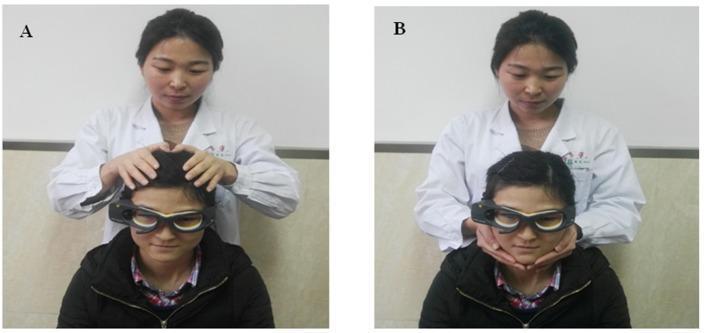
- Examiner places hands on the patient's head
- A small, fast, unpredictable head thrust is given in the plane of the horizontal semicircular canal (or vertical canals)
- The goggles track both the head velocity and eye velocity simultaneously
This is essentially the bedside Halmagyi-Curthoys Head Impulse Test, but made quantitative with goggles.
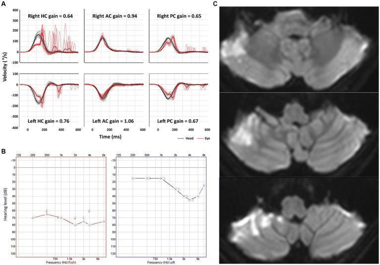
The Key Measurement: VOR GAIN
VOR Gain = Eye velocity / Head velocity
| VOR Gain | Meaning |
|---|
| ~1.0 (normal) | Eyes move at same speed as head - perfect compensation |
| <1.0 (reduced) | Eyes don't keep up - vestibular dysfunction on that side |
| 0 | No eye movement at all - complete vestibular loss |
Normal gain = approximately 1
When the head rotates toward the impaired side, gain drops below 1 - the eyes lag behind and a catch-up saccade is seen.
The Saccades - THE Most Important Finding
There are TWO types of corrective saccades:
| Type | Timing | What it means |
|---|
| Overt (catch-up) saccades | AFTER head rotation ends - visible to naked eye | Acute/uncompensated lesion |
| Covert saccades | DURING head rotation - invisible to naked eye, only vHIT can catch it | Compensated (chronic) lesion |
Memory trick: Overt = Out in the open = Acute. Covert = Covered/hidden = Compensated/chronic.
Clinical Significance of vHIT
- Reduced gain + overt saccades toward one side = peripheral vestibular hypofunction on that side (e.g., vestibular neuritis, labyrinthitis)
- Normal gain bilaterally = normal horizontal canal VOR
- vHIT can also test superior and posterior vertical canals on newer devices
- In AICA infarction - reduced gain in ipsilateral horizontal AND posterior canals (HINTS exam context)
Limitations of vHIT
- Goggle slippage - if goggles slip on the skin during the impulse, it gives a false reading
- Artifact - with very fast impulses (>200-300°/s), artifacts distort the signal
- Maximum reliable head impulse velocity is only 150-200°/s
- Cannot test low-frequency vestibular function (this is where rotational chair excels)
- KJ Lee's Essential Otolaryngology, p. 353
PART 2: ROTATIONAL CHAIR TEST (RCT)
What is it, in simple terms?
The patient sits in a motorized, computer-controlled chair inside a light-proof booth and the chair rotates. Eye movements are recorded by ENG/VNG. This tests the VOR across a range of frequencies - something calorics and vHIT cannot do.
Think of it as giving the vestibular system a workout at different "speeds" (frequencies) of head rotation, from very slow to moderately fast.
Why is it Needed? (Principle and Rationale)
- Caloric test tests only ONE frequency (very low, ~0.003 Hz) - it mimics very slow movements
- vHIT tests very HIGH frequency (brief impulses, ~2-5 Hz)
- Rotational chair fills the gap by testing 0.01 to 2 Hz - the mid-range frequencies that are most physiologically relevant for daily head movements
- Head rotation is the natural stimulus for the VOR - far more physiologic than cold/warm water in the ear
- KJ Lee's Essential Otolaryngology; Shambaugh Surgery of the Ear
Patient Setup
- Patient sits in the chair with chin pitched 30° nose-down - this puts the horizontal semicircular canals in the exact plane of rotation (maximizing their stimulation)
- Complete darkness or eyes open with no visual target (to eliminate visual fixation suppression)
- Eye movements recorded by IR video goggles (VNG)
Types of Rotational Tests
| Test Type | What Happens | What It Measures |
|---|
| Sinusoidal Harmonic Acceleration (SHA) | Chair oscillates back and forth in a sine wave at multiple frequencies (0.01-0.64 Hz) | Gain, Phase, Symmetry across frequencies |
| Velocity Step Test (Constant Angular Acceleration) | Chair spins at constant speed (e.g. 60°/s), then STOPS abruptly | Time constant of VOR decay (velocity storage) |
| Off-Vertical Axis Rotation (OVAR) | Chair tilted slightly and rotated | Tests otolithic organs (utricle/saccule) |
The Three Key Measurements
These are the "3 Golden Parameters" examiners love to ask about:
1. GAIN
- Formula: Peak slow-phase eye velocity ÷ Peak chair velocity
- Normal: Approximately 1 at higher frequencies; slightly lower (<1) at very low frequencies (0.01 Hz) is acceptable
- Abnormal: Reduced bilateral gain = bilateral vestibular hypofunction
- Key point: In unilateral lesions, gain may be NORMAL if the brain has compensated - this is a limitation of RCT
2. PHASE
- The timing relationship between chair movement and eye movement
- In a normal person, eyes lag slightly behind the head at low frequencies (~30° phase lead is normal)
- Abnormal: Exaggerated phase lead at low frequencies
- What it reflects: Loss of velocity storage mechanism (VOR time constant shortened)
- Can be caused by both peripheral (labyrinth loss) and central (vestibular nucleus damage) lesions
3. SYMMETRY (Asymmetry)
- Compares left vs. right slow-phase velocities
- Shows which side is weaker in the acute phase of unilateral lesion
- Recovers over time (returns toward normal with compensation) - useful for monitoring recovery
Memory trick for the 3 parameters: G.P.S. - Gain, Phase, Symmetry
The Rotational Chair Abnormality Pattern
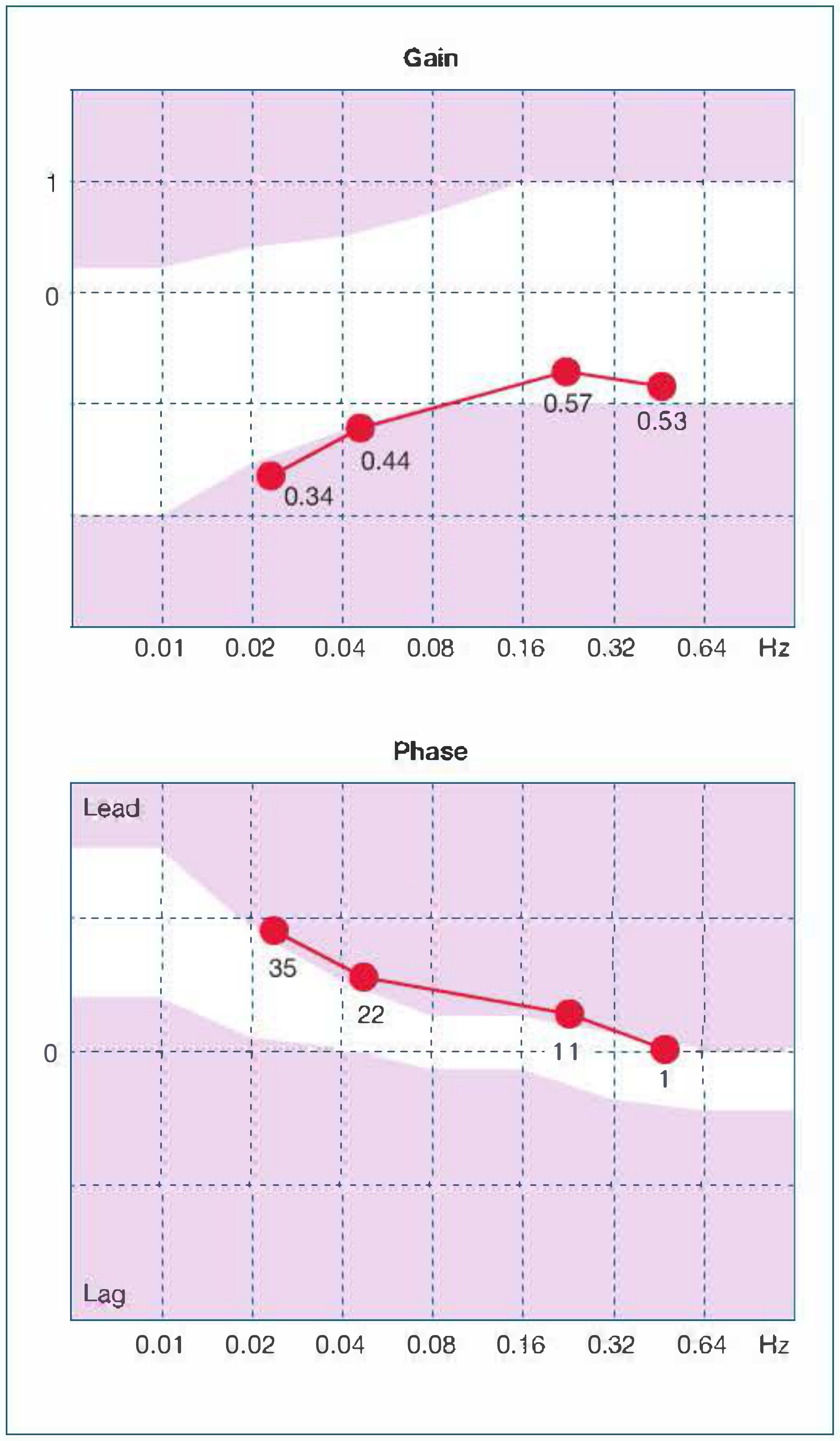
This graph from Shambaugh Surgery of the Ear shows a bilaterally deficient patient - gain points fall below the white normal band, and phase lead is exaggerated above the normal band at lower frequencies (0.01-0.04 Hz).
The Velocity Step Test - Simplified
- Chair accelerates to 60°/s and holds constant speed
- Patient initially feels rotation, then it fades (as velocity storage discharges)
- Chair suddenly STOPS
- Patient feels sudden rotation in the opposite direction - eyes show post-rotatory nystagmus
- The time constant = time for eye velocity to fall to 37% of its initial value
- Normal: ~15-20 seconds
- Reduced in vestibular loss (shorter time constant)
- Shambaugh Surgery of the Ear, p. 150
Key Indications for Rotational Chair Testing
This is what examiners will ask: "When do you do RCT?"
| Indication | Reasoning |
|---|
| Bilateral absent/reduced calorics | Caloric can't differentiate "no response = bilateral loss" from artifact; RCT confirms if vestibular function is truly absent |
| Monitoring vestibular compensation | Symmetry parameter recovers over time - useful for tracking |
| Ototoxicity monitoring (e.g., aminoglycosides, cisplatin) | Serial RCT tracks progressive bilateral vestibular damage |
| Pediatric patients/cannot cooperate with caloric | RCT is more tolerable and reproducible |
| When caloric is equivocal or contradictory | RCT tests different frequency range than caloric |
RCT vs Caloric - Key Exam Table
| Feature | Caloric Test | Rotational Chair |
|---|
| Frequency tested | ~0.003 Hz (very low) | 0.01-2 Hz (wider range) |
| Ears tested | One at a time | Both simultaneously |
| Physiologic | Artificial (thermal) | Natural stimulus (rotation) |
| Sensitivity for bilateral loss | Can miss if both ears equally reduced | Better - gold standard |
| Localization | Good (unilateral vs bilateral) | Poor for lateralization |
| Compensation effect | Less affected | May normalize after compensation |
| Cost/availability | Widely available | Expensive, academic centers only |
RCT vs vHIT - Comparison Table
| Feature | vHIT | Rotational Chair |
|---|
| Frequency | High (~2-5 Hz impulses) | Low-mid (0.01-2 Hz) |
| Type of stimulus | Unpredictable, passive head thrust | Predictable, motorized rotation |
| Canals tested | Lateral, superior, posterior | Primarily lateral (horizontal) |
| Covert saccades | Detected (compensated lesion) | Not assessed |
| Bilateral loss | Shows reduced gain both sides | Best test - confirms bilateral loss |
| Cost | Affordable, portable | Expensive, large equipment |
| Velocity storage | Not measured | Measured via phase/time constant |
A Conceptual Summary in Plain Words
vHIT = Speed camera for the VOR
Measures how fast the eyes respond to a quick head jerk. Like a speed gun catching whether your eyes "keep up" with your head. If they don't keep up, a catch-up saccade is seen. Simple, quick, bedside-friendly with goggles.
Rotational Chair = Treadmill test for the VOR
Puts the vestibular system through a full range of frequencies. Like a treadmill ECG stress test vs. a resting ECG - gives you information no other test can provide, especially for bilateral vestibular loss.
High-Yield Exam Points (Summary)
vHIT:
- Goggles with >250 Hz camera + gyroscope
- Tests horizontal SCC VOR (and vertical canals on some devices)
- Normal gain ~1; reduced gain = ipsilateral vestibular hypofunction
- Overt saccades = acute/uncompensated; Covert saccades = chronic/compensated
- Based on Halmagyi head thrust maneuver
Rotational Chair:
- Tests VOR at 0.01-2 Hz (more physiologic than calorics)
- Head tilted 30° nose-down to align horizontal canals
- 3 parameters: Gain, Phase, Symmetry (GPS)
- Best use: bilateral vestibular loss confirmation
- Velocity step test measures VOR time constant
- OVAR tests otoliths
- Limitation: expensive, may normalize with central compensation
Sources: KJ Lee's Essential Otolaryngology (p. 352-353), Shambaugh Surgery of the Ear (p. 150, 262-263), Cummings Otolaryngology Head and Neck Surgery (p. 3188-3189)

![A multi-panel medical figure presenting data from five patients (P1–P5) with Wernicke’s encephalopathy. The left panels display quantitative video head impulse test (vHIT) results for the right and left horizontal semicircular canals. These graphs plot angular velocity against time, showing head velocity (black traces) and inverted eye velocity (red traces). Reduced vestibulo-ocular reflex (VOR) gains (ranging from 0.58 to 0.88) and overt compensatory catch-up saccades are evident in all patients. The right panels show corresponding brain MRI findings (Fluid-Attenuated Inversion Recovery [FLAIR] images). The MRIs (P1'–P5') demonstrate characteristic bilateral, symmetrical hyperintense lesions (indicated by white arrows) in the medial vestibular nucleus of the medulla, the periaqueductal gray matter, the hypothalamus, and the periventricular regions of the thalamus. This combined presentation illustrates the clinical correlation between acute vestibular dysfunction (bilateral vestibulopathy) and typical thalamic/brainstem neuroanatomical involvement in Wernicke’s encephalopathy, likely secondary to thiamine deficiency.](https://cdn.orris.care/cdss_images/pmc_clinical_VQA_b4a2f0d8061011b7e5524d921e47575f7b60d2daf3843077b7188a539ba55f52.jpg)