
Jaundice biochemistry
jaundice bilirubin metabolism biochemistry diagram
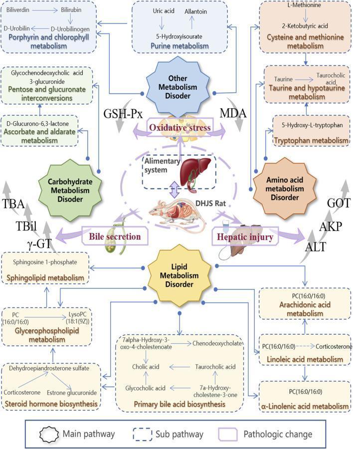
A metabolic network diagram illustrating the pathophysiological pathways of the Dampness-Heat Jaundice Syndrome (DHJS) model. The illustration is centered on a 'DHJS Rat' and the 'Alimentary system' (liver and gallbladder), with four octagonal 'Main pathway' boxes representing Carbohydrate, Lipid, Amino Acid, and Other Metabolism Disorders. Radiating from these nodes are numerous 'Sub-pathway' dashed boxes containing specific KEGG-based metabolic pathways, including Steroid Hormone Biosynthesis, Primary Bile Acid Biosynthesis, and Glycerophospholipid Metabolism. Key metabolites mentioned include bilirubin, cholic acid, corticosterone, and LysoPC. The diagram highlights 'Pathologic changes' such as Oxidative Stress and Hepatic Injury, indicated by thick purple arrows. Clinical biomarkers including TBil, TBA, ̳̳γ-GT, ALT, and MDA are shown with upward gray arrows, signifying elevation in the diseased state. This integrative map demonstrates the complex biochemical dysregulation across multiple systems associated with hepatic injury and bile secretion disorders.
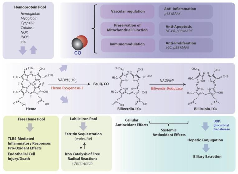
A detailed pathophysiology diagram illustrating the metabolic pathway of heme degradation and its systemic physiological implications. The central panel displays the biochemical conversion of Heme to Biliverdin-IXα by the rate-limiting enzyme Heme Oxygenase-1 (requiring NADPH and 3O2), which releases carbon monoxide (CO) and ferrous iron (Fe II). Subsequently, Biliverdin-IXα is reduced to Bilirubin-IXα by Biliverdin Reductase using NADP(H). The diagram maps the downstream outcomes of these byproducts: CO is linked to vascular regulation, mitochondrial preservation, and immunomodulation through signaling molecules like p38 MAPK, NF-κB, and sGC. The 'Labile Iron Pool' is shown bifurcating into protective Ferritin sequestration or detrimental free radical catalysis. Bilirubin is associated with cellular and systemic antioxidant effects before undergoing hepatic conjugation via UDP-glucuronyl transferase for biliary excretion. Additionally, a 'Free Heme Pool' is noted for its role in TLR4-mediated inflammatory responses and endothelial injury. This clinical illustration serves as a comprehensive guide for medical students and researchers studying oxidative stress, heme metabolism, and cytoprotective mechanisms.
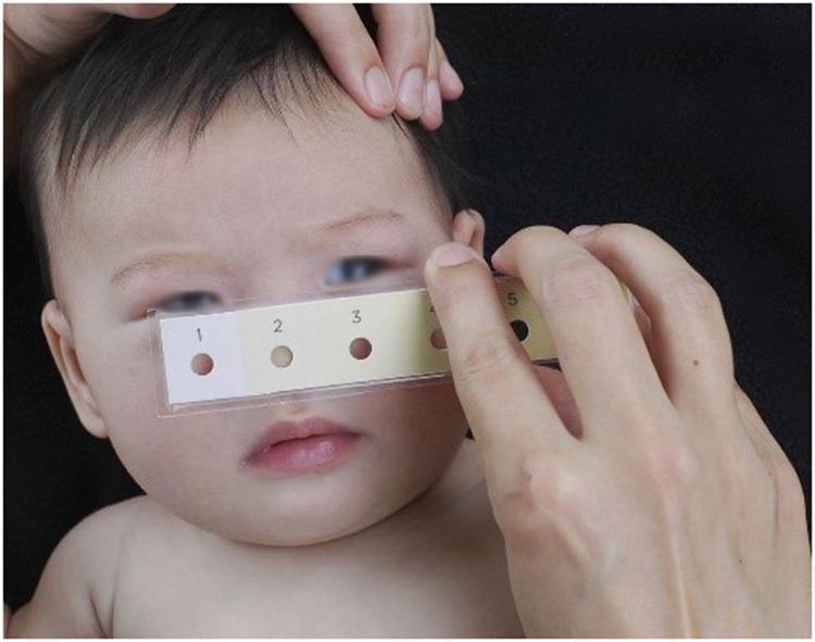
Clinical photograph of a neonate undergoing a non-invasive screening for neonatal jaundice (hyperbilirubinemia) using a Bili-ruler. The image shows a healthcare provider holding a rectangular, transparent-sleeved Bili-ruler against the infant's face. The ruler features a standardized color progression strip with six numbered patches (1–6). Each color patch increases in yellow intensity and contains a central circular aperture (hole). These apertures allow the clinician to compare the underlying skin tone directly against the calibrated reference colors to estimate bilirubin levels. The neonate demonstrates visible icterus (yellowish discoloration) of the skin and sclera. This diagnostic tool is designed as a low-cost, point-of-care alternative to electronic transcutaneous bilirubinometers (TcB) and serum testing, following the principle of cephalocaudal progression of jaundice. It is used in clinical settings to determine the need for further laboratory testing, phototherapy, or management of physiological and pathological jaundice.
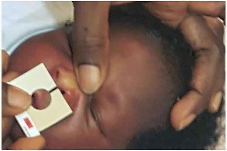
A clinical photograph depicting the assessment of neonatal jaundice in a dark-skinned infant using a Bilistrip™ device. The newborn's face is shown with visible yellowish icterus on the bridge of the nose and the periocular skin. An adult's hand holds the Bilistrip™, a 3.5 x 3.0 cm rectangular handheld tool, against the infant's mid-face. The device features a 1.3 cm central circular aperture used to isolate the skin for visual comparison against two calibrated shades of yellow on the strip. This visual screening method, modeled after the Ingram Icterometer, is used for point-of-care detection of hyperbilirubinemia in newborns. The educational focus is on clinical jaundice screening techniques and the visual manifestations of icterus in neonates with higher skin melanin content. The image demonstrates the application of a non-invasive diagnostic aid in a clinical or home-based triage setting for determining the need for serum bilirubin testing or phototherapy.
Jaundice - Biochemistry
1. Normal Bilirubin Production
- 85% comes from breakdown of senescent red blood cells (RBC lifespan ~120 days)
- 15% comes from myoglobin, cytochromes, and ineffective erythropoiesis ("early-labelled" bilirubin)
Step-by-step pathway:
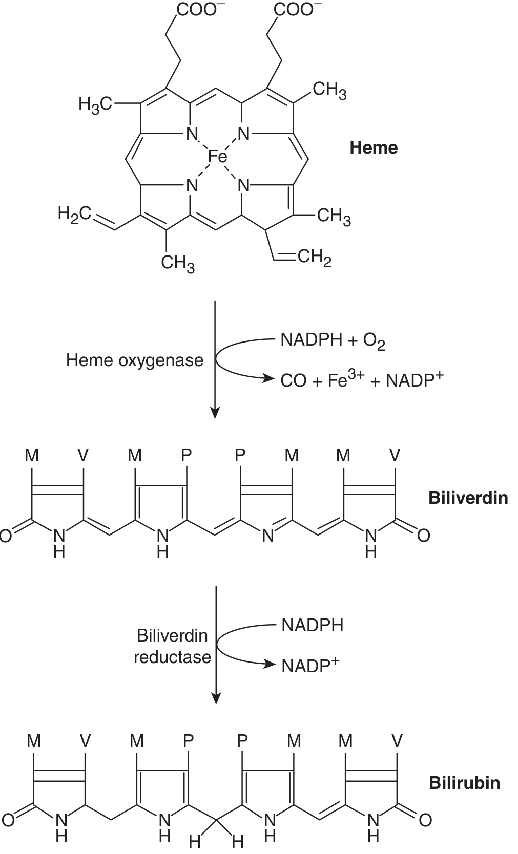
- Heme oxygenase (microsomal; requires NADPH + O₂) opens the porphyrin ring of heme to produce biliverdin-IXα + CO + Fe²⁺
- The CO released is transported as carboxyhemoglobin (clinically useful marker of hemolysis)
- Iron (Fe²⁺) enters the labile iron pool or is stored as ferritin
- Biliverdin reductase (NADPH-dependent) reduces biliverdin → unconjugated bilirubin (UCB)
2. Transport in Blood
- Unconjugated bilirubin (UCB) is tightly bound to albumin in plasma
- This albumin-UCB complex cannot be filtered at the glomerulus → not excreted in urine
- The half-life of UCB is only ~5 minutes (cleared very rapidly by the liver)
3. Hepatic Handling - Four Key Steps
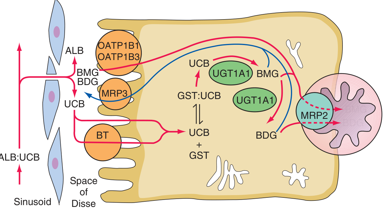
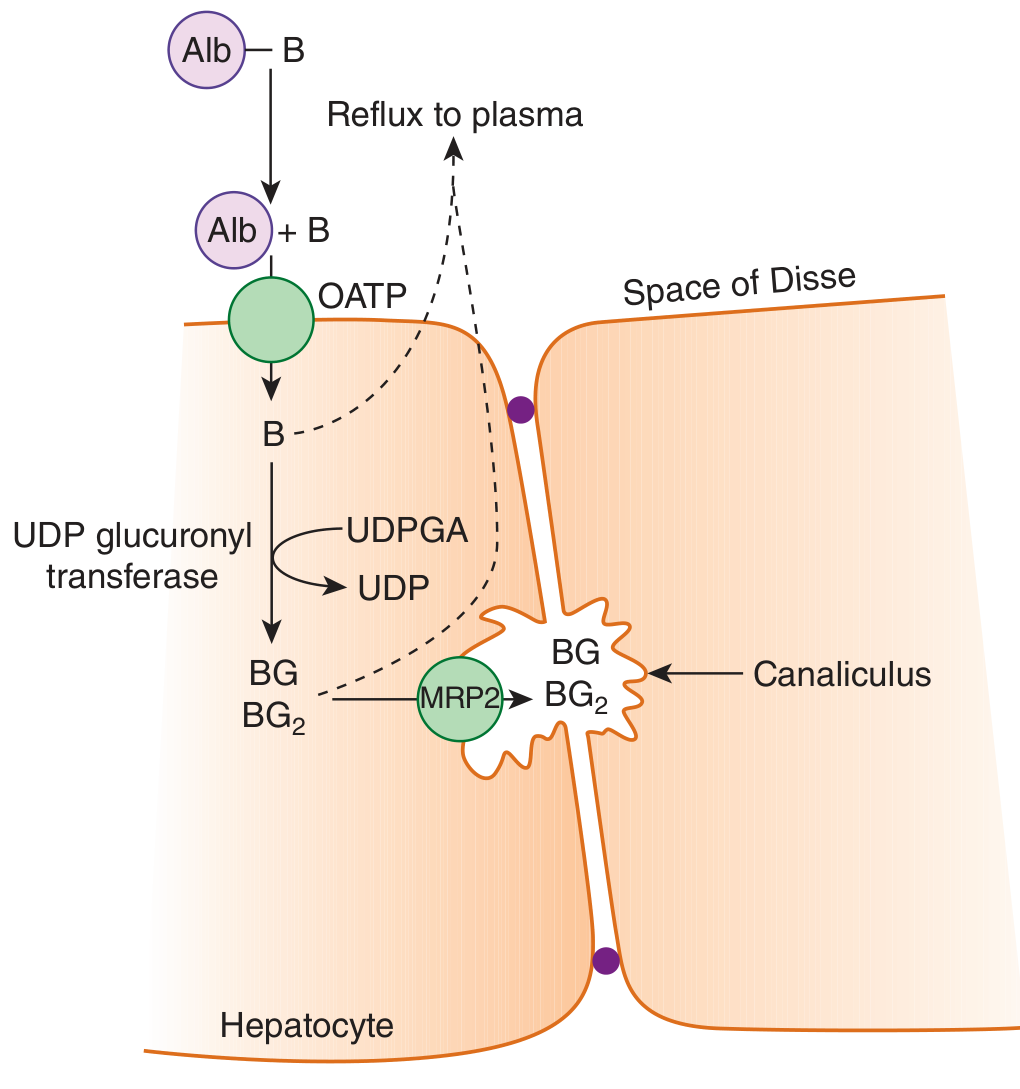
Step 1 - Hepatocellular Uptake
- Albumin-bound bilirubin passes through endothelial cell fenestrae into the Space of Disse
- Bilirubin dissociates from albumin and enters hepatocytes via OATP (organic anion transporting polypeptides) - specifically OATP1B1 and OATP1B3
- This is a carrier-mediated, facilitated process
Step 2 - Intracellular Binding
- Inside the hepatocyte, UCB binds to cytoplasmic glutathione-S-transferases (GST), formerly called ligandins
- This prevents back-diffusion of bilirubin out of the cell
Step 3 - Conjugation
- UCB is conjugated with glucuronic acid (one or two molecules) by UDP-glucuronosyltransferase (UGT1A1), located in the smooth endoplasmic reticulum
- Each bilirubin molecule reacts with two uridine diphosphoglucuronic acid (UDPGA) molecules → bilirubin diglucuronide (BDG) (the predominant form) + bilirubin monoglucuronide (BMG)
- Conjugated bilirubin is now water-soluble
- The UGT1A1 gene is on chromosome 2 and encodes a large UGT1 gene complex
Step 4 - Canalicular Secretion (Rate-limiting step)
- Conjugated bilirubin is actively transported across the canalicular membrane into bile by MRP2 (multidrug resistance-associated protein 2) against a concentration gradient
- A small fraction refluxes back into plasma via MRP3 → taken up again by OATP1B1/1B3 (enterohepatic-like cycling)
The rate-limiting step in the entire hepatic bilirubin pathway is canalicular secretion (MRP2-mediated).
4. Intestinal Fate and Enterohepatic Circulation
| Location | Process |
|---|---|
| Small intestine | Conjugated bilirubin passes through unchanged (intestinal mucosa is impermeable to conjugated bilirubin) |
| Colon | Gut bacteria deconjugate bilirubin and reduce it to urobilinogen (colorless) |
| Colon/Feces | Urobilinogen is oxidized to stercobilin → gives feces their brown color |
| Portal reabsorption | ~20% urobilinogen is reabsorbed into portal blood (enterohepatic circulation) |
| Liver | Most reabsorbed urobilinogen is re-excreted into bile |
| Kidney | Small amount of urobilinogen escapes into systemic circulation → excreted in urine as urobilin (yellow) |
5. Delta (δ)-Bilirubin
- A proportion of conjugated bilirubin covalently bonds to albumin in serum → called δ-bilirubin
- Half-life = half-life of albumin (~3 weeks)
- Explains the delayed fall in serum bilirubin even after resolution of liver injury or biliary obstruction - clinically important
- The "direct bilirubin" assay measures δ-bilirubin + conjugated bilirubin + a small fraction of UCB (hence "direct" ≠ "conjugated" exactly)
6. Renal Excretion
| Form | Urine? | Reason |
|---|---|---|
| Unconjugated bilirubin | NO | Too tightly albumin-bound; cannot be filtered; no tubular secretion mechanism |
| Conjugated bilirubin | YES | Loosely bound to albumin → glomerular filtration possible ("bilirubinuria" = dark urine) |
| Urobilinogen | YES (small amount) | Water-soluble; filtered and excreted as urobilin |
7. Classification of Jaundice
A. Pre-hepatic (Hemolytic) Jaundice
- Mechanism: Excess bilirubin production overwhelms hepatic conjugation capacity
- Type: Predominantly unconjugated hyperbilirubinemia
- Bilirubin level: Rarely exceeds 4 mg/dL from hemolysis alone (the bone marrow can sustain only an ~8-fold increase in erythrocyte production). Higher values suggest concurrent hepatic dysfunction
- Direct fraction: ≤15% of total bilirubin
- Urine: No bilirubinuria; urobilinogen increased
- Stool: Dark (excess stercobilin)
- Causes: Hereditary spherocytosis, G6PD deficiency, sickle cell disease, autoimmune hemolytic anemia, large hematomas, rhabdomyolysis
- Prolonged hemolysis → pigment gallstones (bilirubin stones, not cholesterol)
B. Hepatic (Hepatocellular) Jaundice
- Mechanism: Impaired uptake, conjugation, or secretion by damaged hepatocytes
- Type: Mixed (both conjugated and unconjugated) hyperbilirubinemia
- UGT activity is well preserved until advanced liver disease; jaundice is therefore a hallmark of severe hepatocellular injury
- Urine: Bilirubinuria (dark urine); urobilinogen variable
- Stool: Pale (reduced stercobilin)
- Causes: Viral hepatitis, alcoholic hepatitis, cirrhosis, drug-induced liver injury
- Hepatocellular injury causes mixed picture because: impaired conjugation + direct release of bilirubin from damaged cells + impaired canalicular secretion all occur together
C. Post-hepatic (Obstructive/Cholestatic) Jaundice
- Mechanism: Obstruction of bile flow → conjugated bilirubin regurgitates into blood
- Type: Conjugated (direct) hyperbilirubinemia
- Urine: Bilirubinuria (dark urine, "Coca-Cola urine"); urobilinogen absent/reduced
- Stool: Pale/clay-colored (no stercobilin reaching gut)
- Causes: Gallstones in CBD, carcinoma of head of pancreas, cholangiocarcinoma, primary sclerosing cholangitis
8. Inherited Disorders of Bilirubin Metabolism
| Disorder | Defect | Bilirubin type | Severity |
|---|---|---|---|
| Gilbert syndrome | Mild reduction in UGT1A1 (TATA box promoter variant) | Unconjugated | Benign; intermittent jaundice with fasting/stress |
| Crigler-Najjar type I | Complete absence of UGT1A1 | Unconjugated | Severe; kernicterus; fatal without liver transplant |
| Crigler-Najjar type II (Arias) | Partial UGT1A1 deficiency | Unconjugated | Moderate; responds to phenobarbitone |
| Dubin-Johnson syndrome | Defect of canalicular MRP2 transport | Conjugated | Benign; black liver (melanin-like pigment) |
| Rotor syndrome | Defect in sinusoidal reuptake (OATP1B1/1B3) of conjugated bilirubin | Conjugated | Benign; no pigment in liver |
9. Biochemical Markers - Summary Table
| Parameter | Pre-hepatic | Hepatic | Post-hepatic |
|---|---|---|---|
| Serum bilirubin (total) | Elevated | Elevated | Elevated |
| Conjugated bilirubin | Normal/slight ↑ | Elevated | Markedly elevated |
| Unconjugated bilirubin | Markedly elevated | Elevated | Normal/slight ↑ |
| Urine bilirubin | Absent | Present | Present |
| Urine urobilinogen | Increased | Variable | Absent/very low |
| Stool color | Dark | Pale | Pale/clay |
| ALT/AST | Normal | Markedly elevated | Mildly elevated |
| ALP | Normal | Mildly elevated | Markedly elevated |
| PT/INR | Normal | Prolonged | Prolonged (Vit K-responsive) |
10. Key Biochemical Points to Remember
- Heme oxygenase is the rate-limiting enzyme of heme catabolism
- UGT1A1 on chromosome 2 is the key conjugating enzyme; reduced in Gilbert's, absent in CN-I
- MRP2 is the rate-limiting step for hepatic bilirubin elimination
- UCB cannot be excreted in urine; only conjugated bilirubin appears in urine
- δ-bilirubin explains persistent jaundice despite clinical recovery
- Hemolysis alone rarely raises bilirubin above 4 mg/dL
- In sepsis, inflammatory cytokines rapidly downregulate MRP2 → conjugated hyperbilirubinemia without hepatocellular injury
- Phototherapy in neonatal jaundice works by converting insoluble UCB into water-soluble photoisomers (lumirubin) that can be directly excreted without conjugation