
https://youtu.be/SVbRrPqMe8s?si=E1y6g1kRpFa50SQa/generatebestnotes on the link given above for 1st year mbbs student act as a professor and /teachme each and every point in /deepdive don't miss even a single point explain with the help of images Provide USMLE type questions on this topic with /3levels /doughtkiller.
Reading File
analyse_media
The video is about Olfactory System (Smell / CN I) - covering anatomy, physiology, and clinical correlations for 1st year MBBS. Let me now pull comprehensive textbook content to build the best notes.
Finding Sources
Finding Sources
Finding Sources
Now let me pull the actual textbook content from the best sources in parallel:
Reading File
Reading File
Reading File
Reading File
Excellent! Now let me fetch the key diagrams and get additional content on transduction and pathways:
Loading Image
Loading Image
Loading Image
Loading Image
Excellent! I have all 4 key diagrams. Now let me get the olfactory pathway content and the Guyton content, plus any image for the central olfactory pathway:
Reading File
Reading File
Finding Sources
Finding Sources
I now have everything I need. Let me also fetch one more key image (Guyton's olfactory bulb diagram) and get the Foster Kennedy syndrome detail:
Loading Image
Finding Sources
I now have all the material needed. Let me compile the comprehensive professor-style notes.---
🧠 THE OLFACTORY SYSTEM (CN I) - SMELL
Professor's Deep Dive Notes for 1st Year MBBS
Sources: Costanzo Physiology 7e | Guyton & Hall Medical Physiology | Gray's Anatomy for Students | Junqueira's Histology 17e | Neuroanatomy through Clinical Cases 3e | Adams & Victor's Neurology 12e
📌 OVERVIEW - WHY THIS MATTERS
The olfactory system is the only sensory pathway that bypasses the thalamus and projects directly to the cortex. It is the most phylogenetically ancient sensory system, and it is uniquely tied to memory and emotion via direct connections to the limbic system. That is why a smell can instantly trigger a vivid memory - this is called the Proust phenomenon.
Professor's Tip: CN I is the most commonly tested cranial nerve in MBBS exams and the USMLE, especially for its clinical correlations with head trauma, COVID-19, Parkinson's disease, and Kallmann syndrome.
PART 1 - ANATOMY OF THE OLFACTORY EPITHELIUM
1.1 Location
The olfactory epithelium (also called olfactory membrane or olfactory neuroepithelium) is located in the superior part of the nasal cavity - specifically:
- The roof of the nasal cavity
- Along the superior nasal concha (turbinate) bilaterally
- Over the superior nasal septum
Total surface area: approximately 5 cm² in humans (about 10 cm² in some references, as this includes folded areas). Despite this tiny area, we can detect over 10,000 different odors.
1.2 The Three Cell Types of the Olfactory Epithelium
This epithelium is a pseudostratified columnar epithelium containing three cell types. Memorize all three - they are high-yield for histology MCQs.
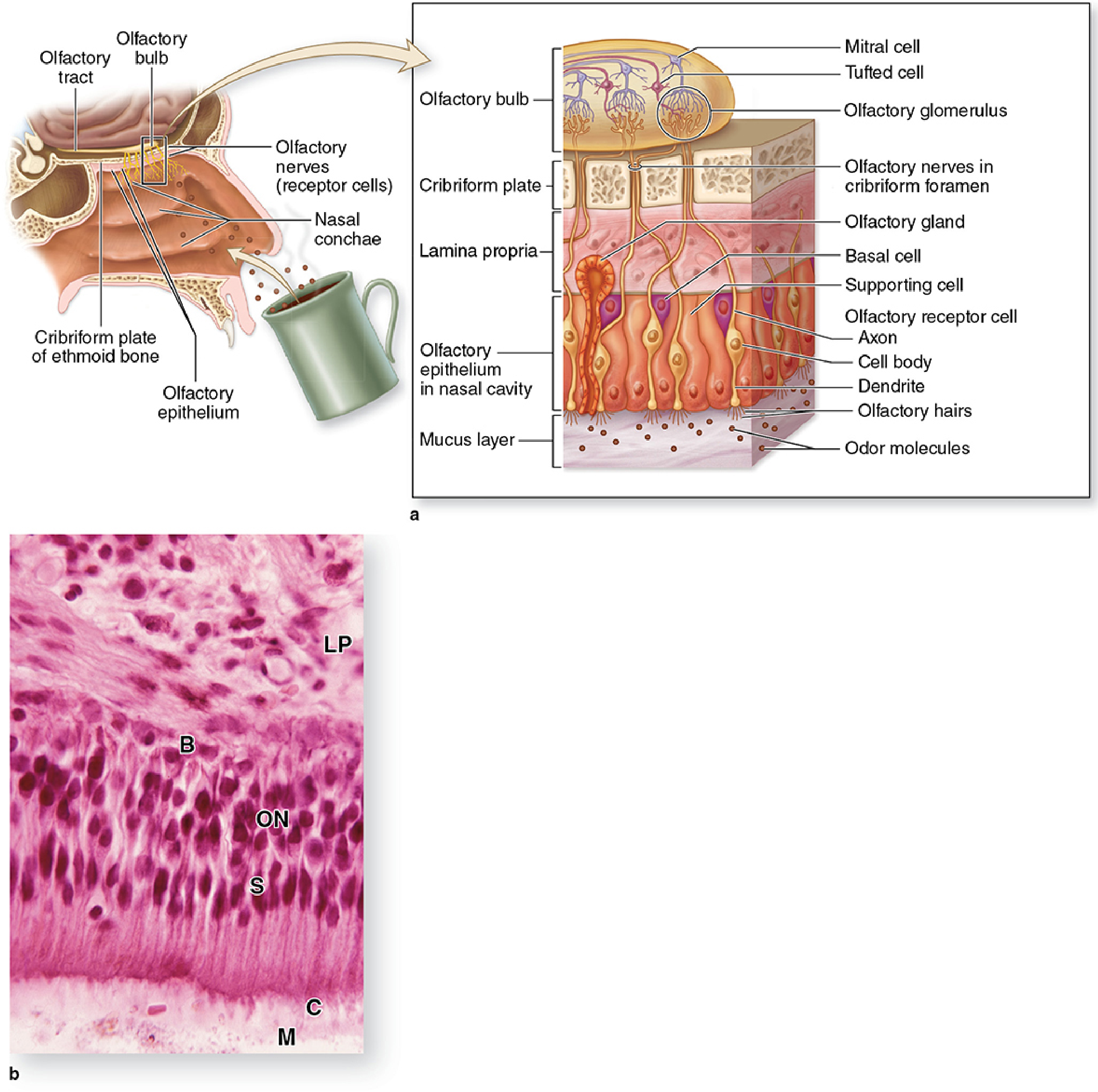
Cell Type 1: Olfactory Receptor Neurons (ORNs) - The Detectors
These are the most important cells. Key facts:
| Feature | Detail |
|---|---|
| Cell type | Bipolar neuron (derived from CNS, not PNS!) |
| Number | ~100 million in the olfactory epithelium |
| Location | Nuclei form an irregular row in the middle of the epithelium |
| Apical pole | Forms a dendritic knob projecting into the mucus layer |
| Cilia | 4-25 long, non-motile cilia per cell (up to 200 μm long, 0.3 μm diameter) |
| Function of cilia | Bear odorant receptor proteins - this is where smell is detected |
| Axon | Unmyelinated (Group C fibers) - the smallest, slowest fibers in the nervous system |
| Axon destination | Through the cribriform plate → olfactory bulb |
Professor says: Remember - ORNs are bipolar neurons with a dendrite going UP (to smell odorants) and an axon going DOWN (to the brain). They are the only neurons in the human body that are regularly replaced throughout life - unique continuous neurogenesis from basal stem cells!
Cell Type 2: Sustentacular Cells (Supporting Cells)
- Tall columnar cells with broad apices and narrow bases
- Nuclei at the apex (this distinguishes them from ORNs whose nuclei are in the middle layer)
- Project microvilli into the mucus layer
- Bound to ORNs via apical junctional complexes
- Function like astrocytes - they regulate the microenvironment around ORNs, provide metabolic support
- High-yield in COVID-19: Sustentacular cells express ACE2 receptors and are the primary target of SARS-CoV-2, causing anosmia. The virus damages sustentacular cells first, which disrupts the microenvironment for ORNs, causing loss of smell even without direct neuronal infection.
Cell Type 3: Basal Cells - The Stem Cells
- Small, spherical or cone-shaped cells near the basal lamina
- These are the undifferentiated stem cells of the olfactory epithelium
- Undergo continuous mitosis → give rise to new ORNs every 1-2 months and sustentacular cells less frequently
- This is why olfaction can recover after damage (unlike most CNS neurons)
Bowman's Glands (in the lamina propria): Serous glands that secrete the mucus layer covering the epithelium. Odorants must dissolve in this mucus before they can bind to receptors. Hydrophobic odorants need special odorant-binding proteins to carry them through the aqueous mucus.
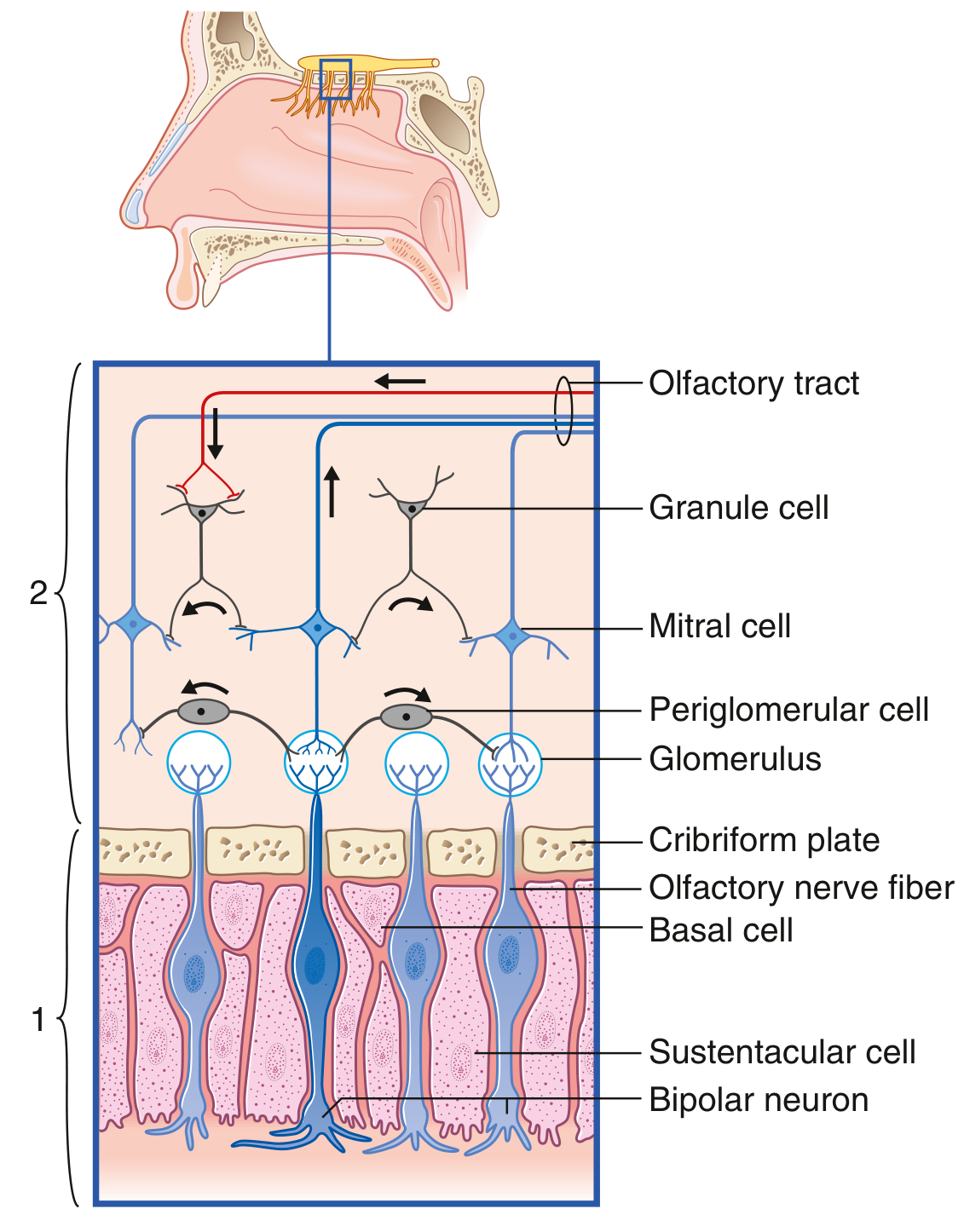
PART 2 - OLFACTORY TRANSDUCTION (Signal Conversion)
2.1 The Signal Cascade - Step by Step
This is the conversion of a chemical signal → electrical signal. It is a classic G-protein coupled receptor (GPCR) cascade.
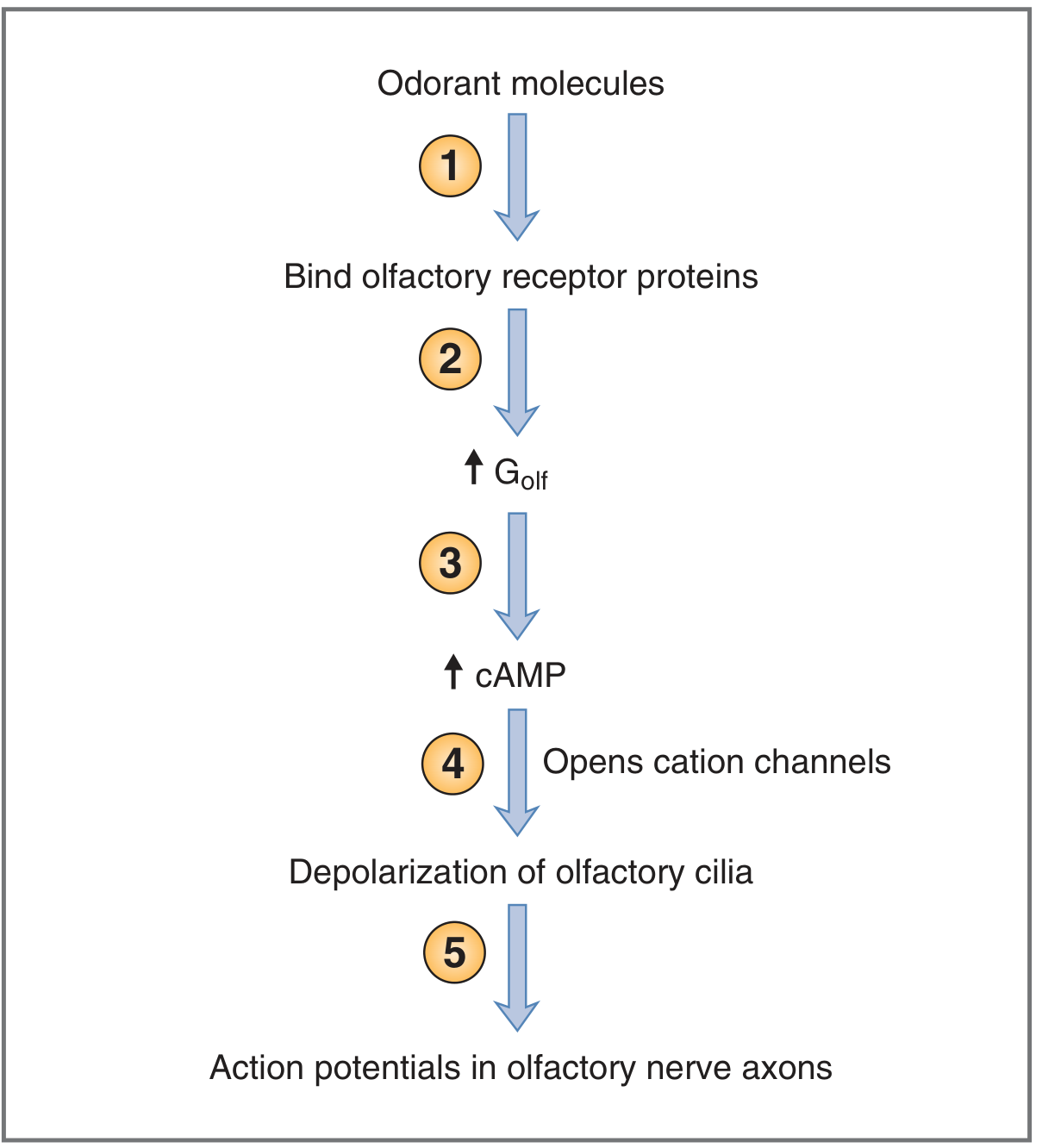
Step-by-step breakdown:
Step 1: Odorant molecule enters the nasal cavity, dissolves in the mucus layer (or carried by odorant-binding proteins if hydrophobic), and binds to olfactory receptor proteins on the cilia of ORNs.
Step 2: Olfactory receptor proteins are 7-transmembrane GPCRs (each threads through the membrane 7 times, folds in/out). There are at least 1,000 different olfactory receptor proteins in humans - each encoded by a different gene. This is the largest gene family in the human genome (~3% of all human genes!). The receptor activates a special G-protein called G_olf (alpha subunit breaks away).
Step 3: G_olf activates adenylyl cyclase (attached to the inside of the ciliary membrane). Adenylyl cyclase converts ATP → cAMP. Intracellular cAMP levels rise rapidly. This amplifies the signal enormously.
Step 4: Elevated cAMP opens cyclic nucleotide-gated (CNG) cation channels in the ciliary membrane. These channels are permeable to Na⁺, K⁺, and Ca²⁺. Na⁺ flows in (down its concentration gradient) → membrane depolarization.
Step 5: Depolarization reaches threshold → action potentials are generated and propagate along the unmyelinated olfactory axons toward the olfactory bulb.
Memory trick: Golf → Adenylyl cyclase → cAMP → Cation channels → Depolarization → Action potential = "GACCA" (like a golfing acronym!)
2.2 Odorant Receptor Specificity and Combinatorial Coding
This is a concept worth understanding deeply:
- Each olfactory receptor protein is NOT dedicated to a single odorant
- One receptor can respond to several different odorants (but with different strengths)
- One odorant activates multiple different receptor proteins in a unique pattern
- The CNS reads this pattern of activation across many receptors - called across-fiber pattern code or combinatorial coding
- Each odorant produces a unique "odor map" on the glomeruli of the olfactory bulb
- This is how we distinguish thousands of odors using only ~1,000 receptor proteins
Professor's analogy: Think of it like a piano. Individual keys (receptors) can be combined in thousands of combinations (chords = odorants). You recognize a "chord" not by a single key, but by the whole pattern.
PART 3 - THE OLFACTORY BULB
3.1 Structure
The olfactory bulb is an oval structure that sits on the cribriform plate of the ethmoid bone, within the olfactory groove of the anterior cranial fossa.
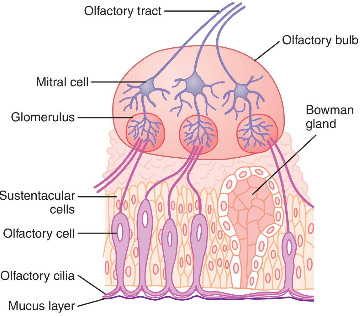
3.2 Key Cells of the Olfactory Bulb
| Cell | Function |
|---|---|
| Mitral cells | Principal output neurons of the olfactory bulb (2nd order neurons). Triangular/pyramid-shaped. Apical dendrites synapse with ORN axons in glomeruli. |
| Tufted cells | Similar to mitral cells, also output neurons |
| Granule cells | Inhibitory interneurons (axon-less). Make dendrodendritic synapses → provide lateral inhibition to sharpen odor discrimination |
| Periglomerular cells | Inhibitory interneurons surrounding each glomerulus |
3.3 The Glomerulus - Key Relay Point
- A glomerulus is a synaptic cluster where ORN axons synapse with mitral cell apical dendrites
- ~1,000 ORN axons converge onto 1 mitral cell in each glomerulus
- This convergence amplifies the signal and sharpens specificity
- ORNs expressing the same receptor protein all converge on the same 2-3 glomeruli in the olfactory bulb regardless of where in the epithelium they originate - this creates a precise "odor map"
PART 4 - THE OLFACTORY PATHWAY (Central Connections)
4.1 The Pathway - Neuron by Neuron
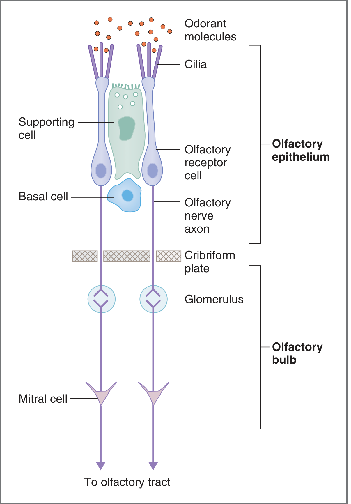
1st Order Neuron (Primary Afferent):
- Olfactory receptor neurons (ORNs) in the olfactory epithelium
- Unmyelinated axons bundle together to form ~20 olfactory filaments (fila olfactoria)
- These filaments pierce through the cribriform plate (multiple small foramina) of the ethmoid bone
- Together, they constitute Cranial Nerve I (Olfactory Nerve)
- Synapse in the glomeruli of the olfactory bulb
⚠️ Important: CN I is NOT a single nerve trunk - it is a collection of ~20 delicate unmyelinated filaments. This is why it is so vulnerable to shear injury in head trauma.
2nd Order Neuron:
- Mitral cells (and tufted cells) of the olfactory bulb
- Their axons form the Olfactory Tract (a white matter tract, not truly a nerve)
- The olfactory tract runs in the olfactory sulcus of the frontal lobe
Olfactory Tract Divisions:
As the tract approaches the anterior perforated substance, it divides into:
| Tract | Destination |
|---|---|
| Lateral olfactory stria | Primary olfactory cortex (piriform cortex, periamygdaloid cortex, olfactory tubercle, entorhinal cortex) |
| Medial olfactory stria | Septal area (crosses in anterior commissure → contralateral olfactory bulb → provides lateral inhibition across sides) |
4.2 Primary Olfactory Cortex
- Piriform cortex (prepiriform cortex) - the main site; named for its pear shape in some animals
- Periamygdaloid cortex - just rostral/dorsal to the amygdala
- Olfactory tubercle
- Entorhinal cortex (area 28)
Key concept: Olfaction is the ONLY sense that does NOT relay through the thalamus before reaching the primary cortex. All other senses (vision, hearing, touch, taste) go through the thalamus first. Olfaction projects DIRECTLY from olfactory bulb → primary olfactory cortex.
Exam pearl: "Which sensory pathway bypasses the thalamus?" → Answer: Olfaction (CN I)
4.3 Connections to the Limbic System and Beyond
From the primary olfactory cortex, fibers project to:
- Amygdala (corticomedial nucleus) → emotional responses to smells (fear, appetite, sexual attraction)
- Hippocampus (via entorhinal cortex) → olfactory memory (why smells so powerfully trigger memories)
- Hypothalamus (via medial forebrain bundle) → autonomic responses: salivation, gastric contraction, arousal
- Orbitofrontal cortex (relays via thalamus at this stage) → conscious appreciation of smell, flavor perception
- Reticular formation → arousal
PART 5 - SPECIAL FEATURES OF THE OLFACTORY SYSTEM
5.1 Unique Neurogenesis
The olfactory receptor neurons are replaced every 1-2 months throughout life:
- Basal stem cells → new ORNs → new axons grow through cribriform plate → re-establish connections in olfactory bulb
- This is one of the few examples of adult neurogenesis in humans
- Clinically: This is why olfactory loss from mild mucosal damage (e.g., a cold) is temporary and recoverable
5.2 Adaptation (Olfactory Fatigue)
You stop noticing a persistent smell after a while - olfactory adaptation. This occurs at the receptor level (receptor desensitization) and at higher cortical levels.
5.3 Olfaction and Flavor
When you think you are tasting food, ~80% of what you call "flavor" is actually smell via retronasal olfaction (odorants from food travel from the mouth UP through the posterior nasopharynx to the olfactory epithelium). This is why:
- Food tastes bland when you have a blocked nose (nasal congestion)
- Patients with bilateral anosmia complain of "loss of taste" even though their taste buds are fine
PART 6 - CLINICAL CORRELATIONS (High-Yield)
6.1 Anosmia vs. Hyposmia
| Term | Meaning |
|---|---|
| Anosmia | Complete absence of smell |
| Hyposmia | Reduced sense of smell |
| Hyperosmia | Increased sensitivity to smell |
| Parosmia | Distorted smell perception |
| Phantosmia | Smelling something that isn't there (olfactory hallucination) |
Testing: Always test each nostril separately with a non-irritating substance (coffee, vanilla, cloves - NOT ammonia, which stimulates CN V/trigeminal, not CN I!). Unilateral anosmia is often not noticed by patients.
6.2 Head Trauma → Anosmia
Mechanism: The olfactory nerve filaments pass through multiple tiny foramina in the cribriform plate of the ethmoid bone. Frontal/occipital impact causes the brain to shift suddenly → the delicate unmyelinated filaments are sheared at the level of the cribriform plate.
- Most common after frontal or occipital head injury (brain moves, filaments tear)
- Often permanent (only ~10% improve)
- More common when trauma is associated with loss of consciousness
- May also cause CSF rhinorrhea (cerebrospinal fluid leak through the cribriform plate → clear watery discharge from nose, positive β-2 transferrin test)
6.3 COVID-19 and Anosmia
Mechanism (Pathophysiology):
- SARS-CoV-2 enters cells via the ACE2 receptor
- ACE2 is highly expressed on sustentacular cells (NOT olfactory neurons themselves)
- Virus infects and destroys sustentacular cells → disrupts the microenvironment of ORNs → ORNs lose function
- This explains why COVID-19 anosmia is often rapid in onset (days) and why recovery is possible as sustentacular cells regenerate
- In some patients (Long COVID), anosmia/parosmia persists for months due to ongoing inflammation or incomplete regeneration
6.4 Kallmann Syndrome
A genetic condition - high-yield for MBBS and USMLE:
- Definition: Hypogonadotropic hypogonadism + anosmia
- Pathophysiology: GnRH-secreting neurons normally originate in the olfactory placode (embryological origin - same as the olfactory epithelium) and migrate along the olfactory nerves through the cribriform plate → through the olfactory bulb → hypothalamus. In Kallmann syndrome, this migration fails. Both the GnRH neurons and the olfactory bulbs fail to develop properly.
- Clinical features: Absent puberty, infertility, anosmia (patient may be unaware), small testes, possibly renal agenesis, cleft lip, bimanual synkinesia
- Inheritance: X-linked (most common, KAL1/ANOS1 gene), autosomal dominant, or autosomal recessive
- Incidence: 1:30,000
6.5 Neurodegenerative Diseases
Anosmia/hyposmia is an early and sometimes pre-clinical feature of:
| Disease | Mechanism |
|---|---|
| Parkinson's disease | Lewy bodies in olfactory bulb and piriform cortex (often precede motor symptoms by years) |
| Alzheimer's disease | Amyloid pathology in entorhinal cortex and olfactory areas |
Olfactory testing is being explored as a biomarker for early neurodegenerative disease.
6.6 Meningioma of the Olfactory Groove → Foster Kennedy Syndrome
- A meningioma grows from the dura in the olfactory groove (anterior cranial fossa floor)
- As it enlarges, it compresses the structures around it
- Foster Kennedy Syndrome (classic triad):
- Ipsilateral anosmia (direct compression of olfactory nerve/bulb/tract on the side of the tumor)
- Ipsilateral optic atrophy (direct pressure on ipsilateral optic nerve → retinal ganglion cell death → atrophy)
- Contralateral papilledema (raised intracranial pressure from the growing mass → increased CSF pressure → disc swelling on the OTHER side)
Pseudo-Foster Kennedy Syndrome: Same appearance (one disc atrophied, other edematous) BUT caused by sequential anterior ischemic optic neuropathy - NOT a tumor. No anosmia.
6.7 Other Causes of Anosmia
- Upper respiratory tract infections (common cold) - mucosal inflammation/congestion, usually temporary
- Nasal polyps or sinusitis - obstructive (conductive anosmia)
- Intranasal cocaine use - damages olfactory epithelium directly
- Zinc deficiency - impairs olfactory function
- Congenital - true isolated congenital anosmia (rare)
- Basal meningitis, sarcoidosis - granulomatous inflammation at skull base
- Frontal lobe tumors (meningiomas, metastases) at the base
PART 7 - QUICK SUMMARY CHART
NASAL CAVITY (olfactory epithelium)
         ↓  [1st Neuron: ORN - unmyelinated axons through cribriform plate]
OLFACTORY BULB (glomeruli → mitral cells)
         ↓  [2nd Neuron: Mitral cell axons → olfactory tract]
OLFACTORY TRACT divides into:
         ├── Lateral olfactory stria → PRIMARY OLFACTORY CORTEX (piriform, periamygdaloid)
         │                           → Amygdala → emotional response
         │                           → Hippocampus → memory
         │                           → Hypothalamus → autonomic responses
         └── Medial olfactory stria → Septal area → anterior commissure → contralateral bulb

KEY FEATURE: NO THALAMIC RELAY (unique among senses!)
🎯 USMLE-STYLE QUESTIONS: 3 LEVELS + DOUBT KILLER EXPLANATIONS
🟢 LEVEL 1 - BASIC KNOWLEDGE (Foundation)
Q1. A student is studying the olfactory epithelium. Which of the following is TRUE about the olfactory receptor neurons?
- A) They are multipolar neurons
- B) Their axons are heavily myelinated and conduct impulses rapidly
- C) They are replaced continuously from basal stem cell precursors
- D) They relay through the thalamus before reaching the cortex
- E) Their cell nuclei are located at the apex of the epithelium
✅ Answer: C
Doubt Killer Explanation:
- (A) WRONG - ORNs are bipolar neurons, not multipolar. One dendrite goes up to the mucosa, one axon goes down.
- (B) WRONG - ORNs have unmyelinated axons (Group C fibers). They are the smallest, slowest fibers in the nervous system. This is why olfactory processing is somewhat slow.
- (C) CORRECT - Basal cells at the base of the olfactory epithelium are stem cells that continuously replace ORNs every ~1-2 months. This is unique adult neurogenesis.
- (D) WRONG - This is the #1 trap answer. Olfaction is the ONLY sense that does NOT relay through the thalamus. It goes directly from the olfactory bulb to the primary olfactory cortex.
- (E) WRONG - Sustentacular cells have apical nuclei. ORN nuclei are in the middle layer of the pseudostratified epithelium.
Q2. A 7-year-old boy is brought by his parents because he has never entered puberty. Workup reveals hypogonadotropic hypogonadism. On further questioning, the child cannot smell anything. What is the most likely diagnosis?
- A) Klinefelter syndrome
- B) Prader-Willi syndrome
- C) Kallmann syndrome
- D) Congenital hypothyroidism
- E) Isolated GnRH deficiency
✅ Answer: C
Doubt Killer Explanation:
The combination of hypogonadotropic hypogonadism + anosmia is the classic hallmark of Kallmann syndrome. The mechanism is failure of migration of GnRH neurons (which originate in the olfactory placode) through the cribriform plate to the hypothalamus. The olfactory bulbs also fail to develop → anosmia.
- (A) Klinefelter (47,XXY) causes hypergonadotropic hypogonadism (high LH/FSH) not hypogonadotropic, and NO anosmia.
- (B) Prader-Willi causes hypogonadism but NOT anosmia - associated with hyperphagia, obesity.
- (E) Isolated GnRH deficiency would NOT have anosmia - that differentiates Kallmann from normosmic hypogonadotropic hypogonadism.
Q3. The second step in olfactory signal transduction, after odorant binding to the receptor protein, is activation of which molecule?
- A) Phospholipase C
- B) G_olf protein
- C) Protein kinase A
- D) IP3 receptor
- E) Voltage-gated Na⁺ channel
✅ Answer: B
Doubt Killer Explanation:
The cascade goes: Odorant → receptor protein (GPCR) → G_olf (activated) → adenylyl cyclase → ↑cAMP → opens CNG cation channels → Na⁺/Ca²⁺ influx → depolarization → action potential.
- G_olf is the specific olfactory G protein (analogous to Gs in other GPCR systems but specific to olfaction).
- Phospholipase C / IP3 is used in the taste system (bitter/sweet/umami) - a common trap!
Q4. Where do the axons of mitral cells project after leaving the olfactory bulb?
- A) Thalamus (ventral posteromedial nucleus)
- B) Primary olfactory cortex directly via olfactory tract
- C) Hypothalamus via the fornix
- D) Olfactory epithelium (feedback)
- E) Cerebellum
✅ Answer: B
Doubt Killer Explanation:
Mitral cell axons form the olfactory tract. This tract divides into lateral and medial olfactory striae. The lateral stria projects to the primary olfactory cortex (piriform cortex, etc.) WITHOUT any thalamic relay. This is what makes olfaction unique among all senses. The thalamus (VPM nucleus - option A) is where taste fibers relay, not smell.
🟡 LEVEL 2 - APPLIED PHYSIOLOGY (Intermediate)
Q5. A 25-year-old woman is recovering from a severe motor vehicle accident in which she had a significant frontal head injury. She now complains that "food has no taste." Examination reveals that her taste is intact but she cannot detect any odors. Which structure was most likely damaged?
- A) Cribriform plate of the ethmoid bone
- B) Superior orbital fissure
- C) Jugular foramen
- D) Internal acoustic meatus
- E) Carotid canal
✅ Answer: A
Doubt Killer Explanation:
This question tests two key points simultaneously:
- Why she thinks she lost taste: Because ~80% of what we experience as "flavor" is actually retronasal olfaction. When the nose can't smell, food seems flavorless. This is why she complains of "loss of taste" but her taste buds and gustatory cranial nerves are normal.
- Why she lost smell: Frontal head trauma causes the brain to slam forward and shift suddenly. The delicate unmyelinated olfactory nerve filaments are sheared at the cribriform plate of the ethmoid bone where they pass through multiple tiny foramina. The ethmoid bone is thin and fragile, and these filaments are exquisitely susceptible to this shear force.
The other foramina listed (superior orbital fissure, jugular foramen, etc.) do not carry the olfactory nerve.
Q6. A researcher blocks the cyclic nucleotide-gated (CNG) channels in the cilia of olfactory receptor neurons in a mouse. What would be the expected result?
- A) Increased odor perception due to receptor hypersensitivity
- B) No effect because an alternative pathway exists via IP3
- C) Loss of ability to transduce odorant signals into electrical activity
- D) Increased cAMP accumulation causing continuous depolarization
- E) Selective loss of only sweet odors
✅ Answer: C
Doubt Killer Explanation:
CNG channels are the essential final step that converts the chemical message (cAMP) into an electrical message (depolarization). If these channels are blocked:
- cAMP accumulates (it cannot open its ion channels)
- No cation influx occurs
- No depolarization → no action potential → no odor signal transmitted to the brain
- Result: Anosmia (loss of smell transduction)
- (B) is tempting but wrong: the alternative IP3 pathway is used in the vomeronasal organ (pheromone detection) and in some bitter taste receptors, not in the main olfactory transduction pathway in humans.
Q7. In the olfactory bulb, approximately 1,000 olfactory receptor axons converge onto one mitral cell in a structure called a glomerulus. The granule cells and periglomerular cells in the olfactory bulb make inhibitory dendrodendritic synapses on mitral cells. What is the functional significance of this inhibitory circuit?
- A) It prevents olfactory adaptation
- B) It provides lateral inhibition that sharpens odor discrimination
- C) It amplifies weak olfactory signals
- D) It prevents action potentials from reaching the cortex
- E) It regulates the number of basal stem cells
✅ Answer: B
Doubt Killer Explanation:
This is analogous to lateral inhibition in the retina (horizontal cells) or somatosensory system (inhibitory interneurons). Granule cells and periglomerular cells inhibit neighboring mitral cells. When a particular odorant strongly activates certain glomeruli, the inhibitory interneurons suppress the activity of adjacent, less-activated mitral cells. This contrast enhancement makes the signal more distinct, sharpening our ability to discriminate between similar odors. Without this, odors would all blur together. This is how the olfactory bulb acts as more than just a relay - it is a signal processor.
Q8. A 60-year-old man presents with a 6-month history of bilateral loss of smell that preceded his recent development of a mild resting tremor of the right hand. What is the most likely explanation for his anosmia?
- A) Bilateral cribriform plate fractures
- B) Olfactory groove meningioma
- C) Lewy body pathology in the olfactory bulb and piriform cortex
- D) COVID-19 infection 6 months ago
- E) Bilateral nasal polyps
✅ Answer: C
Doubt Killer Explanation:
The key here is the sequence: anosmia PRECEDED the tremor. This is the classic story of Parkinson's disease in which olfactory dysfunction (hyposmia/anosmia) is a pre-motor, prodromal symptom that can precede motor symptoms by years. The pathological mechanism is Lewy body (alpha-synuclein) deposition in the olfactory bulb and piriform cortex as part of the Braak staging of Parkinson's disease (Braak stages 1-2 involve the olfactory bulb and brainstem before cortex/motor areas).
- (B) Meningioma would typically be unilateral initially and would also cause other frontal lobe signs or Foster Kennedy syndrome.
- (D) COVID-19 anosmia typically resolves over weeks to months; also, COVID-19 does not cause Parkinson's disease tremor.
🔴 LEVEL 3 - CLINICAL INTEGRATION (Advanced/USMLE Step 1 Difficulty)
Q9. A 55-year-old woman presents with a 2-year history of progressive anosmia on the right side, which she noticed when she developed loss of smell of perfume. On fundoscopic examination, the right optic disc appears pale with clear disc margins, while the left optic disc appears swollen. An MRI shows a 4-cm extra-axial mass in the right olfactory groove. Which of the following best describes the mechanism of the left optic disc finding?
- A) Direct compression of the left optic nerve by the tumor
- B) Increased intracranial pressure transmitted through the optic nerve sheath causing disc swelling
- C) Ipsilateral optic nerve infarction
- D) Bilateral optic neuritis due to an autoimmune process
- E) Demyelination of the left optic nerve
✅ Answer: B
Doubt Killer Explanation:
This is Foster Kennedy Syndrome from an olfactory groove meningioma. The triad:
- Right anosmia = direct tumor compression of right CN I/olfactory tract
- Right optic atrophy (pale disc, clear margins) = direct compression of right optic nerve → retinal ganglion cell death → optic disc pallor
- Left papilledema (swollen disc) = the growing mass increases intracranial pressure → this elevated ICP is transmitted through the CSF that surrounds the LEFT optic nerve in its sheath → the elevated pressure impairs axoplasmic flow in left optic nerve axons → disc edema (papilledema) on that side
Why not the right disc too? Because the right optic nerve is already compressed/atrophied - it cannot swell in response to raised ICP. The left disc, being normal/intact, responds to the ICP elevation by swelling.
Pseudo-Foster Kennedy has the same fundoscopic picture but NO anosmia and no mass - caused by sequential ischemic optic neuropathy.
Q10. A 19-year-old male college student is evaluated for delayed puberty. He reports having no testicular growth, absent pubic hair, and a high-pitched voice. His testosterone is undetectable, LH and FSH are low. He mentions he has never been able to smell anything his whole life but assumed everyone was the same. Genetic analysis reveals a mutation in the ANOS1 gene on the X chromosome. Which embryological event failed in this patient?
- A) Neural crest cell migration from the rhombencephalon
- B) Neuronal migration from the olfactory placode to the hypothalamus
- C) Sonic hedgehog signaling in the telencephalon
- D) Closure of the anterior neuropore
- E) Migration of germ cells from the yolk sac to the gonads
✅ Answer: B
Doubt Killer Explanation:
This is Kallmann syndrome (X-linked, ANOS1/KAL1 gene mutation). Here is the embryological story:
- Early in fetal development, GnRH-secreting neurons are born in the olfactory placode (an ectodermal thickening in the facial region)
- These neurons must migrate along the olfactory nerve pathway: olfactory placode → cribriform plate → olfactory bulb → forebrain → hypothalamus
- The ANOS1 gene encodes anosmin-1, a cell adhesion/migration guidance molecule expressed in the cribriform plate area
- When ANOS1 is mutated, anosmin-1 is absent → the migrating GnRH neurons and olfactory nerve fibers cannot complete their journey
- Result: (1) No GnRH neurons in hypothalamus → no LH/FSH → no testosterone → no puberty; (2) Olfactory bulbs fail to develop → anosmia
- The patient has lived his whole life anosmic without knowing it because most anosmia since birth goes unrecognized.
(A) Neural crest from rhombencephalon forms facial/pharyngeal arch structures - not relevant here. (E) Germ cell migration from yolk sac is a separate process affecting gonads directly.
Q11. A researcher discovers a new compound that is identical in structure to odorant molecule X, except that it has lost its ability to bind to the olfactory receptor protein. When administered intranasally, this compound completely blocks the cilia of all olfactory receptor neurons. The receptor proteins, G_olf, adenylyl cyclase, CNG channels, and all downstream components remain structurally and functionally intact. Which of the following would be expected?
- A) Hyperosmia due to upregulation of adenylyl cyclase
- B) Anosmia, because odorant binding to the receptor is the initiating step of transduction
- C) Normal smell, because the G protein cascade is intact
- D) Selective loss of high-concentration odorant detection only
- E) Parosmia, because distorted signals are sent from unbound receptors
✅ Answer: B
Doubt Killer Explanation:
This question tests understanding of the cascade principle: if the initiating trigger is blocked, the entire downstream cascade cannot proceed, regardless of how functional each downstream component is. The olfactory transduction cascade is:
Odorant binding → G_olf → adenylyl cyclase → ↑cAMP → CNG channels open → depolarization → AP
If odorant molecules cannot bind to the receptor protein (Step 1), no G_olf is activated, no cAMP is produced, no CNG channels open, no depolarization occurs. The fact that all downstream components are intact is irrelevant - without the initial trigger, the cascade cannot start. This results in complete anosmia.
(C) is the most tempting wrong answer - students assume that because the G protein cascade works, smell must work. But the cascade has NO trigger to activate it without odorant-receptor binding.
Q12. During a neuroscience practical, a medical student stimulates the olfactory bulb of an experimental animal bilaterally at the level where the olfactory tract divides into medial and lateral striae. The animal is then presented with food odors. Based on your knowledge of olfactory neuroanatomy, which deficit would you predict?
- A) No deficit - the animal will smell normally
- B) Loss of emotional response to food but normal odor detection
- C) Loss of ALL odor perception bilaterally (complete anosmia)
- D) Loss of only left-sided odor detection
- E) Loss of ability to localize odors but normal detection
✅ Answer: C
Doubt Killer Explanation:
The olfactory tract travels from the olfactory bulb before dividing at the level of the anterior perforated substance into the lateral olfactory stria (to primary olfactory cortex) and medial olfactory stria (to septal area/commissure). If the stimulation disrupts the tract at the point of division bilaterally, BOTH lateral striae are affected → no signals reach the primary olfactory cortex bilaterally → complete anosmia.
Note: Unlike visual or auditory pathways, each olfactory system serves primarily the IPSILATERAL nostril. The medial stria does cross to the contralateral bulb, but lesions ABOVE the bulb (in the tract) are effectively ipsilateral. Bilateral tract disruption = bilateral anosmia.
(B) is tempting because the amygdala (emotional responses) receives olfactory input via the lateral stria. But if the lateral stria is cut, BOTH odor detection AND emotional responses are lost - you cannot detect the odor in the first place.
📊 QUICK REVISION TABLE - HIGH-YIELD FACTS
| Fact | Answer |
|---|---|
| Type of neuron - ORN | Bipolar |
| Myelination of olfactory axons | Unmyelinated (slowest in NS) |
| G protein in olfactory transduction | G_olf |
| Second messenger in olfactory transduction | cAMP |
| Number of olfactory receptor protein types | ~1,000 |
| Gene family size of olfactory receptors | Largest in human genome (~3%) |
| Olfactory epithelium surface area | ~5 cm² |
| ORN turnover rate | Every 1-2 months |
| 2nd order neuron in olfactory pathway | Mitral cell |
| Synapse site in olfactory bulb | Glomerulus |
| Primary olfactory cortex (main area) | Piriform cortex |
| Does olfaction relay through thalamus? | NO (unique) |
| Bone with cribriform plate | Ethmoid bone |
| Clinical syndrome: anosmia + hypogonadism | Kallmann syndrome |
| Clinical syndrome: anosmia + ipsilateral optic atrophy + contralateral papilledema | Foster Kennedy syndrome |
| COVID-19 anosmia - cell target | Sustentacular cells (ACE2+) |
| Pre-motor anosmia in neurodegenerative disease | Parkinson's disease (Lewy bodies) |
| Anosmia testing substance (NOT this!) | Ammonia (stimulates CN V, not CN I) |
Sources: Costanzo Physiology 7e, p.105 | Guyton & Hall Medical Physiology (Olfactory Membrane chapter) | Gray's Anatomy for Students (Olfactory System section) | Junqueira's Basic Histology 17e, p.859-861 | Neuroanatomy through Clinical Cases 3e, pp.822-836, 2663-2675 | Adams & Victor's Principles of Neurology 12e
This is a shared conversation. Sign in to Orris to start your own chat.