
Development of heart in short
development of heart embryology cardiac formation diagram
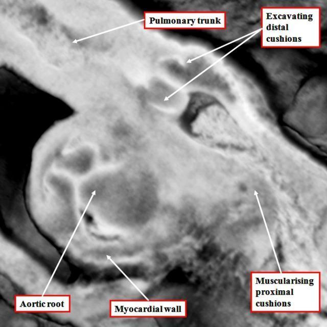
A high-magnification diagnostic image in grayscale showing the embryological development of the human cardiac outflow tract. The image identifies the early morphological stages of arterial valve formation within the intermediate component of the outflow tract. Key labeled structures include the pulmonary trunk and aortic root, illustrating the ongoing septation process. A significant educational feature is the 'excavating distal cushions,' which represent the primordial leaflets of the arterial valves undergoing tissue remodeling. The 'myocardial wall' is clearly visible, forming a myocardial turret that still encases the intermediate outflow tract at this stage. Additionally, the 'muscularising proximal cushions' are highlighted, demonstrating the transition of the cardiac jelly into muscular tissue that will eventually contribute to the ventricular infundibulum. This visual content serves as an educational tool for embryology and pediatric cardiology, focusing on the second heart field's contribution to heart development and the complex transition from simple cushions to structured semilunar valves.
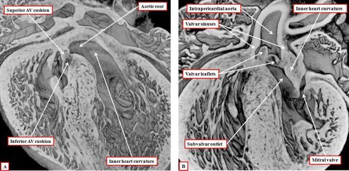
Two high-resolution episcopic data set images (A and B) illustrating the cardiac embryology of mouse hearts at the 15th day of development, used as models for human cardiac morphogenesis. Panel A demonstrates the atrioventricular (AV) region, highlighting the superior and inferior AV cushions as they approximate to close the persisting embryonic interventricular communication. The inner heart curvature is visible adjacent to the developing aortic root. Panel B shows the heart subsequent to foramen closure, emphasizing the spatial relationship between the outflow tract and the left ventricle. Labeled structures include the intrapericardial aorta, valvar sinuses, valvar leaflets, and the subvalvar outlet. The inner heart curvature is clearly interposed between the developing aortic valve leaflets and the mitral valve, illustrating the transitional stage before the formation of adult aortic-to-mitral fibrous continuity. These images provide critical educational insight into the remodeling of the proximal outflow tract and the septation of the ventricular chambers during cardiogenesis.
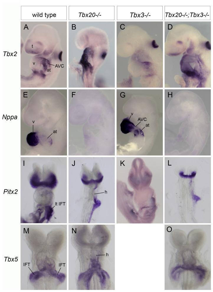
This Comparison Chart displays whole-mount in situ hybridization (ISH) results from mouse embryos at stages E8.5 to E9.25, illustrating the developmental genetics of cardiac chamber formation and patterning. The panel compares four genotypes across rows representing different cardiac marker genes: Tbx2, Nppa, Pitx2, and Tbx5. The vertical columns represent wild type, Tbx20-/-, Tbx3-/-, and double homozygous Tbx20-/-; Tbx3-/- mutants. Key observations include: 1) Tbx2 expression, normally restricted to the atrioventricular canal (AVC), becomes ectopically expanded throughout the heart in Tbx20-/- and double mutants. 2) Nppa (NPPA), a marker for chamber myocardium, is severely down-regulated in Tbx20-/- and double mutants, indicating a failure of chamber differentiation, while being precociously upregulated in the atrium of Tbx3-/- mutants. 3) Pitx2 and Tbx5 expression patterns remain relatively preserved across genotypes at E9.25. Labeled structures include the atrium (at), ventricle (v), telencephalon (t), and inflow tract (IFT). This material is designed for advanced medical education in embryology and molecular cardiology, focusing on transcriptional regulation of heart development.
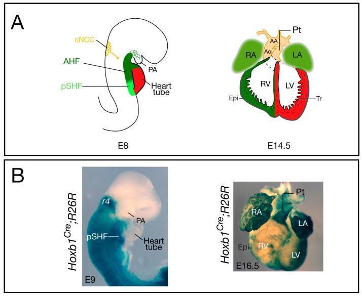
Educational diagram and clinical photographs illustrating murine cardiovascular development and genetic lineage tracing. Panel A provides anatomical diagrams of heart development stages E8 and E14.5. At E8, it identifies the anterior heart field (AHF), posterior second heart field (pSHF), cardiac neural crest cells (cNCC), and the early heart tube. At E14.5, it shows the septated four-chambered heart including the right atrium (RA), left atrium (LA), right ventricle (RV), and left ventricle (LV), alongside the aortic arch (AA) and pulmonary trunk (Pt). Panel B presents clinical photographs of Hoxb1Cre;R26R mouse embryos using X-gal staining for lineage tracing. At E9, staining demonstrates Hoxb1+ cell localization in the pSHF. By E16.5, the ventral view of the heart shows β-galactosidase activity distributed across both atria, the sub-pulmonary myocardium, and the epicardium (Epi). This visual comparison highlights the spatial patterning and subsequent contribution of specific progenitor populations to the mature cardiac architecture, including the trabeculae (Tr) and interventricular structures.
Development of the Heart
1. Origin and Timing
- First Heart Field (FHF / Primary Heart Field, PHF) - forms the initial left ventricular myocardium and early heart tube (days 16-18; controlled by Id gene)
- Second Heart Field (SHF) - pharyngeal mesoderm anterior to the heart tube; contributes to the right ventricle, outflow tract, and atria. Key genes: Hes-1, MEF2C, Pitx-2, dHAND, eHAND
2. Formation of the Heart Tube (Week 3)
- Bilateral endocardial progenitor cells separate from mesoderm → form paired endocardial heart tubes
- Lateral embryonic folding brings the two tubes together, and they fuse craniocaudally into a single primitive heart tube
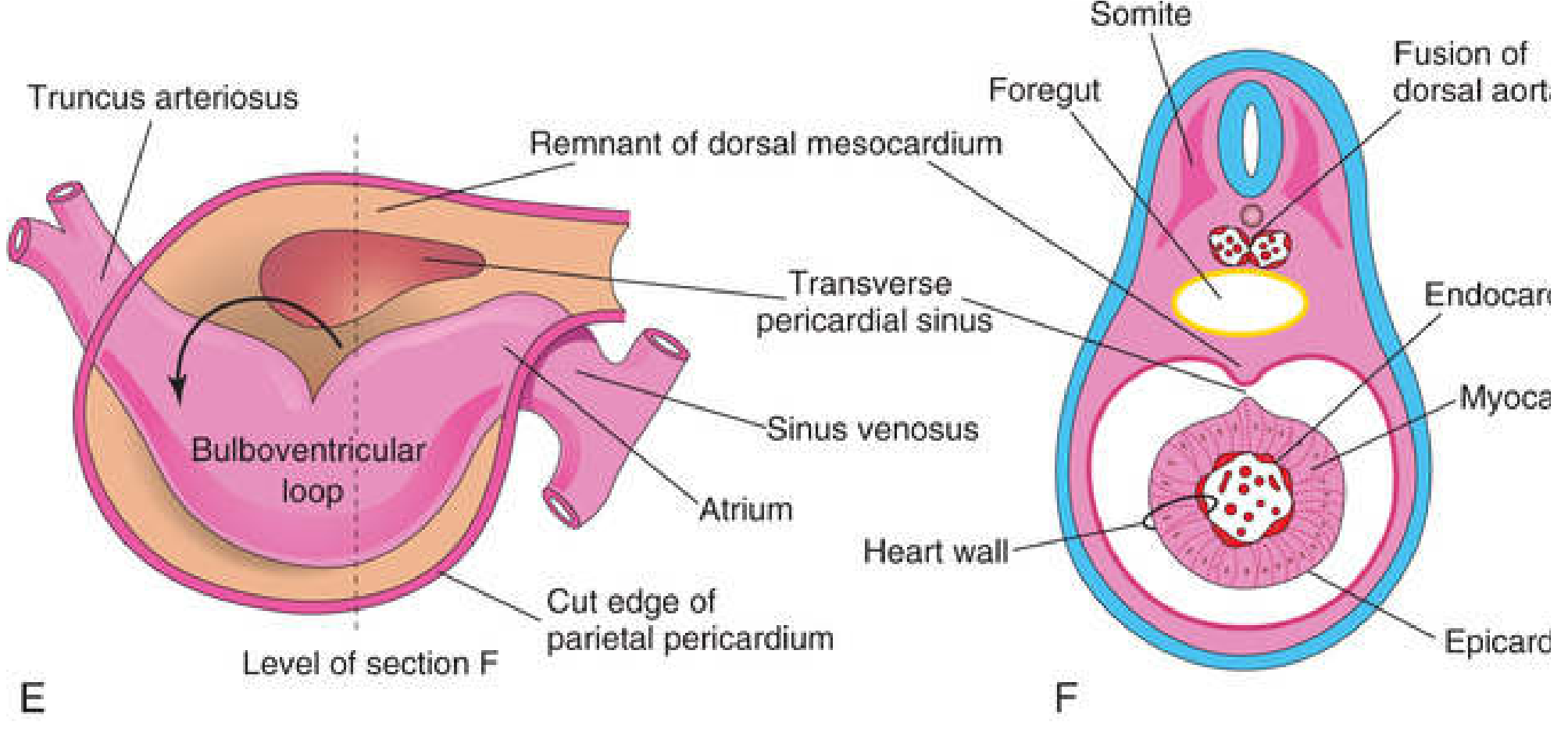
- The tube is suspended in the pericardial cavity by the dorsal mesocardium (which later degenerates, forming the transverse pericardial sinus)
- The wall of the tube at this stage has three layers:
- Inner endothelium (future endocardium)
- Gelatinous cardiac jelly (connective tissue matrix)
- Outer primordial myocardium → becomes myocardium + epicardium
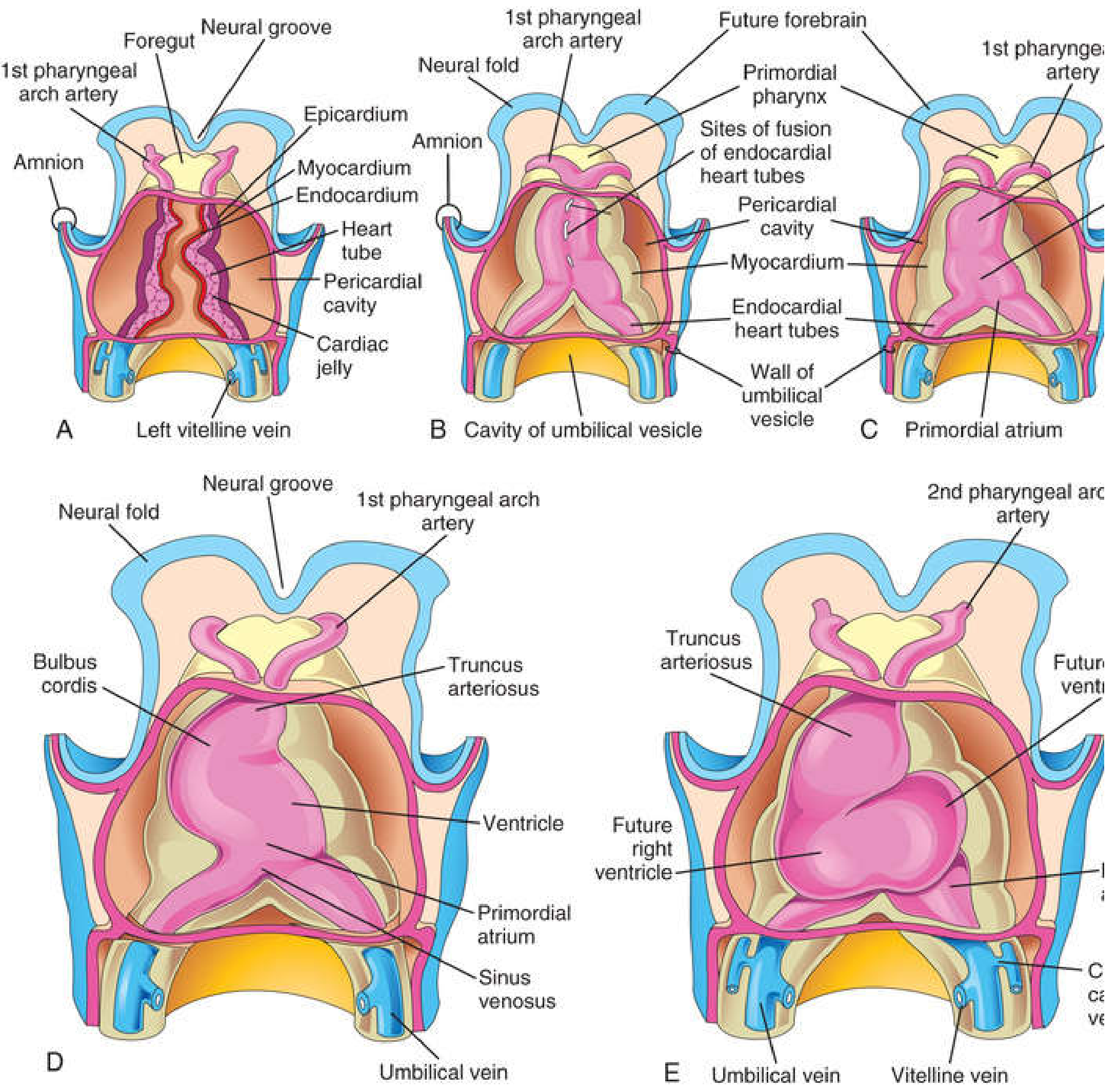
3. Regions of the Primitive Heart Tube (Craniocaudal)
| Region | Adult Derivative |
|---|---|
| Truncus arteriosus | Ascending aorta + pulmonary trunk |
| Bulbus cordis | Smooth parts of right & left ventricles (conus arteriosus) |
| Primitive ventricle | Trabeculated parts of ventricles |
| Primitive atrium | Trabeculated parts of both atria |
| Sinus venosus | Right horn → smooth part of right atrium (sinus venarum); left horn → coronary sinus |
4. Cardiac Looping (Day 23-28)
- Bends to the right → D-loop (dextro-loop, rightward) forming an S-shape
- This positions the bulbus cordis and truncus to the right and anteriorly
- The primitive atrium shifts posterosuperiorly; the sinus venosus shifts posteroinferiorly
- Abnormal looping (e.g., L-loop) causes dextrocardia or situs inversus
5. Partitioning of the Heart (Weeks 4-8)
Atrioventricular Canal Division
- Endocardial cushions form in the dorsal and ventral walls of the AVC (days 26-35)
- They fuse → divide the AVC into right and left AV channels
- Contribute to mitral and tricuspid valves and the interventricular septum
Atrial Septation
- Septum primum grows down from roof of atrium toward the endocardial cushions
- Before it fuses, it leaves a gap - ostium primum
- As ostium primum closes, ostium secundum appears in the upper septum primum
- Septum secundum grows to the right of septum primum, leaving an opening - the foramen ovale
- In fetal life, blood passes right→left through the foramen ovale; at birth, increased left atrial pressure closes it → fossa ovalis
Ventricular Septation
- Muscular interventricular septum (IVS) grows upward from the floor of the ventricle
- A gap remains - the interventricular foramen (IVF)
- Closed by the membranous IVS (formed by fusion of AV cushions + truncal ridges)
- Failure = VSD (most common congenital heart defect)
Division of the Outflow Tract (Truncus Arteriosus)
- Neural crest cells migrate into the truncus and form conotruncal (spiral) ridges
- Ridges spiral 180° and fuse → aorticopulmonary septum dividing truncus into aorta and pulmonary trunk
- Failure → Persistent truncus arteriosus; abnormal spiraling → Transposition of Great Arteries (TGA)
6. Development of the Conducting System
- Sinoatrial node (SAN) → Atrioventricular node (AVN) → Bundle of His → Bundle branches → Purkinje fibers
- Transcription factor TBX3 inhibits differentiation of these cells into ventricular muscle, allowing them to become conduction tissue
- Autonomic nerve fibers (sympathetic/parasympathetic) innervate the SAN to regulate rate, but the conducting cells themselves are intrinsic
7. Fate of the Sinus Venosus
- Right horn → incorporated into right atrium as the sinus venarum (smooth-walled part); right sinovenous valve → crista terminalis + valve of IVC + valve of coronary sinus
- Left horn → coronary sinus
- Left common cardinal vein → oblique vein of left atrium (Marshall)
8. Key Congenital Defects - Summary Table (Langman's)
| Vulnerable Stage | Birth Defect |
|---|---|
| PHF (days 16-18) | ASD, VSD, TGA, dextrocardia |
| Heart tube looping (days 22-28) | Dextrocardia |
| AV endocardial cushions (days 26-35) | VSD, mitral/tricuspid valve defects |
| SHF (days 22-28) | Tetralogy of Fallot, TGA, pulmonary stenosis |
| Outflow tract (days 36-49) | Common truncus arteriosus |
| Aortic arches (days 22-42) | Interrupted aortic arch type B |
Quick Timeline Summary
| Day | Event |
|---|---|
| Day 18 | Bilateral heart tubes form from splanchnic mesoderm |
| Day 22-23 | Tubes fuse; heart begins beating |
| Day 23-28 | Cardiac looping (D-loop) |
| Week 4-5 | Endocardial cushion formation; AVC partitioning begins |
| Week 5-8 | Atrial, ventricular, and outflow tract septation complete |