
Melasma
Reading File
Finding Sources
Searching PubMed
melasma treatment
Reading File
Reading File
Reading File
Reading File
Reading File
Loading Image
Here is a comprehensive overview of melasma drawn from Fitzpatrick's Dermatology, Dermatology 2-Volume Set 5e, and Andrews' Diseases of the Skin.
Melasma (Chloasma / "Mask of Pregnancy")
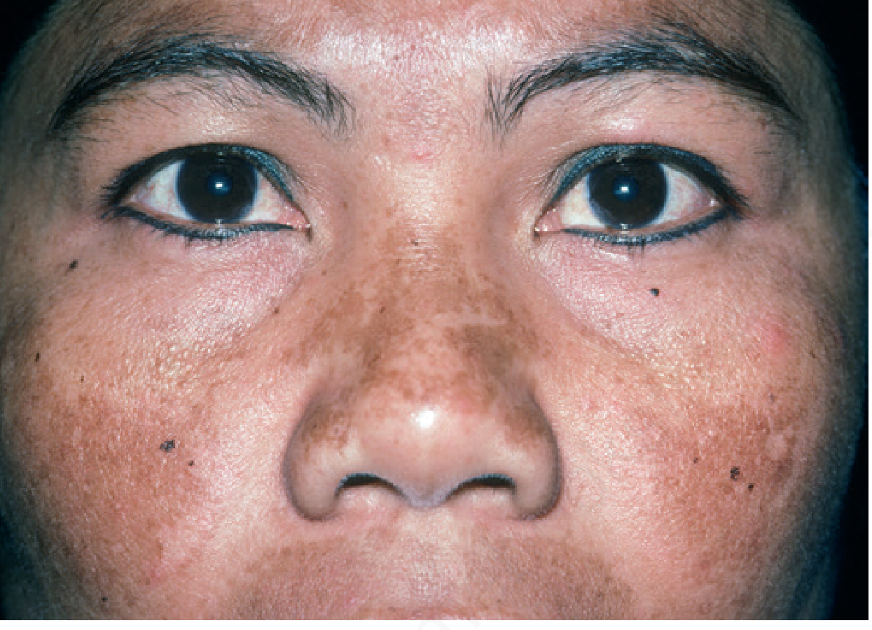
Definition & Epidemiology
Melasma is a common acquired disorder of hyperpigmentation characterized by symmetric, irregularly bordered brown patches occurring predominantly on sun-exposed skin, especially the face. Key epidemiologic facts:
-
90% of patients are women
- Most prevalent in Fitzpatrick skin types III-V: Hispanic, East/West/Southeast Asian, Middle Eastern, and Black individuals
- Subtle melasma (by UV light exam) seen in up to 30% of middle-aged Asian women
- Men are also affected, especially those from Central America
- The forearms are the second most common site after the face
Pathogenesis
The exact pathogenesis is not fully understood, but two major triggers are established:
1. UV / Sun Exposure (Primary Trigger)
- Melasma predominantly affects sun-exposed areas and worsens in summer
- Patients have a lower minimal erythema dose (MED) to UV light
- Affected skin shows increased solar elastosis compared to adjacent unaffected facial skin
- Melasma-affected skin has reduced WIF-1 (a Wnt antagonist), leading to increased Wnt signaling, which stimulates melanogenesis
- Number of melanocytic nevi correlates with melasma risk
2. Female Hormones (Second Trigger)
- Melasma occurs frequently during pregnancy, with oral contraceptive (OC) use, and hormone replacement therapy (HRT)
- Discontinuing OCs/HRT rarely clears the pigmentation - it can persist for years
- Melasma of pregnancy typically clears within months of delivery
- Also seen in other endocrinologic disorders and with phenytoin and finasteride therapy
Clinical Features
Morphology: Well-demarcated, tan-to-dark brown patches, sharply demarcated borders
Distribution patterns (3 classic facial types):
| Pattern | Location |
|---|---|
| Centrofacial (most common) | Forehead, cheeks, upper lip, nose, chin |
| Malar | Cheeks and nose |
| Mandibular | Ramus of mandible |
Most patients have multiple patterns, making classification less useful therapeutically.
Classification by Melanin Location
| Type | Location of Melanin | Wood's Lamp | Response to Treatment |
|---|---|---|---|
| Epidermal | Basal and suprabasal layers | Enhanced (accentuated) | Better |
| Dermal | Within melanophages | Not enhanced | Poor |
| Mixed | Both layers | Variable | Intermediate |
Note: Most cases show both epidermal and dermal melanin on histology and confocal microscopy. Dermal deposits respond poorly to topical agents regardless of Wood's lamp findings - a therapeutic trial should be offered to all patients.
Differential Diagnosis
| Condition | Key Distinguishing Features |
|---|---|
| Postinflammatory hyperpigmentation | History of inflammation; less irregular outline |
| Drug-induced hyperpigmentation | History of doxycycline, amiodarone, etc.; less patterned |
| Riehl melanosis (pigmented contact dermatitis) | Sites of cosmetic application; brown-grey due to dermal melanin |
| Acquired bilateral nevus of Ota (Horii nevus) | Asian women; brown-grey to blue macules; nasal predominance |
| Lichen planus pigmentosus | Temporal/preauricular onset; coexistent LP in ~20% |
| Exogenous ochronosis | History of HQ use; progressive darkening; banana-shaped yellow-brown deposits on histology |
| Erythema dyschromicum persans | Slate-grey to blue-brown; involves sun-protected areas |
Treatment
Treatment must be multimodal. Results take up to 6 months to appreciate. Recurrence is the norm even with adequate treatment.
Universal Recommendations (All Patients)
- Broad-spectrum sunscreen SPF ≥30 daily (ideally with zinc oxide or titanium dioxide as physical blocker)
- Sun-protective hats and clothing
- Camouflage makeup with iron oxide (blocks visible light, which also drives pigmentation)
- Avoid tanning beds
- Discontinue oral contraceptives if possible
First-Line Topical Therapies
- Triple combination (Kligman's formula): Hydroquinone (HQ) + retinoid (tretinoin) + topical corticosteroid - the most effective topical regimen available
- Use 4% HQ (daily, typically bedtime); OTC formulations contain 2%
- Twice-weekly application is effective for maintenance
- Side effects of overuse: fixed erythema, telangiectasias, acneiform eruptions, hypertrichosis; exogenous ochronosis from HQ overuse
- Typically used daily for 2-4 months, then tapered to 1-2x/week
- Azelaic acid 15-20%
- Tretinoin alone (less effective than HQ but beneficial)
Adjunctive Topical Therapies
- L-ascorbic acid 10-15%
- Kojic acid 1-4%
- Tranexamic acid 2-5%
- Niacinamide 4%
Adjunctive Oral Therapies
- Tranexamic acid 250 mg BID for 8-12 weeks - screen patients for thromboembolic risk factors before use
Second-Line Therapies
- Chemical peels: Glycolic acid (start 30%, increase as tolerated) or salicylic acid (20-30%) every 4-6 weeks
- Peels alone do not reproducibly enhance 4% HQ and can cause hyperpigmentation if irritation occurs
Third-Line Therapies (Procedural)
- Non-ablative fractional laser - improvement in 60% of skin types III-V with 75-100% improvement; may enhance HQ absorption via microscopic channels
- Intense pulsed light (IPL) - can improve melasma but has high relapse rate
- Low-fluence Q-switched lasers - Q-switched Nd:YAG can paradoxically increase pigmentation
- Picosecond lasers
- Er:YAG resurfacing - only temporary improvement, often followed by problematic postinflammatory hyperpigmentation
- Pulsed dye laser - may enhance topical combination treatment
⚠️ All light-based therapies should be approached with caution in darker skin types - complications include hyperpigmentation, hypopigmentation, irritation, and scarring. Always perform a test spot first.
Long-Term Maintenance
- Daily sunscreen + sun protection
- Topical retinoid
- Topical alpha-hydroxy acid (e.g., glycolic acid cream)
- Rotating adjunctive topicals: L-ascorbic acid, azelaic acid, kojic acid, tranexamic acid
Recent Evidence (2024-2025)
A 2025 systematic review on glutathione as a skin-lightening agent in melasma (Sarkar et al., Int J Dermatol, 2025; PMID 39444151) evaluated oral and topical glutathione, finding emerging but still limited evidence for its role in melasma management.
Sources: Andrews' Diseases of the Skin, p. 993-994 | Dermatology 2-Volume Set 5e, p. 1343, 2859 | Fitzpatrick's Dermatology
Can you give me final prescription and with company name
Searching the Web
melasma prescription drugs brand names India hydroquinone tretinoin azelaic acid tranexamic acid 2025
Here is a practical melasma prescription with commonly available brand names (India-focused, with international brands noted where relevant):
⚠️ Disclaimer: This is for educational/clinical reference only. Prescriptions must be individualized by a qualified dermatologist based on patient history, skin type, and contraindications.
Melasma - Sample Prescription
Rx
STEP 1 - Sun Protection (Mandatory, All Patients)
| Drug | Brand (India) | Dose/Instructions |
|---|---|---|
| Sunscreen SPF 50+ PA++++ (zinc oxide + titanium dioxide) | Lotus Safe Sun, Bioderma Photoderm MAX, La Shield, Neutrogena Ultra Sheer, Episoft AC | Apply 30 min before going out, reapply every 2-3 hours |
STEP 2 - First-Line: Triple Combination Cream (Gold Standard)
Kligman's Formula = Hydroquinone 4% + Tretinoin 0.025-0.05% + Fluocinolone acetonide 0.01%
| Drug | Brand (India) | Dose |
|---|---|---|
| HQ 4% + Tretinoin 0.025% + Fluocinolone 0.01% (Triple combo) | Tri-Luma (Galderma), Pigmanorm, Melaquin, Lustra AF | Apply at bedtime to affected areas only; daily x 8 weeks, then 2x/week for maintenance |
| OR: Hydroquinone 4% alone | Melanorm HC (Sun Pharma), Melalite Forte (Elder), HQ-4 | Apply at bedtime |
STEP 3 - Adjunctive Topicals (Add-on or Alternative to HQ)
| Drug | Brand (India) | Dose |
|---|---|---|
| Azelaic acid 15-20% | Aziderm 20% (Cipla), Azclear, Finacea (Bayer) | Apply BD (twice daily) |
| Kojic acid 2-4% | Kojivit (Micro Labs), Kojic (various), Melacare Forte (contains kojic + HQ) | Apply once daily at night |
| Tranexamic acid 2-5% (topical) | Transamin cream, Tran-Q cream, Pigment cream | Apply BD |
| Niacinamide 4-5% | Dot & Key, Minimalist 10% Niacinamide, Cetaphil Bright | Apply AM |
| Vitamin C (L-ascorbic acid 10-15%) | Evion C, Skinceuticals CE Ferulic, Dot & Key Vitamin C | Apply in the morning |
STEP 4 - Oral Therapy (for moderate-severe or refractory cases)
| Drug | Brand (India) | Dose |
|---|---|---|
| Tranexamic acid | Traxyl (Cipla), Pause (Elder), Txamic | 250 mg BD x 8-12 weeks - Screen for thromboembolism risk before starting |
STEP 5 - Procedural (Second/Third Line)
| Procedure | Details |
|---|---|
| Glycolic acid peel 30-70% | Every 4-6 weeks x 4-6 sessions |
| Salicylic acid peel 20-30% | Safer in darker skin; every 4-6 weeks |
| Non-ablative fractional laser (1550nm Fraxel / 1540 nm) | 3-5 sessions; test patch mandatory |
| Q-switched Nd:YAG low fluence | Use with caution - risk of rebound hyperpigmentation |
| IPL | High relapse rate; caution in skin types IV-VI |
Key Prescribing Notes
- Tretinoin is Category C in pregnancy - avoid. Use azelaic acid (safer) instead.
- Hydroquinone is Schedule H in India - requires a valid prescription; do NOT use >4-6 months continuously to avoid exogenous ochronosis.
- Triple combination cream: use class 5-7 topical steroid (mild-moderate strength like fluocinolone 0.01% or hydrocortisone 1%); avoid potent steroids.
- Tranexamic acid oral - screen for DVT/PE risk, cardiac disease, or ongoing anticoagulant use before prescribing.
- Always use iron oxide-containing makeup alongside sunscreen - visible light also drives melanogenesis.
- Results take 3-6 months to appreciate. Set realistic patient expectations. Recurrence after stopping treatment is common.
- Do a test patch before any laser/peel in skin type IV-VI.
Commonly used combination in Indian dermatology practice:
- Melalite Forte (HQ 4%) at night + Aziderm 20% AM + Traxyl 250 mg BD x 8 weeks + daily SPF 50+ sunscreen
Face wash for this which contain vitamin c nicanamide
Searching the Web
face wash vitamin C niacinamide melasma hyperpigmentation India brand names 2025
Here is a curated list of face washes containing Vitamin C and/or Niacinamide, suitable for melasma/hyperpigmentation patients:
Face Washes for Melasma - Vitamin C & Niacinamide
💡 Note on face washes: Active ingredients like Vitamin C and Niacinamide have limited contact time in a cleanser (rinse-off product), so their benefit is less than serums/creams. However, they help prep skin, gently brighten, and reduce irritation from other actives.
Indian Brands
| Brand / Product | Key Ingredients | Best For | Approx. Price |
|---|---|---|---|
| Minimalist 10% Vitamin C Face Wash | Ethyl Ascorbic Acid (stable Vit C 10%) + Ferulic Acid | Dull skin, dark spots, melasma | ₹299 |
| Plum Bright Years Vitamin C Face Wash | Vitamin C + Green Tea | Brightening, sensitive skin | ₹200-250 |
| Mamaearth Vitamin C Face Wash | Vitamin C + Turmeric | Brightening, everyday use | ₹199 |
| WOW Skin Science Vitamin C Face Wash | Vitamin C + Hyaluronic Acid | Brightening + hydration | ₹249 |
| Dot & Key Vitamin C + E Brightening Face Wash | Vitamin C + Vitamin E | Antioxidant protection + brightening | ₹395 |
| Pilgrim Vitamin C Face Wash | Vitamin C + Hyaluronic Acid | Brightening, all skin types | ₹349 |
| Deconstruct Brightening Face Wash | Niacinamide 1% + Vitamin C | Pigmentation, oily/combination skin | ₹349 |
| Suganda Vitamin C Face Wash | Vitamin C (stable form) + Niacinamide | Sensitive + pigmented skin | ₹450 |
| MCaffeine Vitamin C Face Wash | Vitamin C + Caffeine | Brightening + de-puffing | ₹299 |
Dermatologist/Pharma Grade (India)
| Brand / Product | Key Ingredients | Company | Notes |
|---|---|---|---|
| Cetaphil Bright Healthy Radiance Face Wash | Niacinamide + Vitamin C precursor | Galderma | Gentle, dermat-recommended |
| Sebamed Brightening Face Wash | Niacinamide + Vitamin C | Sebapharma (Germany) | pH 5.5, good for sensitive skin |
| Bioderma Pigmentbio Foaming Cream | Vitamin C + Niacinamide + Kojic acid | Bioderma (France) | Specifically formulated for hyperpigmentation/melasma |
| La Roche-Posay Vitamin C Face Wash | Vitamin C derivative | L'Oréal | Gentle, fragrance-free |
Top Pick for Melasma
🏆 Best Overall: Bioderma Pigmentbio Foaming Cream - specifically designed for hyperpigmented skin, contains Vitamin C + Niacinamide + Kojic acid (all 3 anti-pigmentation actives in one cleanser). Available at pharmacies and online (₹900-1100).
🏆 Best Budget: Minimalist 10% Vitamin C Face Wash - highest stable Vitamin C concentration in a cleanser at an affordable price point. Widely available online.
How to Use (Melasma Routine)
| Time | Step | Product |
|---|---|---|
| Morning | Face wash | Vitamin C + Niacinamide face wash (any above) |
| Serum | Vitamin C serum 10-15% | |
| Sunscreen | SPF 50+ with PA++++ and iron oxide | |
| Night | Face wash | Same gentle face wash |
| Treatment | Triple combo cream / HQ 4% / Azelaic acid | |
| Moisturizer | Light moisturizer |
Key tip: Since face wash contact time is short, pair it with a leave-on Vitamin C serum in the morning and Niacinamide serum/cream at night for maximum benefit on melasma.
This is a shared conversation. Sign in to Orris to start your own chat.