
seminar on skeletal dysplacia
- Definition and classification
- Normal bone growth and genetic basis
- Common disorders: achondroplasia, osteogenesis imperfecta, thanatophoric dysplasia, multiple epiphyseal dysplasia, and mucopolysaccharidoses
- Clinical features and red flags
- Radiological approach and characteristic patterns
- Diagnostic pathway: family history, examination, imaging, genetic testing
- Complications and multidisciplinary management
- Prenatal diagnosis, genetic counseling, and ethical issues
- Case discussion and take-home messages
Choose the seminar format and depth. · Who is the intended audience?
- Definition and classification
- Normal bone growth and genetic basis
- Common disorders: achondroplasia, osteogenesis imperfecta, thanatophoric dysplasia, multiple epiphyseal dysplasia, and mucopolysaccharidoses
- Clinical features and red flags
- Radiological approach and characteristic patterns
- Diagnostic pathway: family history, examination, imaging, genetic testing
- Complications and multidisciplinary management
- Prenatal diagnosis, genetic counseling, and ethical issues
- Case discussion and take-home messages
- 10-slide student seminar
- 15-slide detailed seminar
- Written seminar notes
- PowerPoint with speaker notes
Write detailed seminar notes for medical students
skeletal dysplasia AND diagnosis
ACMG practice resource skeletal dysplasia genetic evaluation guidelines 2024
https://pmc.ncbi.nlm.nih.gov/articles/PMC3111030
- Core concepts: definition, terminology, epidemiology, normal endochondral ossification
- Genetic and pathophysiological basis: inheritance, de novo mutations, FGFR3 and collagen disorders
- Clinical classification: disproportionate short stature, rhizomelia, mesomelia, acromelia, trunk-predominant disorders
- Common disorders: achondroplasia, osteogenesis imperfecta, thanatophoric dysplasia, hypochondroplasia, diastrophic dysplasia, spondyloepiphyseal dysplasia
- Clinical and radiological evaluation: history, examination, skeletal survey, pattern recognition, molecular testing
- Prenatal diagnosis: ultrasound clues, lethal versus nonlethal disorders, counseling
- Management: multidisciplinary care, complications, surgery, rehabilitation, genetic counseling
- Case discussion and examination revision
Choose the section for detailed seminar notes.
- Definition and classification
- Normal bone growth and genetic basis
- Common disorders: achondroplasia, osteogenesis imperfecta, thanatophoric dysplasia, multiple epiphyseal dysplasia, and mucopolysaccharidoses
- Clinical features and red flags
- Radiological approach and characteristic patterns
- Diagnostic pathway: family history, examination, imaging, genetic testing
- Complications and multidisciplinary management
- Prenatal diagnosis, genetic counseling, and ethical issues
- Case discussion and take-home messages
- 10-slide student seminar
- 15-slide detailed seminar
- Written seminar notes
- PowerPoint with speaker notes
- Core concepts: definition, terminology, epidemiology, normal endochondral ossification
- Genetic and pathophysiological basis: inheritance, de novo mutations, FGFR3 and collagen disorders
- Clinical classification: disproportionate short stature, rhizomelia, mesomelia, acromelia, trunk-predominant disorders
- Common disorders: achondroplasia, osteogenesis imperfecta, thanatophoric dysplasia, hypochondroplasia, diastrophic dysplasia, spondyloepiphyseal dysplasia
- Clinical and radiological evaluation: history, examination, skeletal survey, pattern recognition, molecular testing
- Prenatal diagnosis: ultrasound clues, lethal versus nonlethal disorders, counseling
- Management: multidisciplinary care, complications, surgery, rehabilitation, genetic counseling
- Case discussion and examination revision
skeletal dysplasia AND diagnosis
GeneReviews achondroplasia clinical features management FGFR3
skeletal dysplasia radiograph achondroplasia characteristic findings
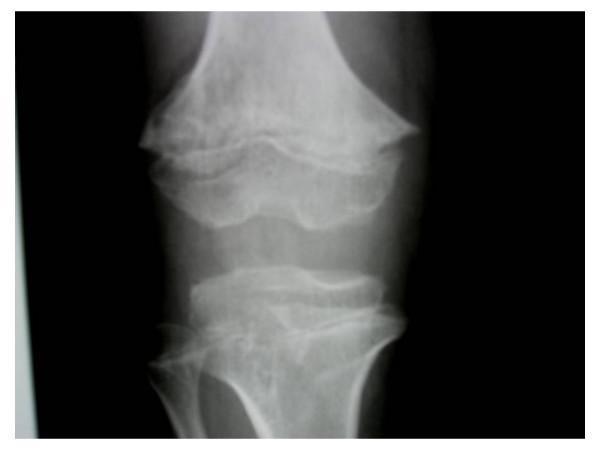
This anteroposterior radiograph of the knee demonstrates skeletal dysplasia characterized by significant metaphyseal irregularities in both the distal femur and proximal tibia. Key diagnostic features include prominent metaphyseal cupping and an abnormal trabecular pattern containing multiple small, enchondroma-like lucent lesions. The internal architecture of the metaphyses shows characteristic rings, arcs, and dense punctate foci, consistent with cartilaginous matrix calcification. These findings are typical of enchondroma-like metaphyseal dysplasia, often associated with systemic skeletal conditions like achondroplasia or Ollier disease. The visual focus is on the junction between the diaphysis and epiphysis, highlighting the expansion and morphological distortion of the growth zones.
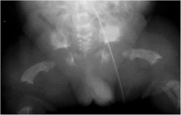
Anteroposterior (AP) radiograph of the pelvis and proximal femora of a neonate demonstrating characteristic findings of thanatophoric dysplasia, type I. The image reveals significant skeletal abnormalities. The femora are markedly shortened and robust, displaying a distinctive bowed appearance often referred to as 'telephone receiver' femora. The pelvis exhibits a squared-off morphology with small, horizontal acetabular roofs. There is prominent narrowing of the sacrosciatic notch, reflecting severe endochondral ossification defects. While these features share qualitative similarities with achondroplasia, they are quantitatively more severe in this presentation. The lumbar spine shows some evidence of platyspondyly (flat vertebral bodies). This diagnostic image is essential for medical education regarding lethal skeletal dysplasias and the radiographic differentiation between FGFR3-related conditions.
achondroplasia AND vosoritide
achondroplasia international consensus management foramen magnum sleep apnea GeneReviews
Seminar Notes: Skeletal Dysplasia
Learning objectives
- Define skeletal dysplasia and distinguish it from dysostosis and metabolic bone disease.
- Describe the genetic and developmental basis of skeletal dysplasias.
- Classify limb shortening patterns and recognize major clinical clues.
- Discuss the features of common skeletal dysplasias.
- Outline a systematic clinical, radiological, and genetic diagnostic approach.
- Identify lethal skeletal dysplasias in the prenatal period.
- Explain principles of multidisciplinary management and genetic counseling.
1. Introduction and definition
- Disproportionate short stature
- Limb shortening or deformity
- Abnormal skull, spine, chest, and pelvic development
- Fractures and bone fragility
- Joint laxity or early osteoarthritis
- Neurological, respiratory, hearing, visual, and dental complications
Important terminology
| Term | Meaning |
|---|---|
| Skeletal dysplasia | Generalized intrinsic disorder of bone and cartilage growth |
| Osteochondrodysplasia | Another term for skeletal dysplasia |
| Dysostosis | Localized developmental malformation of one or more bones, usually nonprogressive, for example cleidocranial dysostosis |
| Dwarfism / short stature | Descriptive terms, not diagnostic labels |
| Disproportionate short stature | Trunk and limbs are affected unequally, strongly suggesting skeletal dysplasia |
| Proportionate short stature | Height, trunk, and limbs are reduced proportionately; endocrine, nutritional, systemic, or chromosomal causes are more likely |
2. Normal bone formation and pathogenesis
Types of ossification
A. Intramembranous ossification
- Flat bones of skull
- Parts of clavicle
- Mandible
B. Endochondral ossification
- Long bones
- Vertebrae
- Pelvis
- Base of skull
Major genetic mechanisms
-
Extracellular matrix protein defects
- Type I collagen: osteogenesis imperfecta
- Type II collagen: spondyloepiphyseal dysplasia congenita and related disorders
- Cartilage oligomeric matrix protein: pseudoachondroplasia, some multiple epiphyseal dysplasias
-
Signal transduction defects
- FGFR3 gain-of-function variants: achondroplasia, hypochondroplasia, thanatophoric dysplasia
-
Enzyme or metabolic defects
- ALPL variants: hypophosphatasia
- Lysosomal storage disorders such as mucopolysaccharidoses can produce dysostosis multiplex
-
Transcription-factor defects
- RUNX2: cleidocranial dysplasia
- SOX9: campomelic dysplasia
-
Ciliary dysfunction
- Some short-rib thoracic dysplasias and syndromic skeletal disorders
3. Classification
- Primary tissue involved: bone, cartilage, or both
- Radiological site involved: epiphysis, metaphysis, diaphysis, spine
- Pattern of short stature
- Molecular cause
- Lethal versus nonlethal phenotype
A. Classification by limb segment involved
| Pattern | Segment mainly shortened | Typical examples |
|---|---|---|
| Rhizomelia | Proximal segment: humerus and femur | Achondroplasia |
| Mesomelia | Middle segment: radius-ulna and tibia-fibula | Leri-Weill dyschondrosteosis |
| Acromelia | Distal segment: hands and feet | Acrodysostosis |
| Micromelia | Entire limb is markedly short | Thanatophoric dysplasia |
| Brachydactyly | Digits are short | Several syndromic and isolated disorders |
B. Classification by anatomical region
| Type | Main site of involvement | Examples |
|---|---|---|
| Epiphyseal dysplasia | Epiphyses | Multiple epiphyseal dysplasia |
| Metaphyseal dysplasia | Metaphyses | Schmid metaphyseal dysplasia |
| Diaphyseal dysplasia | Shafts of long bones | Camurati-Engelmann disease |
| Spondylo-dysplasia | Vertebral bodies | Spondyloepiphyseal dysplasia congenita |
| Craniofacial dysplasia | Skull and face | Cleidocranial dysplasia |
4. Clinical approach to a suspected skeletal dysplasia
History
- Antenatal ultrasound findings: short limbs, bowed long bones, small chest, fractures, polyhydramnios
- Birth length, head circumference, and neonatal respiratory problems
- Growth pattern and developmental milestones
- Recurrent fractures or low-trauma fractures
- Bone pain, joint pain, waddling gait, or reduced mobility
- Hearing loss, visual problems, recurrent otitis media
- Snoring, sleep apnea, cyanotic spells, or respiratory infections
- Family history of short stature, fractures, limb deformity, early osteoarthritis, miscarriages, stillbirths, and consanguinity
- Parental heights and body proportions
Examination
- Height or length, weight, and head circumference plotted on appropriate growth charts
- Upper-to-lower segment ratio
- Arm span
- Sitting height
- Limb proportions
- Skull shape and frontal bossing
- Midface hypoplasia
- Chest size and shape
- Spine: kyphosis, scoliosis, lumbar lordosis
- Limb alignment: genu varum or genu valgum
- Hand pattern: brachydactyly, trident hand, polydactyly
- Joint hypermobility or contractures
- Dentition, scleral color, hearing, and vision
Red flags
- Disproportionate short stature
- Macrocephaly with frontal bossing
- Rhizomelic limb shortening
- Recurrent fractures without adequate trauma
- Blue sclerae or dentinogenesis imperfecta
- Progressive limb bowing
- Narrow thorax or respiratory distress in a newborn
- Kyphosis, neurological deficits, or sleep-disordered breathing
- Family history consistent with Mendelian inheritance
5. Radiological evaluation
- Skull
- Spine: AP and lateral views
- Chest
- Pelvis
- Upper limbs and hands
- Lower limbs and feet
Radiological pattern-recognition questions
- Is mineralization normal, reduced, or increased?
- Are the long bones short, bowed, broad, narrow, or fractured?
- Are abnormalities mainly epiphyseal, metaphyseal, or diaphyseal?
- Is there platyspondyly?
- Is the pelvis abnormal?
- Is there narrowing of interpedicular distance in the lumbar spine?
- Is the thorax small or narrow?
- Are hands and feet affected?
- Are there extraskeletal findings such as craniosynostosis or polydactyly?
6. Common skeletal dysplasias
A. Achondroplasia
Definition and genetics
- Usually caused by a pathogenic gain-of-function variant in FGFR3
- Autosomal dominant inheritance
- Most affected children have average-height parents because the variant is de novo
- FGFR3 activation inhibits chondrocyte proliferation at the growth plate, impairing endochondral ossification
Clinical features
- Disproportionate short stature
- Rhizomelic shortening of proximal limbs
- Macrocephaly with frontal bossing
- Midface hypoplasia and depressed nasal bridge
- Short fingers with separation between middle and ring fingers: trident hand
- Lumbar hyperlordosis
- Thoracolumbar kyphosis in infancy
- Genu varum
- Limited elbow extension
- Normal intelligence in most individuals
Radiological features
- Short long bones, especially proximal segments
- Metaphyseal flaring
- Short, broad pelvis with horizontal acetabula
- Narrowing of lumbar interpedicular distance caudally
- Short pedicles with spinal canal narrowing
- Small foramen magnum
Major complications
- Foramen magnum stenosis and cervicomedullary compression
- Central and obstructive sleep apnea
- Hydrocephalus or ventriculomegaly
- Recurrent otitis media and conductive hearing loss
- Thoracolumbar kyphosis
- Lumbar spinal stenosis, usually later in life
- Obesity
- Genu varum
Management
- Lifelong multidisciplinary follow-up
- Monitor head circumference, neurological status, and developmental progress
- Screen for sleep-disordered breathing
- Hearing assessment and treatment of middle-ear disease
- Monitor kyphosis, genu varum, and spinal stenosis
- Neurosurgical evaluation for symptomatic foramen magnum compression
- Orthopedic treatment for limb alignment or spinal deformity when indicated
- Genetic counseling and psychosocial support
B. Thanatophoric dysplasia
Definition and genetics
Clinical and radiological features
- Severe micromelia
- Very short limbs
- Narrow thorax with short ribs
- Macrocephaly and frontal bossing
- Severe pulmonary hypoplasia
- Platyspondyly
- Hypoplastic pelvis
- Type I: curved femora, classically described as telephone-receiver femora
- Type II: cloverleaf skull may occur
Outcome and management
- Prenatal confirmation where possible
- Detailed counseling regarding prognosis
- Discussion of pregnancy options in accordance with local law and patient values
- Perinatal palliative-care planning when appropriate
- Genetic counseling: recurrence risk is generally low when the causative variant is de novo, but parental testing and individualized counseling are needed
C. Osteogenesis imperfecta
Definition
Genetics and pathology
Clinical features
- Recurrent fractures with minimal trauma
- Bone deformity
- Short stature of variable severity
- Blue sclerae
- Dentinogenesis imperfecta
- Joint hypermobility and ligamentous laxity
- Hearing loss
- Scoliosis
- Family history may be autosomal dominant
Sillence clinical classification
| Type | Main features |
|---|---|
| Type I | Mildest common form, blue sclerae, fractures, often normal or near-normal stature |
| Type II | Severe perinatal lethal form, multiple fractures and marked deformity |
| Type III | Progressively deforming severe form, recurrent fractures and very short stature |
| Type IV | Moderate severity, normal or gray sclerae, variable fractures and deformity |
Radiology
- Generalized osteopenia
- Multiple fractures in different stages of healing
- Bowed long bones
- Wormian bones in skull
- Vertebral compression fractures
- Progressive scoliosis
Management
- Fracture prevention and prompt fracture management
- Physiotherapy, safe mobility, and rehabilitation
- Orthopedic rodding for recurrent fractures or severe deformity
- Dental, hearing, and respiratory assessment
- Bisphosphonates may reduce bone resorption, bone pain, and fracture frequency in selected patients, particularly those with moderate to severe disease. Bailey and Love’s Short Practice of Surgery, osteogenesis imperfecta section.
- Consider nonaccidental injury carefully in infants with unexplained fractures, but do not assume abuse without a full clinical, radiological, biochemical, and genetic assessment.
D. Hypochondroplasia
Features
- Short stature, often recognized later in childhood
- Mild rhizomelic or mesomelic limb shortening
- Less marked craniofacial abnormalities than achondroplasia
- Mild spinal and pelvic radiological changes
- Normal intelligence in many, though developmental or learning issues can occur in some individuals
Key distinction from achondroplasia
E. Diastrophic dysplasia
Genetics
- Usually autosomal recessive
- Caused by pathogenic variants in SLC26A2
Clinical features
- Short stature and limb shortening
- Joint contractures
- Clubfoot
- Hitchhiker thumb
- Scoliosis and kyphosis
- Cleft palate may occur
- Swollen external ears in infancy, later becoming deformed
F. Spondyloepiphyseal dysplasia congenita
Genetics
Features
- Short-trunk dwarfism
- Platyspondyly
- Delayed epiphyseal ossification
- Coxa vara
- Early osteoarthritis
- Myopia and retinal complications
- Hearing loss may occur
G. Cleidocranial dysplasia
Genetics
- Autosomal dominant
- Caused by pathogenic variants in RUNX2
Clinical features
- Delayed closure of fontanelles
- Persistent wide cranial sutures
- Frontal bossing
- Hypoplastic or absent clavicles
- Ability to approximate shoulders anteriorly
- Delayed eruption of permanent teeth and supernumerary teeth
- Short stature may be mild
Key diagnostic clue
7. Prenatal diagnosis
- Femur or humerus length below expected values
- Limb shortening
- Bowed or fractured long bones
- Poor bone mineralization
- Small thorax
- Abnormal skull shape
- Polydactyly
- Clubfoot
- Hydrops or polyhydramnios
Prenatal markers of likely lethality
- Very small thorax relative to abdomen
- Severe micromelia
- Markedly reduced bone mineralization
- Multiple fractures
- Severe pulmonary hypoplasia
- Severe skull, spine, and long-bone abnormalities
Prenatal investigations
- Detailed targeted ultrasound
- Fetal MRI in selected cases
- Amniocentesis or chorionic-villus sampling for genetic testing
- Targeted familial variant testing if a diagnosis is known in a parent
- Skeletal-dysplasia gene panel, exome sequencing, or genome sequencing where available
- Multidisciplinary counseling involving fetal medicine, clinical genetics, neonatology, radiology, and palliative care if required
8. Genetic testing and counseling
Why establish a molecular diagnosis?
- Confirm the clinical and radiological diagnosis
- Clarify prognosis
- Identify risks to the airway, brain, spine, lungs, eyes, hearing, or heart
- Guide surveillance and treatment
- Inform reproductive choices
- Determine recurrence risk
- Enable family testing
Inheritance counseling
| Inheritance pattern | Typical recurrence consideration |
|---|---|
| Autosomal dominant, affected parent | 50% risk in each pregnancy |
| Autosomal recessive, both parents carriers | 25% affected, 50% carrier, 25% unaffected in each pregnancy |
| X-linked | Depends on whether the mother is a carrier and the sex of the fetus |
| De novo dominant variant | Usually low recurrence risk, though gonadal mosaicism means risk is not zero |
Specific point in achondroplasia
- If one parent has heterozygous achondroplasia and the other parent is average height, each pregnancy has a 50% chance of achondroplasia.
- If both parents have achondroplasia, there is a risk of homozygous achondroplasia, which is usually lethal.
9. Principles of management
Members of the team
- Pediatrician
- Clinical geneticist and genetic counselor
- Orthopedic surgeon
- Neurosurgeon
- Pediatric neurologist
- ENT specialist and audiologist
- Ophthalmologist
- Pulmonologist and sleep specialist
- Physiotherapist and occupational therapist
- Dentist
- Psychologist, social worker, and rehabilitation services
- Obstetric and anesthetic teams for pregnancy care in affected adults
General management goals
- Prevent and treat complications.
- Preserve mobility and independence.
- Monitor growth and development using condition-specific charts when available.
- Manage pain, fractures, deformity, and neurological compromise.
- Support education, participation, and mental health.
- Provide respectful counseling that recognizes disability, patient autonomy, and quality of life.
10. Differential diagnosis
- Familial short stature
- Constitutional delay of growth and puberty
- Growth hormone deficiency
- Hypothyroidism
- Chronic malnutrition or systemic disease
- Rickets and vitamin D deficiency
- Renal osteodystrophy
- Hypophosphatasia
- Mucopolysaccharidoses
- Chromosomal disorders
- Nonaccidental injury in the setting of fractures
- Neuromuscular disorders causing secondary deformity
11. Suggested case discussion
Case
Questions
-
What is the likely diagnosis?
Achondroplasia. -
What is the likely molecular defect?
Gain-of-function pathogenic variant in FGFR3. -
What complication must be considered urgently in infancy?
Foramen magnum stenosis with cervicomedullary compression. -
What other surveillance is important?
Sleep-disordered breathing, hearing loss, hydrocephalus, kyphosis, limb alignment, and later spinal stenosis. -
What is the inheritance pattern?
Autosomal dominant, although many cases are de novo.
12. Take-home messages
- Skeletal dysplasias are genetic disorders of bone and cartilage development.
- Disproportionate short stature is a major clinical clue.
- Accurate diagnosis requires careful history, anthropometry, examination, skeletal survey, and molecular testing.
- Achondroplasia is the commonest nonlethal skeletal dysplasia and is caused by FGFR3 activation.
- Thanatophoric dysplasia is a severe lethal FGFR3-related disorder.
- Osteogenesis imperfecta is characterized by bone fragility and is commonly due to defects in type I collagen.
- Management is lifelong, multidisciplinary, and individualized.
- Genetic counseling is essential for affected individuals and families.
Add labeled radiology images and interpretation points
Achondroplasia AP radiograph pelvis lumbar spine shortened long bones interpedicular distance trident hand
Osteogenesis imperfecta radiograph multiple fractures generalized osteopenia bowed long bones
Thanatophoric dysplasia radiograph telephone receiver femurs narrow thorax platyspondyly
skeletal dysplasia AND radiology
skeletal dysplasia radiographic approach achondroplasia osteogenesis imperfecta radiology review
https://pmc.ncbi.nlm.nih.gov/articles/PMC4209426
Add-on Section: Labeled Radiology Images and Interpretation Points
Systematic radiographic reading in suspected skeletal dysplasia
-
Bone density and mineralization
Is it normal, reduced, or increased? Look for poor calvarial mineralization, thin ribs, and fractures. -
Distribution of shortening
Identify rhizomelia, mesomelia, acromelia, or generalized micromelia. -
Long-bone morphology
Look for bowing, widening, metaphyseal flaring, cupping, fractures, or abnormal diaphyses. -
Spine
Assess vertebral height, pedicles, interpedicular distance, scoliosis, and kyphosis. -
Pelvis
Note iliac shape, acetabular orientation, sacrosciatic notch, and proximal femora. -
Skull and thorax
Look for macrocephaly, skull mineralization, foramen magnum narrowing, chest size, rib length, and pulmonary hypoplasia. -
Hands and feet
Identify brachydactyly, trident hand, polydactyly, and phalangeal changes.
Image 1: Achondroplasia
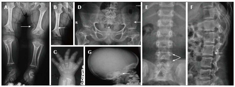
| Label / finding | What to look for | Significance |
|---|---|---|
| Rhizomelic shortening | Humeri and femora are disproportionately short compared with distal segments | Characteristic limb-shortening pattern of achondroplasia |
| Metaphyseal flaring | Widened, flared metaphyses of long bones | Reflects abnormal endochondral ossification |
| Chevron deformity | V-shaped or chevron-like proximal femoral configuration | Supportive radiological feature |
| Trident hand | Increased separation between middle and ring fingers with short proximal phalanges | Classic clinical and radiographic sign |
| Short broad pelvis | Squared iliac wings and small pelvis | Common pelvic pattern |
| Horizontal acetabula | Acetabular roofs are relatively horizontal | Supports diagnosis |
| Narrow sacrosciatic notch | Reduced width of the greater sciatic notch | A helpful pelvic clue |
| Narrowing interpedicular distance | Lumbar pedicle distance decreases from upper to lower lumbar levels | Predisposes to lumbar spinal stenosis |
| Short pedicles and posterior scalloping | Seen on lateral spinal film | Explains narrow spinal canal |
| Foramen magnum narrowing | Best assessed with skull-base imaging and MRI when indicated | Risk of cervicomedullary compression in infancy |
Suggested seminar narration
“Achondroplasia is an FGFR3-related dysplasia with normal bone mineralization but defective endochondral ossification. The radiograph shows rhizomelic shortening, metaphyseal flaring, trident hand, a short broad pelvis, and progressive narrowing of lumbar interpedicular distance. The spinal findings are clinically important because they predispose to spinal stenosis.”
Image 2: Osteogenesis Imperfecta

| Label / finding | What to look for | Significance |
|---|---|---|
| Diffuse osteopenia | Generalized reduction in bone density with thin cortices | Indicates poor bone strength |
| Multiple fractures | Fracture lines in long bones, often at different sites | Typical of bone fragility |
| Callus formation | Peri-fracture new bone formation | Suggests healing fractures, potentially of different ages |
| Bowing deformity | Curved long bones, especially femora and tibiae | Results from repeated fractures and weak bone |
| Codfish vertebrae | Biconcave vertebral bodies due to endplate compression | Vertebral fragility and osteopenia |
| Possible Wormian bones | Accessory sutural skull bones, if skull film is included | Supportive but not diagnostic in isolation |
Suggested seminar narration
“In osteogenesis imperfecta, the first radiographic question is whether mineralization is reduced. Here, diffuse osteopenia, multiple long-bone fractures, bowing, and vertebral compression changes indicate a generalized bone-fragility disorder. These findings should be correlated with blue sclerae, dentinogenesis imperfecta, hearing impairment, family history, and genetic testing for collagen-related disease.”
Image 3: Thanatophoric Dysplasia Type I
| Label / finding | What to look for | Significance |
|---|---|---|
| Severe micromelia | Marked shortening of all limbs | More severe than achondroplasia |
| Telephone-receiver femora | Very short, broad, markedly bowed femora | Hallmark of thanatophoric dysplasia type I |
| Small pelvis | Hypoplastic pelvis with square iliac wings | Severe failure of endochondral bone growth |
| Horizontal acetabular roofs | Acetabula appear flat and horizontal | Similar direction of change to achondroplasia, but much more marked |
| Narrow sacrosciatic notch | Reduced pelvic notch width | Supportive finding |
| Platyspondyly | Flattened vertebral bodies | May be severe, described as wafer-thin in classic cases |
| Narrow thorax with short ribs | Best appreciated on full babygram | Leads to pulmonary hypoplasia and respiratory failure |
Suggested seminar narration
“Thanatophoric dysplasia is also FGFR3-related, but the radiographic abnormalities are much more severe than in achondroplasia. The key clue is the telephone-receiver appearance of the femora. Severe micromelia, a narrow chest, short ribs, and platyspondyly indicate a lethal skeletal dysplasia due to pulmonary hypoplasia.”
Achondroplasia versus thanatophoric dysplasia: radiological comparison
| Feature | Achondroplasia | Thanatophoric dysplasia |
|---|---|---|
| Severity | Nonlethal, variable complications | Usually lethal in neonatal period |
| Long bones | Rhizomelic shortening, relatively straight | Extreme shortening and marked bowing |
| Femora | Short, may show metaphyseal change | Telephone-receiver femora in type I |
| Thorax | May be relatively narrow | Very narrow, often with severe pulmonary hypoplasia |
| Vertebrae | Narrow caudal interpedicular distance, short pedicles | Severe platyspondyly or wafer-thin vertebral bodies |
| Skull | Macrocephaly and foramen magnum narrowing | May show cloverleaf skull, especially type II |
| Pelvis | Short broad pelvis, horizontal acetabula | More severely hypoplastic pelvis with trident configuration |
| Prognosis | Usually normal intelligence and survival with surveillance | High neonatal mortality from respiratory insufficiency |
Short radiology viva points
-
What is the imaging modality of choice for postnatal skeletal dysplasia?
A complete radiographic skeletal survey. -
Which dysplasia shows trident hand and narrowing of lumbar interpedicular distance?
Achondroplasia. -
Which skeletal dysplasia causes telephone-receiver femora?
Thanatophoric dysplasia type I. -
Which disorder shows diffuse osteopenia with recurrent fractures and bone deformity?
Osteogenesis imperfecta. -
What does a narrow thorax in a fetus with severe limb shortening suggest?
Risk of pulmonary hypoplasia and a potentially lethal skeletal dysplasia. -
Which imaging finding in achondroplasia has neurological importance in infancy?
Foramen magnum stenosis, requiring clinical assessment and often MRI based on symptoms and local protocols. -
What is platyspondyly?
Flattening of vertebral bodies. -
What does progressive reduction in lumbar interpedicular distance indicate?
A characteristic achondroplasia-pattern spinal abnormality that contributes to spinal canal stenosis.
Add radiology images for osteogenesis imperfecta and achondroplasia
osteogenesis imperfecta radiograph diffuse osteopenia multiple fractures infant
achondroplasia radiograph pelvis spine trident hand rhizomelia
PMC achondroplasia osteogenesis imperfecta radiographs figures
Radiology Images for Seminar
1. Achondroplasia: characteristic skeletal survey findings
- Rhizomelic shortening: proximal limbs, especially humeri and femora, are disproportionately short.
- Metaphyseal flaring: broad, flared metaphyses of long bones.
- Trident hand: short fingers with increased separation between the middle and ring fingers.
- Pelvis: short broad pelvis, squared iliac wings, horizontal acetabular roofs, and narrow sacrosciatic notch.
- Lumbar spine: progressive reduction of interpedicular distance from L1 to L5, short pedicles, and posterior vertebral scalloping.
- Skull: enlarged calvarium with possible narrowing of the foramen magnum.
Normal mineralization with rhizomelic short-limb dwarfism, characteristic pelvic changes, and caudal interpedicular narrowing indicates achondroplasia.
2. Osteogenesis imperfecta: severe neonatal form
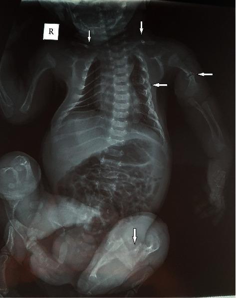
- Generalized osteopenia: diffuse reduction in bone density, with thin cortices and gracile bones.
- Multiple fractures: fractures may involve clavicles, ribs, humeri, femora, and other long bones.
- Fractures at different stages: callus around some fractures indicates healing injury.
- Bowing of long bones: recurrent intrauterine or postnatal fractures cause angulation and deformity.
- Rib changes: thin, beaded, or fractured ribs may result in a small thoracic cage.
- Spine: osteopenia and compression deformities can occur, producing biconcave or “codfish” vertebrae.
- Severe phenotype: multiple fractures with marked deformity in a neonate should suggest severe, often perinatal osteogenesis imperfecta.
Diffuse osteopenia with multiple fractures and bowed long bones is typical of osteogenesis imperfecta, particularly a severe form.
Quick comparison for the slide
| Radiological feature | Achondroplasia | Osteogenesis imperfecta |
|---|---|---|
| Bone density | Usually normal | Reduced, generalized osteopenia |
| Main long-bone abnormality | Short, broad bones with metaphyseal flaring | Thin, fragile bones with fractures and bowing |
| Pelvis | Short broad pelvis, horizontal acetabula | Usually no characteristic achondroplasia-type pelvic pattern |
| Spine | Narrow lumbar interpedicular distance, short pedicles | Osteopenia, compression fractures, codfish vertebrae |
| Major diagnostic clue | Rhizomelia + trident hand + pelvic/spinal pattern | Recurrent fractures + osteopenia + bone deformity |