
Post shoulder instability
Reading File
Posterior shoulder instability (PSI) involves recurrent subluxation or dislocation of the humeral head posteriorly, making up about 2-5% of all shoulder instability cases.
Searching sources...
Finding Sources
Finding Sources
Searching PubMed
"posterior shoulder instability" AND treatment
Good - found a dedicated section in Campbell's Operative Orthopaedics. Let me read the full content.
Reading File
Finding Sources
Reading File
Finding Sources
Reading File
Reading File
Reading File
Finding Sources
Reading File
Reading File
Reading File
Loading Image
I now have comprehensive information from Campbell's Operative Orthopaedics 2026, Miller's Review, and Rockwood & Green's 2025. Let me compile the full answer.
Posterior Shoulder Instability
Overview
Posterior shoulder dislocations and recurrent posterior instability account for approximately 5-10% of all shoulder dislocations - making it far less common than anterior instability but clinically important and frequently missed. - Campbell's Operative Orthopaedics 15th Ed 2026, p. 2908
Classification: ABC System (Moroder & Scheibel)
The ABC system provides a comprehensive, pathophysiology-based framework with treatment implications:
| Group | Type | Subtype | Description | Treatment |
|---|---|---|---|---|
| A - First Time | A1 | Subluxation | No significant bony/soft tissue defects | Conservative |
| A2 | Locked dislocation | Temporary engagement, possible bony defect | Closed/open reduction ± reconstruction | |
| B - Dynamic Recurrent | B1 | Functional | Abnormal muscle activation + hyperlaxity/capsular redundancy | Physical therapy |
| B2 | Structural | Posterior Bankart lesion, glenoid bone loss, reverse Hill-Sachs | Surgery if PT fails | |
| C - Static | C1 | Constitutional | Structural deficiency from birth | Technically demanding surgery |
| C2 | Acquired | Structural defects from injury/neglect | Capsulorrhaphy, bone graft, osteotomy, arthroplasty |
- Rockwood and Green's Fractures in Adults 10th Ed 2025, p. 1268
Etiology and Mechanisms
Traumatic
- Direct blow to the anterior shoulder
- Fall on a forward-flexed, adducted, internally rotated extremity
- High-energy events associated with altered consciousness (seizures, electrical shock, intoxication) - classically associated with acute locked posterior dislocation
Microtraumatic / Atraumatic
- Repetitive posterior capsule loading from:
- Pitching, tennis, swimming (backstroke/breaststroke)
- Weight lifting - especially bench press
- Offensive linemen blocking
- Baseball batters (check swing or pulling an outside pitch - alters mechanics and increases posterior shear forces)
Voluntary Instability
-
Patients with microtraumatic or atraumatic origin may learn to sublux with horizontal adduction + internal rotation
-
Patients who sublux using muscular contraction alone (bland affect) may have psychologic overlay and secondary gain
-
These patients should rarely, if ever, be surgically treated
-
Campbell's Operative Orthopaedics 15th Ed 2026, p. 2908
Pathoanatomy and Bony Lesions
Reverse Hill-Sachs Lesion
- Impaction fracture of the anterosuperior humeral head (the "trough sign" on AP radiograph)
- Created as the posterior glenoid rim engages the front of the humeral head during dislocation
- When >20-25% of articular surface involved: McLaughlin procedure (subscapularis transfer) or Neer modification (lesser tuberosity transfer)
- When >35-40% articular loss: hemiarthroplasty or TSA
Reverse Bankart Lesion
- Posterior labrocapsular avulsion from the posterior glenoid rim
- Most common structural lesion in traumatic recurrent posterior instability
Kim Lesion
- Incomplete and concealed avulsion of the posteroinferior labrum (Type II posteroinferior labral lesion in Kim's classification)
- Associated with posterior and multidirectional instability
- May be subtle or falsely negative on MR arthrography
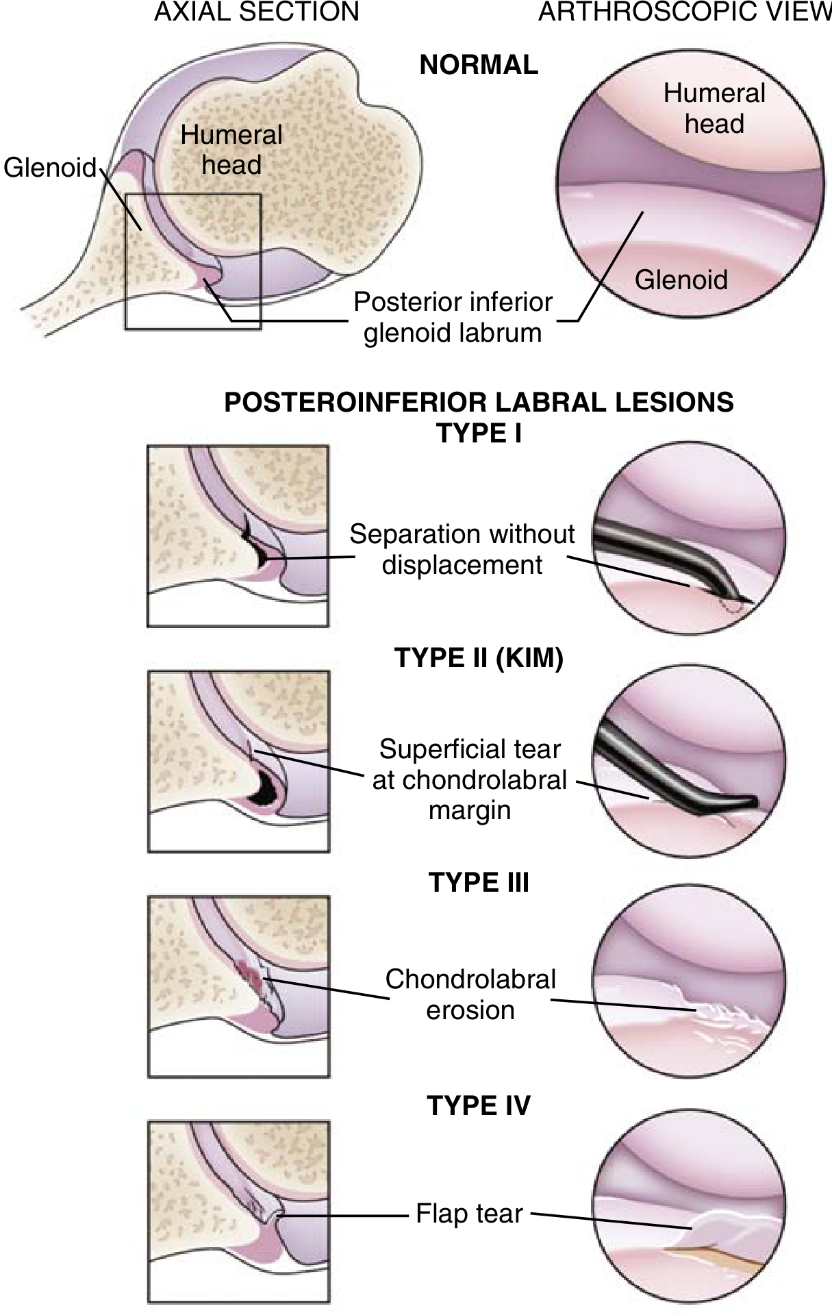
Posteroinferior labral lesion types: Type I (separation without displacement), Type II/Kim (superficial tear at chondrolabral margin), Type III (chondrolabral erosion), Type IV (flap tear). - Miller's Review of Orthopaedics 9th Ed
Glenoid Retroversion
- Contributes significantly only in severe congenital dysplasia or traumatic disruption
- Glenoid osteotomy indicated when retroversion exceeds 10 degrees (Fuchs, Jost, Gerber)
- High complication rates have led to these falling out of favor
Clinical Diagnosis
History
- Pain and apprehension with forward flexion + adduction + internal rotation
- Athletes in overhead sports with posterior-dominant pain
- Clicking or subluxation during specific activities
Key Physical Examination Tests
| Test | Target Lesion | Technique |
|---|---|---|
| Jerk Test | Posterior labral lesion | Axially load, forward flex to 90°, horizontally adduct - positive = clunk |
| Kim Test | Posteroinferior lesion | Axially load in 90° abduction, elevate arm diagonally - positive = pain/clunk |
| Load and Shift | Direction/degree of instability | Seat patient, stabilize scapula, load humerus, translate posteriorly |
| Sulcus Sign | Inferior laxity / MDI component | Inferior traction with arm at side - positive groove = MDI |
The jerk and Kim tests are highly sensitive and specific for their respective lesions. - Miller's Review of Orthopaedics 9th Ed
Imaging
- AP radiograph: Lightbulb sign (humeral head appears rounded/symmetric on AP due to internal rotation in locked dislocation); trough sign
- Axillary lateral: Best view for confirming posterior dislocation - diagnosis is easy with axillary view but missed in ~50% of cases on AP alone
- MRI / MR arthrography: T2-weighted axial with contrast - evaluates capsular deficiency, chondrolabral containment, Kim lesion (may be subtle), rotator interval, and glenoid version
- Kim et al. and Antoniou & Harryman found chondrolabral cavity lesions in >80% of patients with posterior instability
Treatment
Nonoperative (First Line)
- Avoid provocative activities and voluntary subluxation maneuvers
- Strengthening of external rotators and posterior deltoid
- Restore normal range of motion
- In overhead athletes: coaching correction of mechanics
- Most patients with microtraumatic/atraumatic origin respond well, especially those with generalized ligamentous laxity
Patients less likely to respond: traumatic dislocations in contact athletes (football linemen, hockey players, platform divers)
Surgical Indications
Surgery is indicated when:
- Appropriate rehabilitation has failed
- Habitual/voluntary dislocation has been ruled out
- Patient is emotionally stable
- Pain and instability preclude adequate function
- Structural lesion confirmed (positive jerk test, labral pathology, capsular deficiency on MRI)
Additionally, if rotator interval gap >1 cm with external rotation >90° at 0° abduction and positive sulcus sign: rotator interval closure is added
Surgical Options
| Procedure | Indication | Approach |
|---|---|---|
| Arthroscopic posterior capsulolabral repair | B2 recurrent structural instability (reverse Bankart, Kim lesion) | Arthroscopic |
| Inferior capsular shift (Neer/Tibone) | Disabling posterior subluxation; MDI with posterior predominance; overhead athletes | Posterior open |
| Hawkins-Janda capsulorrhaphy | Laborers/contact athletes with recurrent posterior subluxation from capsular deficiency | Open posterior |
| McLaughlin procedure | Reverse Hill-Sachs 20-25% articular involvement | Open |
| Neer modification (lesser tuberosity transfer) | Same as McLaughlin but with subscapularis/tuberosity transfer | Open |
| Posterior glenoid osteotomy | >10° retroversion with severe dysplasia | Open (rarely used) |
| Hemiarthroplasty / TSA | >35-40% humeral head articular loss; end-stage arthritis | Open |
Best surgical results are obtained in patients with recurrent traumatic posterior dislocation, not the more common atraumatic/microtraumatic posterior instability syndromes. - Campbell's Operative Orthopaedics 15th Ed 2026, p. 2909
Postoperative Protocol (Open Posterior Capsular Shift)
- Arm immobilized in neutral rotation, slight abduction x 6 weeks
- ROM with elevation in scapular plane + external rotation begun at 6 weeks
- Avoid elevation >150° and internal rotation exercises x 3 months
- Sports (swimming, throwing): 9 months to 1 year
Causes of Surgical Failure
- Inadequate capsular shift / under-tensioned repair
- Failure to identify and address all labral lesions (especially Kim lesion)
- Unrecognized significant glenoid bone loss (>11-15% increases failure risk)
- Voluntary instability / psychologic overlay not identified preoperatively
- Female sex, reduced glenoid bone width - identified as independent risk factors for failure after arthroscopic posterior capsulolabral repair
Recent Evidence (2024-2026)
- A 2024 systematic review (Ralph et al., PMID 38825224) confirmed generally good outcomes for arthroscopic stabilization of posterior instability
- A 2025 systematic review (Afetse et al., PMID 40738256) found that female sex, reduced glenoid bone width, and glenoid bone loss >11-15% increase failure risk after arthroscopic posterior capsulolabral repair - important for pre-surgical risk stratification
- A 2026 systematic review (Wollenman et al., PMID 40426285) noted significant variability in return-to-play criteria after posterior shoulder soft tissue stabilization - lack of standardized criteria remains a gap
Sources: Campbell's Operative Orthopaedics 15th Ed 2026 | Miller's Review of Orthopaedics 9th Ed | Rockwood and Green's Fractures in Adults 10th Ed 2025
This is a shared conversation. Sign in to Orris to start your own chat.