

Explain in detail all label the short forma
coronary artery anatomy diagram LMS LAD LCX RCA branches
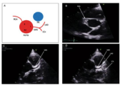
Summary : This figure presents a schematic diagram and three echocardiographic images illustrating the anatomical relationship and visualization of the coronary arteries and aorta. diagram and echocardiographic images: # Panel A: Schematic Diagram : • Shows the anatomical relationship between the aorta (red circle) and pulmonary artery (blue circle). • Labels include: RCA (right coronary artery), LMS (left main stem), LAD (left anterior descending), LCx (left circumflex). • Arrows indicate the direction and origin of the coronary arteries from the aorta. # Panel B: Echocardiographic Image (Short Axis View) : • Displays the aortic root in cross-section. • Labeled structures: RCA (right coronary artery), LMS (left main stem). • The coronary ostia are visible as small openings in the aortic root. # Panel C: Echocardiographic Image (Short Axis View) : • Similar view as Panel B, focused on the LMS (left main stem). • The LMS is labeled and its origin from the aorta is visualized. # Panel D: Echocardiographic Image (Long Axis View) : • Shows the aortic root and left ventricle. • Labeled structures: LMS (left main stem), LAD (left anterior descending), LCx (left circumflex). • The course of the coronary arteries is visualized as they branch from the aorta. # Analysis : • The figure effectively demonstrates the anatomical origins and visualization of the coronary arteries using both schematic and echocardiographic imaging. • The labeled images help correlate anatomical landmarks with ultrasound findings, aiding in the identification of coronary ostia and branches. • No anomalies or abnormal patterns are visible; the images represent typical coronary anatomy.
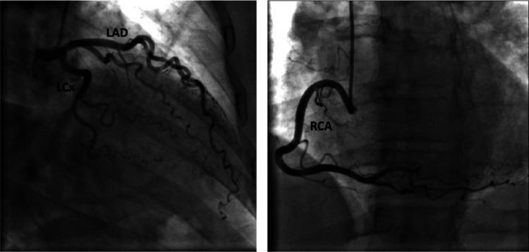
This diagnostic image is a coronary angiogram consisting of two panels demonstrating normal coronary anatomy. The left panel shows the left coronary system with labeled Left Anterior Descending (LAD) and Left Circumflex (LCx) arteries. The LAD is seen coursing anteriorly and inferiorly, giving off diagonal branches, while the LCx travels within the atrioventricular groove, exhibiting normal tapering and branching without evidence of luminal narrowing, stenosis, or occlusion. The right panel illustrates the Right Coronary Artery (RCA) in a characteristic 'C' shape, originating from the right coronary sinus and extending along the posterior interventricular groove. The RCA appears smooth with patent distal flow and visible side branches. This invasive imaging study serves as a clinical reference for angiographically normal epicardial coronary arteries, typically used in cardiology and radiology to rule out obstructive coronary artery disease (CAD) or vasospastic disorders in the setting of chest pain or ischemic ECG changes.
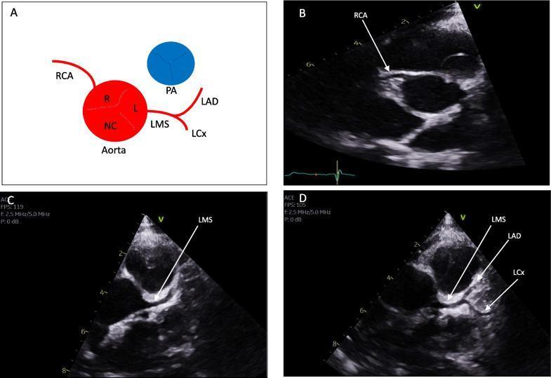
This composite educational graphic details normal human coronary artery anatomy through a schematic diagram and transthoracic echocardiography (TTE) images. Panel A presents a schematic illustrating the aorta with its three sinuses: Right (R), Left (L), and Non-Coronary (NC). It shows the Right Coronary Artery (RCA) originating from the right sinus and the Left Main Stem (LMS) originating from the left sinus, subsequently bifurcating into the Left Anterior Descending (LAD) and Left Circumflex (LCx) arteries. The Pulmonary Artery (PA) is shown in spatial relation to these structures. Panels B, C, and D are clinical diagnostic ultrasound images in the parasternal short-axis (PSAX) view at the level of the aortic valve. Panel B identifies the origin of the RCA. Panel C demonstrates the origin of the LMS from the aortic sinus. Panel D highlights the bifurcation of the LMS into the LAD and LCx. This material serves as a reference for identifying normal coronary ostia and branching patterns, essential for diagnosing anomalous coronary origins or interarterial courses.
Coronary Artery Anatomy - All Labels Explained
🔴 Main Vessels
| Short Form | Full Name | Details |
|---|---|---|
| LMS | Left Main Stem (also: Left Main Coronary Artery) | The origin of the entire left coronary system. Arises from the left aortic sinus (of Valsalva) at the top of the heart. It is a short, wide trunk (usually 5-20 mm long) that quickly bifurcates - or trifurcates - into the LAD and LCX. Disease here is called "left main disease" and is considered very high-risk. |
| LAD | Left Anterior Descending Artery | One of the two main branches of the LMS. Runs down the anterior interventricular groove toward the apex. It supplies the anterior wall of the left ventricle, most of the interventricular septum, and the apex. It is the most commonly affected artery in coronary artery disease. Sometimes called "the widow maker." |
| LCX | Left Circumflex Artery | The other main branch of the LMS. Travels in the left atrioventricular (AV) groove, wrapping around the left side of the heart. Supplies the lateral and posterior walls of the left ventricle and the left atrium. |
| RCA | Right Coronary Artery | Arises from the right aortic sinus. Runs in the right AV groove, travels around the right side of the heart. Supplies the right ventricle, the SA node (in ~60% of people), the AV node (in ~80% of people), and in most people (right-dominant circulation) gives rise to the PDA. |
🔴 Branches of LAD
| Short Form | Full Name | Details |
|---|---|---|
| D1 | First Diagonal Branch | The first major diagonal branch coming off the LAD. Diagonals run across the anterolateral surface of the left ventricle. D1 supplies the upper-mid anterolateral wall. It is an important landmark in coronary anatomy - stenosis here can cause anterior and lateral wall ischemia. |
| D2 | Second Diagonal Branch | The second diagonal branch of the LAD. Runs further down the anterolateral wall, below D1. Supplies the mid-to-lower anterolateral left ventricle. |
🔴 Branches of LCX
| Short Form | Full Name | Details |
|---|---|---|
| OM1 | First Obtuse Marginal Branch | The first major branch of the LCX. "Obtuse marginal" refers to the obtuse (blunt) margin of the heart, which is the left lateral border. OM1 is often the most important branch of the LCX system and supplies the lateral wall of the left ventricle. |
| OM2 | Second Obtuse Marginal Branch | The second obtuse marginal branch off the LCX. Supplies the posterolateral wall of the left ventricle, running below OM1 along the lateral/posterior surface. |
🔴 Branch of RCA
| Short Form | Full Name | Details |
|---|---|---|
| PDA | Posterior Descending Artery (also: Posterior Interventricular Artery) | Runs in the posterior interventricular groove toward the apex. Supplies the posterior third of the interventricular septum, inferior wall of the left ventricle, and posterior right ventricle. Its origin determines coronary dominance: if it arises from the RCA (most common, ~70% of people) = right dominant; if from the LCX = left dominant; if from both = co-dominant. |
Summary Tree
Aorta
├── LMS (Left Main Stem)
│   ├── LAD (Left Anterior Descending)
│   │   ├── D1 (First Diagonal)
│   │   └── D2 (Second Diagonal)
│   └── LCX (Left Circumflex)
│       ├── OM1 (First Obtuse Marginal)
│       └── OM2 (Second Obtuse Marginal)
└── RCA (Right Coronary Artery)
    └── PDA (Posterior Descending Artery)
Clinical Importance at a Glance
- LMS disease - affects all left coronary territory; surgically treated with CABG
- LAD occlusion - anterior STEMI; "widow maker"
- RCA occlusion - inferior STEMI; may cause AV block due to AV node ischemia
- LCX occlusion - lateral/posterior STEMI; often electrically "silent" on standard ECG
- PDA - determines coronary dominance, critical in planning revascularization