
A 21-year-old woman, gravida 1, para 0, aborta 1, comes to the emergency department because of vaginal spotting and lower abdominal pain for 3 hours. She is sexually active and does not use contraception. Menarche was at the age of 13 years. Menses occur at regular 30-day intervals; her last menstrual period was 6 weeks ago. Pelvic examination shows slightly blood-tinged vaginal discharge. There is cervical motion tenderness with a firm cervix and a closed cervical os. The uterus is firm and tender and consistent in size with a 6-week gestation. The right adnexa is tender. Urine pregnancy test is positive. Which of the following is the most likely diagnosis
- Positive pregnancy test with LMP 6 weeks ago
- Closed cervical os (rules out inevitable/incomplete abortion)
- Right adnexal tenderness (classic for tubal ectopic)
- Cervical motion tenderness
- Uterus only 6-week size (not enlarged beyond dates)
- Prior ectopic/abortion history (aborta 1 - risk factor)
ectopic pregnancy tubal sites diagram
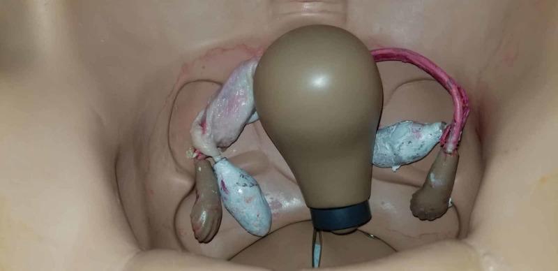
This photograph depicts an anatomical diagram within a gynecologic surgical simulation mannequin, viewed from a superior perspective. The setup illustrates the pelvic cavity with a central, pear-shaped uterine model and bilateral adnexal structures. To simulate a tubal ectopic pregnancy for obstetric surgical training, the mannequin's fallopian tubes and ovaries have been modified with organic and synthetic materials. On the left side of the image, the simulated tube appears pale and translucent with fleshy textures, while the right side features a reddish tubular structure and irregular, bluish-grey masses representing adnexal pathology. These modifications are designed to provide postgraduate resident physicians with a realistic visual and tactile representation of tissue for practicing laparoscopic salpingostomy or salpingectomy. The visual contrast between the tan plastic mannequin and the moist, textured simulated biological tissues emphasizes the target surgical site. This educational resource is used in high-fidelity simulation to bridge the gap between theoretical knowledge of ectopic pregnancy and operative skill acquisition.
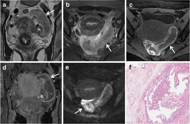
This composite educational resource presents diagnostic imaging and histopathology of a tubal ectopic pregnancy in a 44-year-old female. (a, b) Coronal and axial T2-weighted MR images show a distended, predominantly hypointense sac-like cystic mass in the left pelvic cul-de-sac. (c, d) Axial and coronal fat-suppressed T1-weighted images reveal low-to-intermediate signal intensity within the tubal lumen and a characteristically hyperintense, thickened wall, signifying hematosalpinx. (e) An axial diffusion-weighted image (DWI, b=800 s/mm²) demonstrates restricted diffusion within the lesion, indicated by high signal intensity. The MRI series also highlights the absence of an intrauterine gestational sac. (f) Corresponding light microscopy (H&E stain, 200x) provides histopathological confirmation, showing chorionic villi embedded within the fallopian tube wall. This case illustrates the multi-parametric MRI features of ectopic pregnancy, including hematosalpinx and restricted diffusion, which are critical for differentiating adnexal masses in patients with elevated β-hCG and acute abdominal pain.
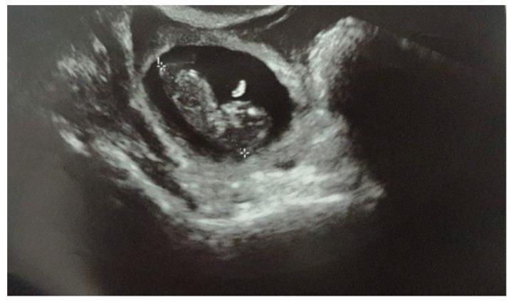
This diagnostic ultrasound image displays a live tubal ectopic pregnancy at a gestational age of approximately 10 weeks and 4 days. The image shows a well-defined, hypoechoic (anechoic) gestational sac located outside the uterine cavity within the adnexal region. Inside the fluid-filled sac, a distinct embryo (fetal pole) is visible with a measurable Crown Rump Length (CRL). An echogenic yolk sac is seen adjacent to the embryo. The surrounding tubal and pelvic tissue exhibits a heterogeneous echotexture characteristic of an ectopic implantation site. Measurement calipers (+) are positioned across the gestational sac to assess dimensions. The image demonstrates key sonographic findings of an advanced extrauterine pregnancy, emphasizing the presence of a gestational sac and fetal pole in the fallopian tube, which is a critical diagnostic indicator in obstetric emergency medicine and radiology.
Diagnosis: Ectopic Pregnancy
How Each Finding Points to Ectopic Pregnancy
| Clinical Finding | Significance |
|---|---|
| Positive urine pregnancy test | Confirms pregnancy |
| LMP 6 weeks ago | Expected gestational age ~6 weeks |
| Closed cervical os | Rules out threatened/inevitable/incomplete abortion (os would be open) |
| Right adnexal tenderness | Classic sign - points to right fallopian tube implantation |
| Cervical motion tenderness | Hallmark of peritoneal irritation from adnexal pathology |
| Uterus only 6-week size | Consistent with an intrauterine pregnancy - but no adnexal mass yet rules out rupture |
| Vaginal spotting + abdominal pain | Classic presenting triad (bleeding + pain + adnexal tenderness) |
| G1P0Ab1 history | Prior pregnancy loss is a risk factor for tubal scarring/ectopic implantation |
Why Not the Alternatives?
- Threatened abortion: Presents with open os, bleeding, but no adnexal tenderness or cervical motion tenderness
- Inevitable/incomplete abortion: Requires an open cervical os
- Corpus luteum cyst rupture: Would not give a positive pregnancy test (though this can co-exist)
- Appendicitis: No pregnancy association; no cervical motion tenderness pattern
- Ovarian torsion: Would not cause a positive pregnancy test per se and lacks the classic ectopic triad
Key Pathophysiology
Immediate Next Steps (Clinical)
- Transvaginal ultrasound - to confirm absence of intrauterine gestational sac and identify adnexal mass
- Quantitative serum beta-hCG - serial levels every 48 hours; in ectopic, rise is abnormally slow (<53% increase in 48 h) or plateaus
- Type and crossmatch - in case rupture occurs requiring emergency surgery
- Surgical/medical management - methotrexate (if unruptured, stable) or surgical salpingostomy/salpingectomy (if ruptured or unstable)
why uterus was is 6 weeks then
Why the Uterus Is 6-Week Size in Ectopic Pregnancy
The Hormone Is the Key
- Maintains the corpus luteum in the ovary
- The corpus luteum keeps producing progesterone
- Progesterone causes the uterus to:
- Develop a decidual reaction (endometrium thickens into decidua)
- Increase in overall size and become soft/boggy
- Become tender due to hormonal stimulation
This Is Actually a Diagnostic Clue
| Finding | Interpretation |
|---|---|
| Uterus 6-week size | Hormonal response to ectopic hCG - expected |
| No sac seen inside uterus | Embryo is NOT inside |
| Adnexal tenderness | Embryo is in the tube |
| Closed os | Uterus is not trying to expel anything |
The "Decidual Cast" Phenomenon
Summary
what if PID was in option too
Ectopic Pregnancy vs PID - How to Distinguish
The Single Killer Finding: Positive Pregnancy Test
PID does not cause a positive pregnancy test.
Full Comparison Table
| Feature | Ectopic Pregnancy | PID |
|---|---|---|
| Pregnancy test | Positive | Negative |
| Cervical motion tenderness | Yes | Yes |
| Adnexal tenderness | Yes (usually unilateral) | Yes (often bilateral) |
| Vaginal discharge | Mild spotting | Purulent/mucopurulent |
| Fever | Usually absent | Often present |
| WBC / ESR | Normal or mildly elevated | Elevated (inflammatory) |
| Closed cervical os | Yes | Yes (no relation to pregnancy) |
| LMP | Missed (6 weeks ago) | Usually normal/recent |
| Uterine size | Slightly enlarged (6 wk) | Normal size |
| Pain character | Often unilateral, crampy | Diffuse, bilateral lower abdomen |
In This Case Specifically
- Positive pregnancy test - eliminates PID as primary diagnosis
- LMP 6 weeks ago (amenorrhea) - PID patients are not pregnant
- Unilateral right adnexal tenderness - PID tends to be bilateral
- No fever, no purulent discharge - against active infection/PID
- Closed os + uterine enlargement - pregnancy-related changes
The Exam Rule
Sexually active woman + missed period + positive hCG + adnexal tenderness + cervical motion tenderness = Ectopic pregnancy FIRST, not PID
One More Point - They Can Coexist
Nulliparity or chronic an-ovulation which has greatest risk of endometrial cancer
Chronic Anovulation > Nulliparity for Endometrial Cancer Risk
The Relative Risk Numbers (from the textbook table)
| Risk Factor | Relative Risk |
|---|---|
| Nulliparity | 2-3x |
| Anovulatory cycles / irregular menses | Higher (mechanism-driven, part of the unopposed estrogen spectrum) |
| Obesity (>50 lb overweight) | 10x |
| Unopposed estrogen therapy | 4-8x |
| Atypical endometrial hyperplasia | 8-29x |
Why Chronic Anovulation Is Worse - The Mechanism
- Follicular phase: Estrogen rises → endometrium proliferates
- Ovulation → corpus luteum forms → progesterone rises
- Progesterone opposes estrogen, differentiates the endometrium, causes secretory transformation, and limits proliferation
- Estrogen is produced continuously from follicles that never ovulate
- No corpus luteum forms → no progesterone
- The endometrium is exposed to unopposed estrogen month after month, year after year
- This drives: normal endometrium → hyperplasia → atypical hyperplasia → endometrial carcinoma (Type I)
Why Nulliparity Is a Risk Factor (but Weaker)
- More lifetime ovulatory cycles (each cycle = some estrogen exposure)
- Possibly underlying anovulation or infertility
- No progesterone-dominant gestational periods (pregnancy is highly protective due to high progesterone)
The Key Conceptual Point
It is not the number of pregnancies that matters most - it is whether progesterone is present to oppose estrogen.
- Berek & Novak's Gynecology, p. 2144
if obesity is also an option then
The Numbers Side by Side
| Risk Factor | Relative Risk |
|---|---|
| Nulliparity | 2-3x |
| Chronic anovulation (PCOS) | ~3-5x (mechanism-based, part of unopposed estrogen) |
| Obesity (21-50 lb overweight) | 3x |
| Obesity (>50 lb overweight) | 10x |
| Unopposed estrogen therapy | 4-8x |
Why Obesity Is So Powerful
- Adipose tissue contains aromatase enzyme
- Aromatase converts androstenedione → estrone (a form of estrogen)
- The more fat tissue, the more peripheral estrogen production
- This estrogen acts on the endometrium without progesterone opposition
- Obese women are also frequently anovulatory (obesity suppresses the HPO axis) - so they often have both mechanisms simultaneously
How to Think About It on an Exam
| Scenario | Best Answer |
|---|---|
| Nulliparity vs anovulation | Anovulation (direct unopposed estrogen) |
| Nulliparity vs obesity | Obesity (higher RR numerically) |
| Anovulation vs mild obesity | Anovulation (comparable, but mechanistically cleaner) |
| Anovulation vs severe obesity | Severe obesity (RR 10 vs ~3-5) |
| All three as options | Obesity (if severe) or anovulation (if exam is testing mechanism) |
The Unifying Principle
Prolonged endometrial exposure to estrogen without progesterone opposition
| Risk Factor | How It Causes Unopposed Estrogen |
|---|---|
| Chronic anovulation | No corpus luteum → no progesterone |
| Obesity | Peripheral aromatization → excess estrone |
| Nulliparity | Fewer progesterone-dominant gestational periods |
Exam Rule of Thumb
- If the question asks "which has the HIGHEST relative risk" and obesity is listed → obesity (severe) wins numerically (RR 10)
- If the question asks "which is the primary/direct hormonal mechanism" → anovulation is the cleaner answer
- If PCOS is mentioned in the stem → anovulation + obesity are usually both present, which is why PCOS patients have disproportionately high endometrial cancer risk even at a young age
autosomal recessive disease in wife only, what chances are children going to get disease
Autosomal Recessive Disease - Wife Affected, Husband Unaffected
Setting Up the Genotypes
- Wife is affected → must be homozygous recessive = aa
- Husband is unaffected → could be AA (normal) or Aa (carrier)
Scenario 1: Husband is AA (homozygous normal)
| a | a | |
|---|---|---|
| A | Aa | Aa |
| A | Aa | Aa |
- 100% of children = Aa (carriers)
- 0% affected
- All children carry one copy but will never show disease
Scenario 2: Husband is Aa (carrier)
| a | a | |
|---|---|---|
| A | Aa | Aa |
| a | aa | aa |
- 50% carriers (Aa)
- 50% affected (aa)
- 0% completely normal (AA)
What the Exam Usually Assumes
Husband is AA (unaffected, non-carrier)
- For most autosomal recessive diseases, the majority of the general population is homozygous normal (AA)
- Carrier frequency in the population depends on disease prevalence (Hardy-Weinberg)
- Without specific information suggesting the husband is a carrier, assume AA
The Exception - Consanguinity or Known Carrier
- Cousin marriage → husband likely Aa → 50% affected children
- Husband's sibling is affected → husband has 2/3 chance of being Aa
- Common disease (e.g., sickle cell in high-prevalence population) → carrier frequency matters
Summary Table
| Husband's Genotype | Children Affected | Children Carriers | Children Normal |
|---|---|---|---|
| AA (assumed if no info) | 0% | 100% | 0% |
| Aa (known carrier) | 50% | 50% | 0% |
| aa (affected) | 100% | 0% | 0% |
Key Rule to Remember
In autosomal recessive disease, an affected person (aa) crossed with a normal non-carrier (AA) produces zero affected children - but every child becomes a carrier.
disease outcome in tay-sach carriers
Tay-Sachs Carriers - Disease Outcome
Short Answer
Why Carriers Are Unaffected
| Genotype | Hex-A Activity | GM2 Accumulation | Clinical Status |
|---|---|---|---|
| AA (normal) | 100% | None | Normal |
| Aa (carrier) | ~50% | None significant | Clinically normal |
| aa (affected) | ~0% | Massive | Fatal disease |
The Special Feature of Tay-Sachs Carriers
- Serum Hex-A assay - carriers show ~50% of normal activity
- Leukocyte Hex-A assay - more reliable, especially in pregnancy
- DNA mutation analysis - gold standard
What Happens in Affected Children (aa) - for Contrast
| Age | Feature |
|---|---|
| Birth - 6 months | Normal development |
| 3-6 months | Motor weakness begins |
| ~1 year | Cherry-red spot on macula (classic finding) |
| Progressive | Blindness, seizures, dementia |
| 2-4 years | Death (no treatment available) |
Carrier x Carrier Cross (Exam Favorite)
| A | a | |
|---|---|---|
| A | AA | Aa |
| a | Aa | aa |
- 25% normal (AA)
- 50% carriers (Aa) - clinically normal
- 25% affected (aa) - fatal Tay-Sachs
Key Point for Exams
Tay-Sachs carriers have no clinical disease. The 50% enzyme activity from one functional allele is completely sufficient. Carriers are only identified biochemically or genetically - never clinically.
A 27-year-old woman, gravida 2, para 2, comes to the physician for advice regarding contraception. She is sexually active with two male partners and does not use contraception. She does not want to become pregnant now. She had a deep venous thrombosis during her second pregnancy, and she is not certain if she would like to have more children in the future. Three months ago, she was treated for Chlamydia trachomatis infection. She currently takes no medications and reports that she has had difficulty remembering to take daily medications in the past. Her blood pressure is 110/70 mm Hg. Physical examination, including pelvic examination, shows no abnormalities. In addition to advising the patient to use condoms, which of the following is the most appropriate recommendation regarding contraception?
Most Appropriate Contraception: Copper IUD (Paragard)
Working Through Each Clinical Constraint
| Patient Factor | What It Eliminates | What It Favors |
|---|---|---|
| DVT during pregnancy | All estrogen-containing methods (combined OCP, patch, ring) - estrogen increases clotting risk | Progestin-only or non-hormonal methods |
| Difficulty remembering daily meds | Combined OCP, progestin-only pill (must be taken same time daily) | Long-acting methods (IUD, implant, injection) |
| Recent Chlamydia (treated, resolved, normal exam) | Active STI would bar IUD - but it is treated and exam is normal, so IUD is now acceptable | IUD is not contraindicated |
| Uncertain about future fertility | Permanent sterilization (tubal ligation) | Reversible long-acting methods |
| Multiple partners | Hormonal-only methods (STI protection needed) | Condoms already being advised; IUD added |
| Normal BP, no other contraindications | Nothing further eliminated | Broad options remain |
Why Each Option Is Wrong
| Option | Reason to Reject |
|---|---|
| Combined OCP (estrogen + progestin) | Contraindicated - DVT history = high VTE risk with estrogen |
| Progestin-only pill (minipill) | Acceptable re: DVT, but eliminated by difficulty with daily compliance |
| Depot medroxyprogesterone (Depo-Provera injection) | Acceptable re: DVT, long-acting - but causes irreversible bone density loss with long-term use and delays return of fertility up to 18 months - poor choice if undecided about future pregnancy |
| Levonorgestrel IUD (Mirena) | Hormonal - generally considered safe re: DVT (low systemic absorption), but copper IUD is more definitively non-hormonal and preferred |
| Tubal ligation | Permanent - patient is undecided about future children |
| Contraceptive implant (Nexplanon) | Acceptable (progestin only, long-acting) but copper IUD is preferred because it is non-hormonal and fully reversible without requiring a procedure to reverse |
Why Copper IUD Wins
- Non-hormonal - zero VTE risk, safe with DVT history
- Long-acting (up to 10 years) - no daily compliance needed
- Fully reversible - fertility returns immediately upon removal
- Highly effective - >99% (among most effective methods available)
- Infection not a current contraindication - prior Chlamydia is treated, exam is normal; IUD insertion is safe now
- Also serves as emergency contraception if inserted within 5 days of unprotected sex
The One Nuance - Recent STI and IUD
- Active, current, untreated STI - do not insert IUD
- Treated, resolved STI with normal exam - IUD insertion is safe
Final Answer
Copper IUD - non-hormonal, long-acting, reversible, no compliance burden, safe after treated STI, and completely avoids the VTE risk that makes estrogen-containing contraceptives dangerous in this patient.
but NBME says depot medroxyprogesterone
Why NBME Picks Depot Medroxyprogesterone
Recent STI (within the past 3 months) = relative contraindication to IUD insertion
NBME's Reasoning Framework for This Question
| Factor | NBME's Interpretation |
|---|---|
| DVT history | Eliminates all estrogen → no combined OCP, patch, ring |
| Daily compliance difficulty | Eliminates daily pills (combined OCP, minipill) |
| Recent Chlamydia + multiple partners | Eliminates IUD (ongoing STI risk = IUD risk for PID/ascending infection) |
| Undecided about future fertility | Eliminates tubal ligation |
| What's left? | Depot medroxyprogesterone |
Why Depo Is Acceptable Here Despite Its Drawbacks
- The patient said she is "not certain" she wants more children - this is not the same as "definitely wants more children soon"
- Depo is progestin-only → no VTE risk → safe with DVT history
- Injection every 3 months → no daily compliance issue
- Does not require IUD insertion → avoids STI/PID risk concern
- Fertility does return eventually (12-18 months after last injection)
The Hierarchy NBME Uses
1. Eliminate estrogen (if VTE/DVT history, migraines with aura, smoker >35)
2. Eliminate daily methods (if compliance is a stated problem)
3. Eliminate IUD (if current or very recent STI, or multiple partners with ongoing STI risk)
4. Eliminate permanent methods (if future fertility uncertain)
5. What remains = the answer
- Step 1: Out → combined OCP, patch, ring
- Step 2: Out → minipill, combined OCP (already gone)
- Step 3: Out → copper IUD, levonorgestrel IUD
- Step 4: Out → tubal ligation
- Remaining: Depot medroxyprogesterone ✓
Where My Reasoning Went Wrong
- Recent STI + multiple partners = ongoing high PID risk = avoid IUD
The Takeaway for Your Exam
| Clinical Reality (CDC) | NBME Test Logic |
|---|---|
| Treated STI = IUD is fine | Recent STI + multiple partners = avoid IUD |
| Copper IUD preferred in VTE history | Depo preferred when IUD is "risky" |
recent STI should be how recent
CDC Medical Eligibility Criteria (Real Clinical Guidelines)
| Condition | CDC Category | Meaning |
|---|---|---|
| Current purulent cervicitis or active Chlamydia/gonorrhea | Category 4 | Absolute contraindication - do NOT insert IUD |
| Past STI, now treated, no current infection | Category 1-2 | IUD is acceptable - no meaningful restriction |
The Practical Clinical Rule
| Situation | IUD Timing |
|---|---|
| Active STI right now | Treat first, then insert |
| STI treated, test-of-cure confirmed | Insert any time after |
| STI treated, no test-of-cure but asymptomatic | Insert after ~1 month |
| High-risk behavior ongoing (multiple partners) | Insert with caution, counsel on condoms |
So Why Did NBME Use 3 Months as a Red Flag?
- Multiple sexual partners - ongoing STI acquisition risk
- Recent STI - suggests high-risk sexual behavior pattern
- Combined together = elevated ongoing PID risk if IUD inserted
This patient has behavioral risk factors suggesting she is likely to acquire another STI, and placing an IUD in that context increases ascending infection/PID risk.
The NBME Heuristic (Not a Guideline Cutoff)
| NBME Signal | Interpretation |
|---|---|
| STI "currently" or "last month" | Clearly too recent → avoid IUD |
| STI "3 months ago" + multiple partners | Recent enough + ongoing risk → avoid IUD |
| STI "2 years ago" + monogamous | Too long ago → IUD acceptable |
| STI history, no ongoing risk | IUD acceptable |
Bottom Line
| Framework | Answer |
|---|---|
| CDC guidelines | No specific waiting period - treat and insert |
| Real clinical practice | Confirm resolution, then insert |
| NBME exam logic | Recent STI + multiple partners = avoid IUD regardless of exact timeframe |