
Rheumatic heart disease nd endocarditis
rheumatic heart disease mitral valve stenosis fish mouth
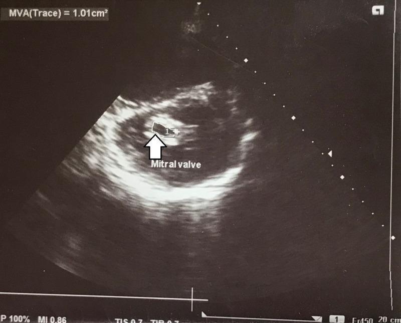
This diagnostic image is a transthoracic echocardiogram (TTE) in a parasternal short-axis view at the level of the mitral valve. The image illustrates severe mitral stenosis, likely of rheumatic etiology, characterized by significantly thickened and echogenic (calcified) mitral valve leaflets. A planimetric measurement of the mitral valve orifice is demonstrated, with the 'MVA(Trace)' value displayed at the top left as 1.01 cm², which falls within the range for severe stenosis. A white arrow and the label 'Mitral valve' highlight the orifice, showing the restricted opening during diastole (the 'fish-mouth' appearance). The image serves as an educational example of cardiac ultrasonography used to quantify valvular disease through direct planimetry. It provides clinical relevance for assessing patients with suspected rheumatic heart disease and correlating hemodynamic data with structural pathology.
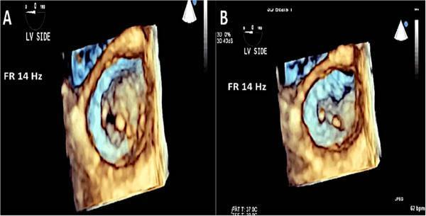
This diagnostic image consists of two panels (A and B) featuring 3D transesophageal echocardiography (TEE) of the mitral valve, captured from the left ventricular (LV) perspective. The imaging demonstrates classic features of rheumatic mitral disease. Panel A shows the mitral valve in diastole, where the orifice appears relatively open and rounded, yet displays thickened leaflet margins. Panel B shows the valve in systole, demonstrating restricted leaflet motion and a characteristic 'fish mouth' or slit-like configuration of the valve orifice, which is typical of commissural fusion and rheumatic remodeling. Technical annotations indicate a frame rate (FR) of 14 Hz, a heart rate of 62 bpm, and a patient temperature (PAT T) of 37.0°C. This comparison illustrates the transition between diastolic opening and systolic restricted closure, highlighting the structural pathology that leads to both mitral stenosis and regurgitation in rheumatic heart disease.
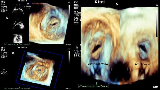
This diagnostic image set consists of 3D transthoracic echocardiography (TTE) frames showcasing severe mitral valve stenosis in a patient with Rheumatic Heart Disease (RHD). Panel A provides an atrial view with corresponding 2D reference planes. Panel B displays a volumetric 3D zoom of the mitral apparatus. Panel C provides a side-by-side comparison of the stenotic valve from the ventricular (left) and atrial (right) perspectives. The imaging demonstrates classic morphological hallmarks of rheumatic mitral stenosis, including significant thickening and calcification of the valve leaflets and commissural fusion. The valvular orifice is markedly reduced, exhibiting the characteristic 'fish-mouth' or 'buttonhole' appearance. The 3D reconstructions highlight the funnel-shaped deformity of the mitral apparatus and the restricted mobility of the fused cusps. These views are essential for assessing valvular area, subvalvular involvement (such as chordae retraction), and suitability for percutaneous mitral balloon valvuloplasty or surgical intervention.
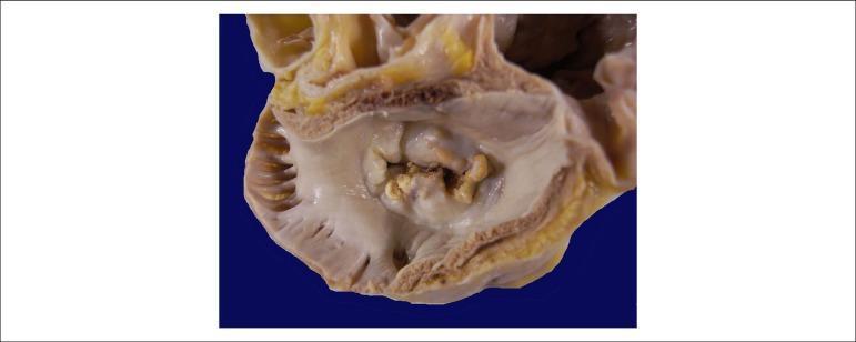
This clinical photograph shows a gross pathological specimen of a human heart, specifically an atrial view of the mitral valve. The image demonstrates hallmark signs of chronic rheumatic heart disease. Key features include significant thickening and fibrosis of the valve leaflets, resulting in a 'fish-mouth' or 'buttonhole' appearance of the valvular orifice. There is evidence of extensive commissural fusion and multiple focal areas of yellowish-white calcification along the leaflet margins and within the valve body. These structural changes characterize severe mitral stenosis, where the restricted opening of the valve impairs blood flow from the left atrium to the left ventricle. The surrounding atrial endocardium appears thickened, consistent with chronic pressure overload. This visual is highly representative for educational purposes in cardiology and pathology to illustrate the long-term sequelae of rheumatic fever on cardiac valves.
infective endocarditis vegetations on heart valve
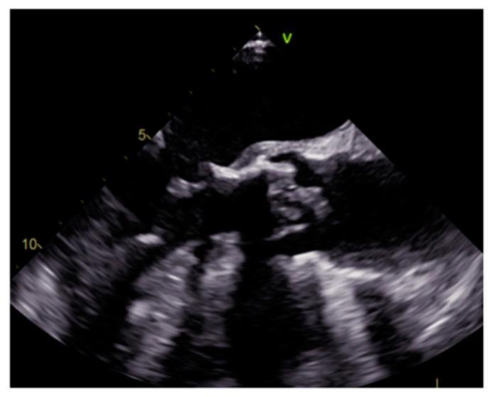
This diagnostic image is a transesophageal echocardiography (TEE) still frame demonstrating prosthetic aortic valve infective endocarditis. The visual focus is on the aortic valve region, where irregular, heterogeneously echogenic masses are visible, representing vegetations. These lesions exhibit lobulated contours and poorly defined borders, appearing more echogenic than the adjacent cardiac tissue. The vegetations are attached to the prosthetic valve leaflets or the perivalvular area, a hallmark of endocarditis in a prosthetic setting. The image is optimized for teaching the diagnostic criteria of infective endocarditis, specifically illustrating the morphology, echogenicity, and typical location of vegetations. The ultrasound highlights the contrast between the smooth, highly echogenic prosthetic material and the irregular, mobile-appearing infectious growths. This content is intended for advanced cardiology and radiology learners studying valvular heart disease and its complications.
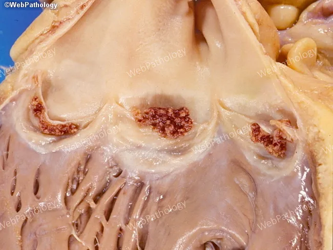
This is a gross pathology photograph of a heart specimen focusing on the right heart outflow tract and pulmonary valve region. The image demonstrates infective endocarditis with visible vegetations adherent to the pulmonic valve leaflets. Vegetations appear irregular, friable, and reddish-brown, measuring small to moderate size along the cusps; surface roughness and protrusions suggest valvular destruction risk. The pulmonary valve leaflets show nodular, irregular thickening with associated endocardial involvement; underlying cusp tissue may be eroded at the base. This right‑sided involvement is characteristic of intravenous drug use with Staphylococcus aureus bacteremia, and carries potential for septic emboli to the lungs. Associated findings may include mild dilation of the right ventricular outflow tract and adjacent endocardial surface. The image is an ex vivo photograph acquired under standard laboratory illumination; no histological staining or contrast enhancement was used. In clinical context, these gross features support a diagnosis of acute infective endocarditis of the pulmonary valve, with high risk of hemodynamic compromise if untreated. Differential considerations include nonbacterial thrombotic endocarditis and carcinoid valvulopathy, though friable infectious vegetations favor bacterial endocarditis. This finding underscores the need for urgent antimicrobial therapy and evaluation for surgical valve replacement in severe cases, especially with ongoing bacteremia.
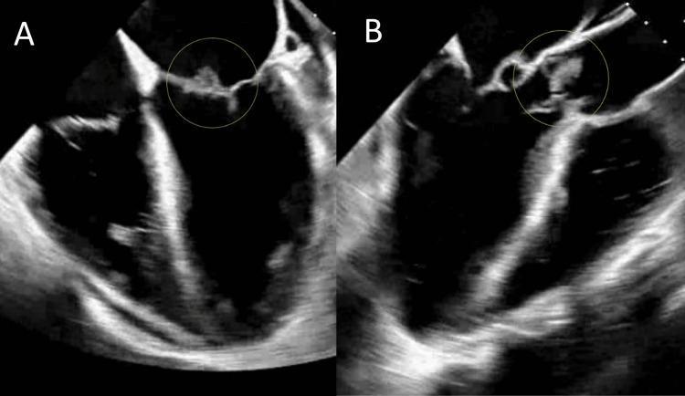
Two side-by-side transesophageal echocardiogram (TEE) images, labeled A and B, demonstrating infective endocarditis with valvular vegetations. Image A shows a grayscale ultrasound view of the mitral valve, with a yellow circle highlighting an irregular, heterogeneously echogenic mass (vegetation) attached to the mitral leaflet. Image B displays the aortic valve region, where a similar yellow circle indicates a lobulated, hyperechoic vegetation adherent to the aortic valve leaflets. Both structures exhibit typical characteristics of cardiac vegetations, including surface irregularities and moderate-to-high echogenicity relative to the myocardium. The surrounding valvular tissue in image B appears more disrupted, consistent with advanced infectious involvement. These diagnostic images are used to illustrate the visual manifestations of infective endocarditis, specifically the identification of vegetations on different heart valves, which are critical for confirming a clinical diagnosis of sepsis secondary to bacteremia.
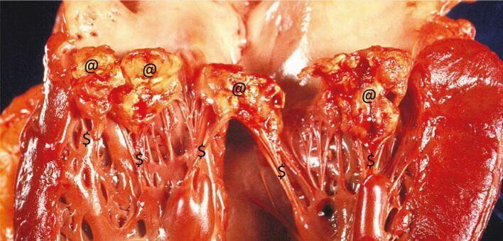
This clinical photograph displays a gross pathological view of a human heart specimen affected by infective endocarditis (IE). The primary focus is the mitral valve apparatus, which exhibits significant structural damage. Large, friable, cauliflower-like vegetations (marked with '@') are visible on the atrial surface and free margins of the valve leaflets. These vegetations appear as irregular, yellowish-tan and reddish masses composed of fibrin, platelets, and bacterial colonies. The underlying mitral valve leaflets show evidence of chronic scarring and acute destruction, including visible perforations and tissue loss. Additionally, the chordae tendineae (marked with '$') are thickened, inflamed, and scarred, which contributes to valvular incompetence. This image illustrates the severe valvular compromise—including obstruction and regurgitation—that occurs in advanced infective endocarditis, often necessitating surgical intervention.
"rheumatic heart disease" AND prevention
Rheumatic Heart Disease and Infective Endocarditis
Rheumatic Heart Disease
- Aschoff bodies (pathognomonic): foci of fibrinoid necrosis surrounded by lymphocytes, plasma cells, and activated macrophages called Anitschkow cells ("caterpillar cells," with slender wavy nuclear chromatin).
- Acute phase: small, warty vegetations (verrucae) along the lines of valve closure; fibrinous pericarditis; interstitial myocarditis.
- Chronic phase: leaflet thickening and fibrosis, commissural fusion, and shortening/fusion of the chordae tendineae - producing the classic "fish-mouth" or "buttonhole" mitral stenosis. Rheumatic heart disease is essentially the only cause of acquired mitral stenosis. The mitral valve is affected in nearly all cases, often together with the aortic valve.
Infective Endocarditis
- Underlying valve abnormalities predisposing to IE: rheumatic heart disease, mitral valve prolapse, bicuspid aortic valve, calcific valvular stenosis. As RHD has declined, mitral valve prolapse has become the leading predisposing lesion in higher-income countries.
- Prosthetic valves account for 10-20% of all IE cases.
- Other risk factors: neutropenia, immunodeficiency, malignancy, diabetes, alcohol/IV drug use, indwelling catheters/pacemaker lines.
- Microbiology: the three most common causes worldwide are staphylococci, streptococci, and enterococci.
- Community-acquired subacute IE (on damaged/deformed valves): 50-60% Streptococcus viridans.
- Healthcare-associated and IV drug use-related acute IE: Staphylococcus aureus - now the leading overall cause in higher-income countries, can attack even normal valves.
- HACEK organisms (Haemophilus, Actinobacillus, Cardiobacterium, Eikenella, Kingella) and fungi are less common causes.
- ~10% of cases are "culture-negative" (prior antibiotics or fastidious organisms).
- Fever is the most consistent finding, though it may be absent in subacute disease in older adults (presenting instead with fatigue, weight loss, flu-like symptoms; splenomegaly common).
- Acute disease: rapid fever, chills, lassitude.
- Murmurs present in ~90% of left-sided lesions.
- Classic embolic/immunologic phenomena: petechiae, splinter hemorrhages, Roth spots (retinal hemorrhages), Janeway lesions (painless palm/sole lesions), Osler nodes (painful fingertip nodules).
- Diagnosis confirmed by positive blood cultures plus echocardiographic evidence of vegetations (modified Duke criteria in clinical practice).
The connection between the two
Recent evidence to be aware of
Rheumatic heart disease nd endocarditis give in point vise notes
Rheumatic Heart Disease & Infective Endocarditis - Quick Notes
RHEUMATIC HEART DISEASE (RHD)
- Cardiac manifestation of rheumatic fever
- Rheumatic fever = acute, immunologically-mediated, multisystem inflammatory disease after Group A β-hemolytic streptococcal infection (pharyngitis mainly, occasionally skin)
- Immune cross-reactivity ("molecular mimicry") between strep M protein and host cardiac antigens
- T cells + antibodies attack myocardium/valves
- Antibody injury via complement activation + Fc-receptor macrophages
- Cytokines from T cells activate macrophages -> Aschoff bodies
- Only 1-3% of infected people develop rheumatic fever -> genetic susceptibility
- Symptom onset delayed 2-3 weeks post-infection (time for immune response; strep no longer present in lesion)
- Aschoff bodies (pathognomonic): fibrinoid necrosis + lymphocytes + plasma cells + Anitschkow ("caterpillar") cells
- Pancarditis - affects all 3 layers:
- Pericardium: fibrinous exudate, resolves without sequelae
- Myocardium: myocarditis, scattered Aschoff bodies
- Endocardium/valves: fibrinoid necrosis + small warty vegetations (verrucae) along closure lines
- Chronic changes: leaflet thickening/fibrosis, commissural fusion, chordae shortening/fusion
- Classic result: "fish-mouth" / "buttonhole" mitral stenosis
- RHD = essentially the ONLY cause of acquired mitral stenosis
- Mitral valve most commonly involved; aortic valve often co-involved
- Carditis worsens cumulatively with recurrent attacks
- Chronic RHD may stay silent for years-decades after acute episode
- Murmurs, cardiac hypertrophy/dilation, CHF
- Arrhythmias - especially atrial fibrillation (with mitral stenosis)
- Thromboembolism from atrial mural thrombi
- Scarred valves -> increased susceptibility to infective endocarditis
- Declining in high-income countries (better hygiene, prompt antibiotic Rx, reduced strain virulence)
- Still leading cause of acquired heart disease in children/young adults in low-income countries
- Mitral valvuloplasty / valve replacement improves outcomes
- Secondary prophylaxis (penicillin) to prevent recurrence
- WHO-backed echo screening programs in at-risk populations
INFECTIVE ENDOCARDITIS (IE)
- Microbial infection of heart valves/endocardium
- Vegetations = thrombotic debris + organisms, often with tissue destruction
- Acute: rapid, destructive, high morbidity/mortality even with treatment
- Subacute: insidious, protracted (weeks-months), usually responds to antibiotics
- Spectrum overlap common; clear distinction not always possible
- Underlying valve disease: RHD, mitral valve prolapse, bicuspid aortic valve, calcific stenosis
- Mitral valve prolapse now leading risk factor (as RHD declines in rich countries)
- Prosthetic valves: 10-20% of all IE cases
- Host factors: neutropenia, immunodeficiency, malignancy, diabetes, alcohol/IV drug use
- Portals of bacteremia: dental/surgical procedures, IV drug injection, occult GI/oral source, catheters/pacemaker lines
- Top 3 causes overall: staphylococci, streptococci, enterococci
- Community-acquired subacute IE: Streptococcus viridans (50-60%) - on already-damaged valves
- Acute/healthcare-associated/IV drug use IE: Staphylococcus aureus - attacks even normal valves, most common cause in high-income countries
- HACEK group (Haemophilus, Actinobacillus, Cardiobacterium, Eikenella, Kingella) - less common
- ~10% culture-negative (prior antibiotics, fastidious organisms)
- Friable, bulky, destructive vegetations (fibrin + inflammatory cells + microbes)
- Aortic & mitral valves most common sites; tricuspid in IV drug users
- Can erode into myocardium -> ring abscess
- Emboli shedding -> septic infarcts, mycotic aneurysms
- Fever = most consistent sign (may be absent in subacute elderly patients)
- Subacute: fatigue, weight loss, flu-like illness, splenomegaly
- Acute: rapid fever, chills, lassitude
- Murmurs in ~90% with left-sided lesions
- Immune/embolic phenomena:
- Petechiae
- Splinter hemorrhages (nail bed)
- Roth spots (retinal hemorrhage)
- Janeway lesions (painless palm/sole)
- Osler nodes (painful fingertip nodules)
- Positive blood cultures + echocardiographic vegetations (modified Duke criteria in practice)
- Immune complex glomerulonephritis (hematuria, albuminuria, renal failure)
- Arrhythmias (myocardial extension)
- Systemic embolization
- Septicemia
- Untreated -> generally fatal; antibiotics ± valve replacement reduce mortality
LINK BETWEEN RHD AND IE
- Rheumatic scarring creates turbulent flow + endothelial damage -> ideal substrate for bacterial seeding during transient bacteremia
- Historic basis for endocarditis prophylaxis in RHD patients
- New/worsening valve dysfunction in a known RHD patient should raise suspicion for superimposed IE
- 2024 systematic review, JAHA (PMID 38390809): preventive interventions (primary/secondary prophylaxis, echo screening) to reduce RHD burden
- 2024 systematic review, Lancet Global Health (PMID 38762298): handheld echo screening for RHD informing WHO guidelines
- Both support secondary penicillin prophylaxis + echo screening as key strategies in low/middle-income settings
Rheumatic heart disease nd endocarditis give in point vise notes bpt level
physiotherapy management precautions rheumatic heart disease infective endocarditis exercise
Rheumatic Heart Disease & Infective Endocarditis - BPT Level Notes
A. RHEUMATIC HEART DISEASE (RHD)
- Chronic heart damage following rheumatic fever (RF)
- RF = autoimmune disease occurring 2-3 weeks after Group A β-hemolytic streptococcal throat infection
- Untreated/inadequately treated strep throat infection
- Body's antibodies against strep cross-react with heart tissue (molecular mimicry)
- Aschoff bodies form in heart tissue (diagnostic feature)
- Repeated attacks -> valve leaflets thicken, scar, and fuse
- Mitral valve most commonly damaged -> mitral stenosis ("fish-mouth" valve)
- Aortic valve second most common
- History of sore throat/fever in childhood
- Joint pain (migratory polyarthritis) - in acute stage
- Breathlessness, easy fatigue
- Palpitations (irregular heartbeat - AF common)
- Chest discomfort
- Swelling of feet (if heart failure develops)
- Murmur on auscultation
- Congestive heart failure
- Atrial fibrillation
- Blood clots -> stroke (embolism)
- Infective endocarditis (secondary infection on damaged valve)
- Antibiotics (penicillin) - treat and prevent recurrence
- Anti-inflammatory drugs (aspirin/NSAIDs) in acute stage
- Diuretics/heart failure medicines if needed
- Surgery: valve repair or replacement in severe cases
- Acute stage: bed rest, avoid exertion (active carditis = exercise contraindicated)
- Chronic stable stage: graded aerobic exercise (walking, cycling) to improve endurance
- Breathing exercises if breathlessness/heart failure present
- Monitor vitals (HR, BP, SpO2) before/during/after exercise
- Educate patient on activity pacing and energy conservation
- Post valve-surgery: chest physiotherapy, early mobilization, sternal precautions
- Avoid vigorous/contact sports if significant valve damage present
B. INFECTIVE ENDOCARDITIS (IE)
- Infection of the inner lining of the heart (endocardium) and heart valves by microorganisms (mostly bacteria)
- Damaged/abnormal valves (RHD, prosthetic valves, mitral valve prolapse)
- IV drug users
- Recent dental/surgical procedures (source of bacteria entering blood)
- Streptococcus viridans - common in damaged valves (slow, subacute IE)
- Staphylococcus aureus - common in IV drug users/healthcare settings (rapid, acute IE)
- Fever (most common and constant sign)
- Chills, weakness, weight loss
- New or changing heart murmur
- Petechiae (small skin spots)
- Splinter hemorrhages (under nails)
- Osler nodes (painful finger/toe nodules)
- Janeway lesions (painless palm/sole spots)
- Roth spots (on retina)
- Enlarged spleen
- Valve destruction -> heart failure
- Emboli -> stroke, organ infarcts
- Kidney damage (glomerulonephritis)
- Can be fatal if untreated
- Blood culture (positive for organism)
- Echocardiography (shows vegetations)
- Prolonged IV antibiotics (weeks)
- Surgery (valve replacement) in severe/destructive cases
- Acute febrile phase: rest, NO active exercise/mobilization until medically stable
- Avoid chest physiotherapy techniques that could dislodge vegetations/emboli during active infection
- Once stable and cleared by physician: gradual reconditioning, breathing exercises, low-intensity aerobic program
- Antibiotic prophylaxis before dental/invasive procedures for high-risk patients (as per physician/AHA advice) - physiotherapists should be aware when planning any procedure with tissue trauma
- Post-cardiac surgery rehab: same principles as valve surgery (early mobilization, chest physio, sternal precautions)
Quick Comparison Table
| Feature | RHD | Infective Endocarditis |
|---|---|---|
| Cause | Autoimmune reaction to strep infection | Direct microbial infection of valve/endocardium |
| Onset | Weeks after strep throat, chronic course | Can be acute (rapid) or subacute (slow) |
| Valve affected most | Mitral (stenosis) | Any valve, often already-damaged one |
| Key sign | Murmur + history of sore throat/joint pain | Fever + new murmur + embolic skin signs |
| Link | Damaged valve from RHD is a risk factor for IE | Occurs on top of RHD-damaged valves |